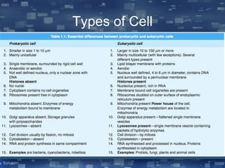
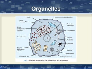
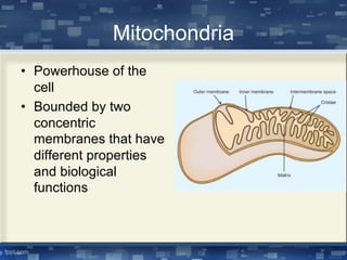
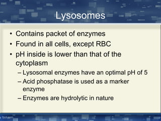
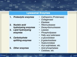
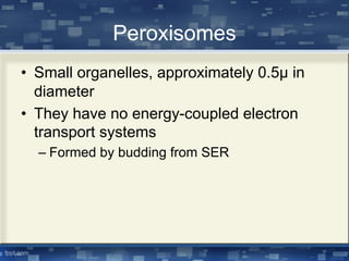
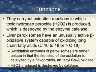
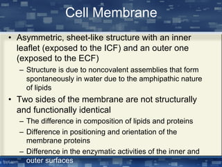
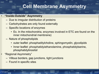
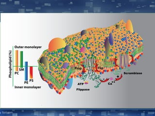
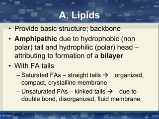
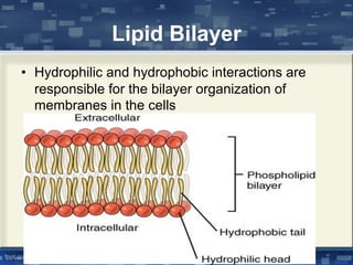
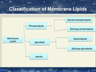
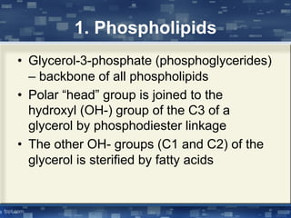
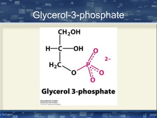
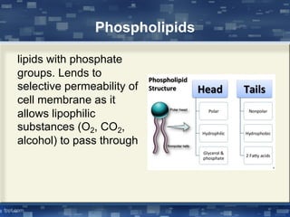
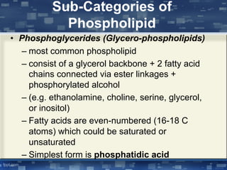
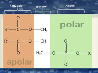
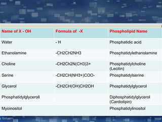
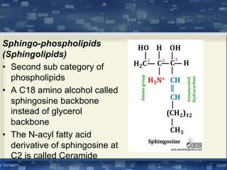
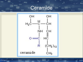
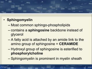
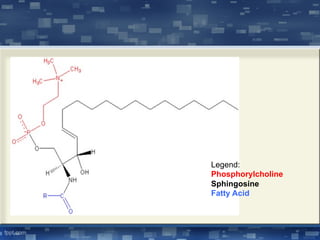
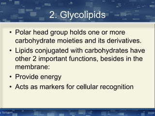
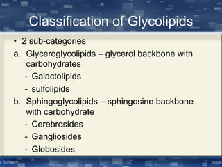
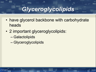
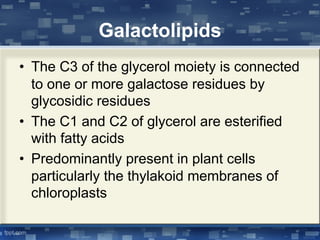
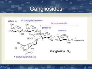
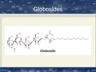
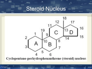
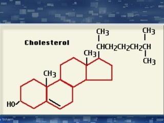
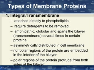
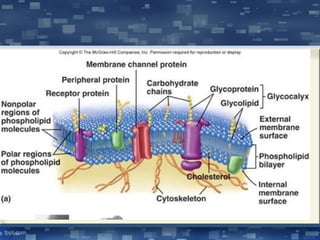
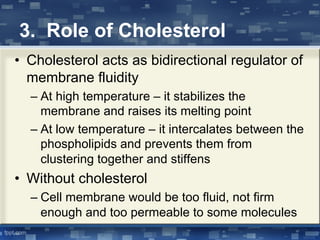
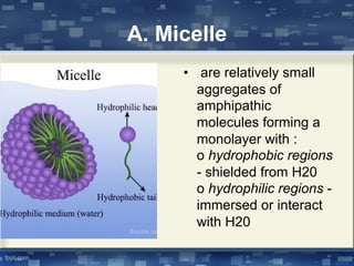
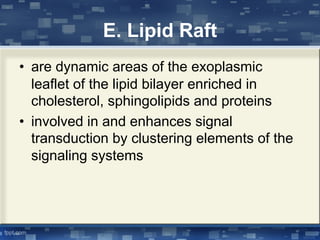
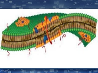
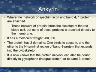
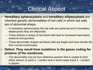
Cell Membrane
Alissa Laurel N. Calderon, MD, FPAFP, MMHA
Cell
• Fundamental unit of life
• Cells make up all living matter
• All cells arise from other cells
• The chemical reactions of an organism ,
both anabolism and catabolism, takes
place in the cell
Types of Cell
Organelles
Nucleus
• Contains more than 95% of the cell’s DNA
• Control center of the cell
• Where DNA replication and RNA
transcription of DNA occur
Parts of a Nucleus
• Nuclear envelope – double membrane structure
that separates the nucleus from the cytoplasm
• Nuclear pore complexes – these are embedded
in the nuclear envelope; control the movement
of proteins and RNAs across the nuclear
envelope
• Nucleoplasm – contains various enzymes such
as DNA polymerases and RNA polymerases for
mRNA and tRNA synthesis
Parts of a Nucleus
• Chromatin – made up of coiled DNA;
stained darkly with certain dyes
• Nucleolus – second dense mass closely
associated with the inner nuclear envelope
– Nonmembranous; contains RNA polymerase,
ATPase, but no DNA polymerase
– Site of synthesis of ribosomal RNA (rRNA)
– Major site where ribosome subunits are
assembled
Mitochondria
• Powerhouse of the
cell
• Bounded by two
concentric
membranes that have
different properties
and biological
functions
Mitochondrial Membrane
• Outer mitochondrial membrane – consists
mostly of phospholipids and cholesterol;
also contains protein porin
– Porin – proteins that form channels that
permits substances with MW <10,000 to
diffuse freely across the mitochondrial
membrane
Mitochondrial Membrane
• Inner mitochondrial membrane
– Very rich in proteins
– Contains high proportion of the phospholipids
cardiolipin
– Impermeable to polar and ionic substances
(as opposed to outer membrane)
– Cristae – inner mitochondrial membrane is
highly folded; tightly packed inward folds are
called cristae
• Mitochondrial matrix – region enclosed by
the inner membrane
– Where enzymes responsible for citric acid
cycle and fatty acid oxidation are located
Endoplasmic Reticulum
• Extends from the cell membrane, coats
the nucleus, surrounds the mitochondria
and appears to connect directly to the
Golgi apparatus
Kinds of ER
• Rough ER – coated with ribosomes; near
the nucleus, it merges with outer
membrane of the nuclear envelope;
synthesizes membrane lipids and
secretory proteins
• Smooth ER – do not have attached
ribosomes; responsible with lipid
synthesis, and modifies and transports
proteins synthesized by RER
Golgi Apparatus
• Unique stack of smooth surfaced compartments
or cisterna
• It is usually closely associated with ER
• Has proximal or cis compartment, a medial
compartment and a distal or trans compartment
• Serves as a unique sorting device that receives
newly synthesized proteins, all containing signal
or transit peptides from the ER
– Those proteins with no signal or transit peptides
regions are rejected by the golgi apparatus without
processing further
– Remains as cytoplasmic protein
Functions
• Proximal or cis side – receives the newly
synthesized proteins by the ER via transfer
vesicles
• Median part – where posttranslational
modification take place
– Where the carbohydrates and lipid precursors
are added to proteins to form glycoprotein and
lipoproteins respectively
• Distal or trans side – release proteins via
modified membranes called secretory vesicles
Lysosomes
• Contains packet of enzymes
• Found in all cells, except RBC
• pH inside is lower than that of the
cytoplasm
– Lysosomal enzymes have an optimal pH of 5
– Acid phosphatase is used as a marker
enzyme
– Enzymes are hydrolytic in nature
Peroxisomes
• Small organelles, approximately 0.5μ in
diameter
• They have no energy-coupled electron
transport systems
– Formed by budding from SER
Functions
• They carryout oxidation reactions in which
toxic hydrogen peroxide (H2O2) is produced,
which is destroyed by the enzyme catalase.
• Liver peroxisomes have an unusually active β-
oxidative system capable of oxidizing long
chain fatty acids (C 16 to 18 or > C 18)
– β-oxidation enzymes of peroxisomes are rather
unique in that the first step of the oxidation is
catalyzed by a flavoprotein, an “acyl Co-A oxidase”
– H2O2 produced is destroyed by catalase.
Biological Membrane system
• Highly dynamic two layers thick sheath like
structures formed by lipids, proteins and
carbohydrates
• Serves as closed boundaries between
different compartments of the cells
• Nuclear membrane: nucleoplasm from
cytoplasm
• Act as barriers to the passage of polar
molecules and ions
Cell Membrane
• Exists in viscous—gel-fluid like—and plastic
structures (ex. RBCs).
• Dynamic—exhibits rapid turnover and lateral
diffusion
• Has a thermodynamically stable and
metabolically active arrangement
• Composed of lipids, proteins and carbohydrates
Functions of Cell Membrane
• Selective barrier – aided by carriers and
channels, allowing exchange between the
cell and the environment
• Permits cell individuality – separates cell
from other cells
• Cell-to-cell interaction – due to hormone-
receptor interactions
• Cell adhesion to basement membrane and
other cells
Functions of Cell Membrane
• Transmembrane signaling – signal
transduction mechanism
• Compartmentalization
• Localize enzymes
• Excitation-response coupling
• Site for energy transduction
Cell Membrane
• Asymmetric, sheet-like structure with an inner
leaflet (exposed to the ICF) and an outer one
(exposed to the ECF)
– Structure is due to noncovalent assemblies that form
spontaneously in water due to the amphipathic nature
of lipids
• Two sides of the membrane are not structurally
and functionally identical
– The difference in composition of lipids and proteins
– Difference in positioning and orientation of the
membrane proteins
– Difference in the enzymatic activities of the inner and
outer surfaces
Cell Membrane Asymmetry
• “Inside-Outside” Asymmetry
– Due to irregular distribution of proteins
– Carbohydrates are only found externally
– Specific locations of enzymes
• Ex. In the mitochondria, enzymes involved in ETC are found on the
inner mitochondrial membrane)
– Nature of phospholipids
• outer leaflet: phosphaditylcholine, sphingomyelin, glycolipids
• Inner leaflet: phosphatidyiethanolamine, phosphatidylserine,
phosphatidylinositol
• “Regional Asymmetry”
– Villous borders, gap junctions, tight junctions
– Found in specific sites
Significance
• Changes in phospholipid asymmetry is
involved in various cell functions: cell
polarity, membrane trafficking and
organelle functions
• Phospholipid asymmetry is generated,
maintained and regulated by lipid
translocation or “flip-flop”
Composition of Cell
Membrane
• Composed of lipids, proteins and
carbohydrates
– 40% - lipids
– 60% - proteins
– 1-10% - carbohydrates
• All membrane carbohydrate is covalently
attached to proteins or lipids
A. Lipids
• Provide basic structure; backbone
• Amphipathic due to hydrophobic (non
polar) tail and hydrophilic (polar) head –
attributing to formation of a bilayer
• With FA tails
– Saturated FAs – straight tails à organized,
compact, crystalline membrane
– Unsaturated FAs – kinked tails à due to
double bond, disorganized, fluid membrane
Lipid Bilayer
• Hydrophilic and hydrophobic interactions are
responsible for the bilayer organization of
membranes in the cells
Classification of Membrane Lipids
Membrane
Lipids
Phospholipids
glycolipid
sterols
Glycero-phospholipids
Sphingo-phopholipids
Galactolipids
Sphingo-glycolipids
1. Phospholipids
• Glycerol-3-phosphate (phosphoglycerides)
– backbone of all phospholipids
• Polar “head” group is joined to the
hydroxyl (OH-) group of the C3 of a
glycerol by phosphodiester linkage
• The other OH- groups (C1 and C2) of the
glycerol is sterified by fatty acids
Glycerol-3-phosphate
Phospholipids
lipids with phosphate
groups. Lends to
selective permeability of
cell membrane as it
allows lipophilic
substances (O2, CO2,
alcohol) to pass through
Sub-Categories of
Phospholipid
• Phosphoglycerides (Glycero-phospholipids)
– most common phospholipid
– consist of a glycerol backbone + 2 fatty acid
chains connected via ester linkages +
phosphorylated alcohol
– (e.g. ethanolamine, choline, serine, glycerol,
or inositol)
– Fatty acids are even-numbered (16-18 C
atoms) which could be saturated or
unsaturated
– Simplest form is phosphatidic acid
Fatty acid glycerol Alcohol
Name of X - OH Formula of -X Phospholipid Name
Water - H Phosphatidic acid
Ethanolamine -CH2CH2NH3 Phosphatidylethanolamine
Choline -CH2Ch2N(CH3)3+ Phosphatidylcholine
(Lecitin)
Serine -CH2CH(NH3+)COO- Phosphatidylserine
Glycerol -CH2CH(OH)CH2OH Phosphatidylglycerol
Phosphatidylglyceroll Diphosphatidylglycerol
(Cardiolipin)
Myoinositol Phosphatidylinositol
Cardiolipin
• Complex glycero-phospholipid
• polar head (-X) is a phosphatidylglycerol
thus structurally, a diphosphatidylglycerol
• An important component of the inner
mitochondrial membrane – constitutes
about 20%
• Lipid marker of the inner mitochondrial
membrane (not found in outer
mitochondrial membrane)
Cardiolipin
Plasmalogen
• C1 of glycerol moiety is linked to fatty acid
via an α,β-unsaturated ether linkage rather
than an ester linkage
• Present in abundance in neurons and cells
of cardiovascular system
Plasmalogen
Sphingo-phospholipids
(Sphingolipids)
• Second sub category of
phospholipids
• A C18 amino alcohol called
sphingosine backbone
instead of glycerol
backbone
• The N-acyl fatty acid
derivative of sphingosine at
C2 is called Ceramide
Ceramide
• Sphingomyelin
– Most common sphingo-phospholipids
– contains a sphingosine backbone instead of
glycerol
– A fatty acid is attached by an amide link to the
amino group of sphingosine = CERAMIDE
– Hydroxyl group of sphingosine is esterified to
phosphorylcholine
– Sphingomyelin is prominent in myelin sheath
Legend:
Phosphorylcholine
Sphingosine
Fatty Acid
2. Glycolipids
• Polar head group holds one or more
carbohydrate moieties and its derivatives.
• Lipids conjugated with carbohydrates have
other 2 important functions, besides in the
membrane:
• Provide energy
• Acts as markers for cellular recognition
Classification of Glycolipids
• 2 sub-categories
a. Glyceroglycolipids – glycerol backbone with
carbohydrates
- Galactolipids
- sulfolipids
b. Sphingoglycolipids – sphingosine backbone
with carbohydrate
- Cerebrosides
- Gangliosides
- Globosides
Glyceroglycolipids
• have glycerol backbone with carbohydrate
heads
• 2 important glyceroglycolipids:
– Galactolipids
– Glyceroglycolipids
Galactolipids
• The C3 of the glycerol moiety is connected
to one or more galactose residues by
glycosidic residues
• The C1 and C2 of glycerol are esterified
with fatty acids
• Predominantly present in plant cells
particularly the thylakoid membranes of
chloroplasts
Sulfolipids
• Membrane glycolipids with sulfur
containing functional groups
• Sulfonated glucose is joined to the C3 of
diacylglycerol in glycosidic linkage
Sphingoglycolipids
• sugar attached to ceramide backbone
rather than glycerol moieties
• found in nerve tissue
a. Cerebrosides
b. Gangliosides
c. Globosides
Cerebrosides
• A ceramide with single sugar residue at
the C1-hydroxyl group
• Sugar residue is either glucose or
galactose
• Lack the phosphate group thus they are
non-ionic (no charge)
• Abundant in cell membranes of nerves
and muscles of animals
– Nerve cells – rich in galactocerebrosides
Cerebrosides
Gangliosides
• Membrane lipid with most complex
structure
• It is a glyco-sphingolipid with many
carbohydrate moieties and one or more
sialic acids linked on the sugar chain
• About 6% of brain lipids are gangliosides
and they are first isolated from the
ganglion of brain cells
– Also abundant in the lipid rafts of plasma
membrane
Gangliosides
Globosides
• With one or more than one sugar as the
side chain of a ceramide
• Differ from gangliosides since they lack
the sialic acid residues
• The carbohydrate moieties are usually a
combination of N-acetyl-galactosamine,D-
glucose or D-galactose
Globosides
3. Sterols
• Third major category of membrane lipids, usually present
in eukaryotoc membranes
• Structurally consist of four fused carbon rings (A, B, C,
D) and a hydrooarbon chain (alkyl side chain)
• Rings A, B, C are 6-carbon rings; D is 5-C structure
• This fused ring structure is called the steroid nucleus
• Cyclopentanoperhydrophenanthrene
• One of its peculiarities of carbon in fused ring
structure – does not allow C-C free rotation even
though they are in single bonds
Steroid Nucleus
Cholesterol
• Most common sterol and intercalates with membrane
phospholipids
• 27-C atom with 4-rings conferring rigidity
• Amphipathic with single polar hydroxyl (-OH) ‘head’ and
non-polar hydrocarbon ‘tail’
• The –OH group and hydrocarbon chain are attached to
the C3 and C17 of steroid nucleus
• Can esterify long chain fatty acids to to form cholesterol
esters such as cholesteryl stearate
• “moderator molecule” moderates
membrane fluidity
Tm – transition temperature; temperature at
which cell membrane become
disorganized;structure undergoes from
ordered to disordered state
B. Proteins
• Amphipathic structures
• Determines membrane function
• Act as pumps, channels, carriers,
receptors, enzymes, structural
components, antigens
MEMBRANE PROTEIN
Membrane proteins have various functions:
1. Transporters
2. Enzymes
3. Cell surface receptors
4. Cell surface identity markers
5. Cell-to-cell adhesion proteins
6. Attachments to the cytoskeleton
Types of Membrane Proteins
1. Integral/Transmembrane
– attached directly to phospholipids
– require detergents to be removed
– amphipathic, globular and spans the bilayer
(transmembrane) several times in certain
proteins
– asymmetrically distributed in cell membrane
– nonpolar regions of the protein are embedded
in the interior of the bilayer
– polar regions of the protein protrude from both
sides of the bilayer
2. Peripheral
- do not interact directly with phospholipids
- attached to integral proteins
- usually found inside the cell
- Are free to move throughout one layer of
the bilayer
- Possess nonpolar regions that are
inserted in the lipid bilayer
C. Carbohydrates
• occur in association with lipids or proteins :
glycolipids or glycoproteins
• mostly found on the external membrane
surface
• functions :
o receptors
o antigens
o confers negative charge to cell
(glycocalyx)
Fluid-Mosaic Model
(Singer & Nicholson)
• universally accepted description of
membrane structure
• “icebergs” (proteins) floating in a “sea” of
phospholipids
• membranes undergo phasic changes from
stiff (gel or crystalline) to fluid state
• both lipids and proteins undergo "rapid
redistribution" in the plane of the
membrane ("lateral diffusion")
Factors Affecting Membrane
Fluidity
1. Lipid composition
- longer and more saturated fatty acid
chains exhibit higher transition temperature
- unsaturated cis bonds tend to increase
membrane fluidity
- presence of cholesterol the moderator
molecule
2. Temperature
• At low temperature – fluidity is less
• As temperature increases –increase fluidity
• The hydrophobic side chains undergo a
transition from ordered state to a disordered
state
• Transition Temperature (Tm) - temperature
at which structure undergoes transition from
ordered to disordered state
3. Role of Cholesterol
• Cholesterol acts as bidirectional regulator of
membrane fluidity
– At high temperature – it stabilizes the
membrane and raises its melting point
– At low temperature – it intercalates between the
phospholipids and prevents them from
clustering together and stiffens
• Without cholesterol
– Cell membrane would be too fluid, not firm
enough and too permeable to some molecules
Importance of Increased
Membrane Fluidity
1. Permeability to water and other
hydrophilic molecule increases
2. Lateral mobility of integral proteins
increases*
• * especially important with proteins
involved in transport and receptor proteins
3. Increased protein diffusion – since some
proteins are internalized, allows for faster
appearance
Artificial Membranes and
Other Special Membrane
Structures
A. Micelle
• are relatively small
aggregates of
amphipathic
molecules forming a
monolayer with :
o hydrophobic regions
- shielded from H20
o hydrophilic regions -
immersed or interact
with H20
• single-layer unlike cell membrane
• arrangement of different regions depends on the
chemical environment where the micelle is
situated
• used in detergents
• clinical application of micelles :
– are formed when bile acids (which are
amphipathic) associate with products of lipid
digestion
– bile acids-formed micelles assist in the
digestion and absorption of fat plus ADEK
B. Liposomes
• Vesicles surrounded with lipid bilayer
• Consist of phospholipids that are of
natural or synthetic origin
• Lipid content can be varied allowing for
examination of varying lipid composition
on certain functions (ie., transport)
• In the study of factors that affect protein
and enzyme function
• May be used for specific drug delivery and
gene therapy
C. Tight Junctions
• Located below the apical surface of
epithelial cells
• Prevents the diffusion of macromolecules
between them
• Composed of proteins occludin, claudins
• Sites of paracellular transport
• Means of attachment
Tight Junctions
• Prevents diffusion of macromolecules
• Allows paracellular transport of water (e.g.
Na+ K+ ATPase)
• Physical connection between cells
D. Gap Junctions
• Low resistance connection between cells
• More functional connection
• Made of connexons (made of connexins)
and are aligned with another cell
• Transports small ions, molecules, and
impulses
• In heart muscles, they are known as
syncytium
Gap Junctions
Desmosomes
• Junctions which act like spot welds between adjacent
epithelial cells
• Involves a complex of proteins, some extend across the
membrane while others anchor the junction within the
cell
• Pin adjacent cells together, ensuring that cells in organs
and tissues that stretch, such as skin and cardiac
muscle, remian connected in an unbroken sheet.
• Cathedrins, specialized adhesion proteins, attach to a
structure called the cytoplasmic plaque which connects
to the intermediate filaments and helps anchor the
junction
Desmosome
E. Lipid Raft
• are dynamic areas of the exoplasmic
leaflet of the lipid bilayer enriched in
cholesterol, sphingolipids and proteins
• involved in and enhances signal
transduction by clustering elements of the
signaling systems
Caveolae
• Derived from lipid rafts
• Usually contain special protein called
caveolin-1, which is involved in the
formation of lipid rafts
• Also takes part in signal transduction
Special Characteristics of Red
Cell Membrane
Integral Protein
• Two major integral proteins in RBC:
– Glycophorin
– Band-3-protein
Glycophorin
• Glycoproteins; contains 60% carbohydrates by weight
• Oligosaccharides are linked to serine, threonine and
asparagine residues
• RBC membrane contains about 6 × 10⁵ glycophorin
molecules.
• The polypeptide chain of glycophorin contains 130 amino
acid residues.
– A sequence of 23 hydrophobic amino acid residues lies within the
non- polar hydrocarbon phase of the phospholipid bilayer, tightly
associated with phospholipids and cholesterol.
– This 23 amino acid residue sequence has an α-helical
conformation.
Functions
• Some of the oligosaccharides of
glycophorin are the M and N blood group
antigens.
• Other carbohydrates bound to glycophorin
are sites through which the influenza virus
becomes attached to red blood cells.
Band-3-Protein
• Integral protein found in red cell membrane.
• It is dimeric having molecular weight of
93,000.
• The polypeptide chain of the dimer is thought
to traverse the membrane about a dozen time.
– Both the C and N terminals of band-3-protein are
on the cytosolic side of the membrane.
– The N-terminal residues extend into the cytosol
and interact with components of the cytoskeleton.
•
Functions
• plays an important role in the function of red
blood cells.
– As red blood cells flow through the capillaries of
the lungs, they exchange bicarbonate anions
(HCO3–.) produced, by the reaction of CO2 and
H2O, for chloride (Cl–) ions.
– This exchange occurs by way of a channel in
band-3-protein, which forms a Pore through the
membrane. .
Peripheral Protein
• The inner face of the red blood cells
membrane is laced with a network of proteins
called cytoskeletons that stabilizes the
membrane and is responsible for the
biconcave shape of the cells:
• The special peripheral membrane proteins
participate in this stability of red cells are:
– Spectrin
– Actin
– Ankyrin and Band 4, 1 protein.
Spectrin
• Spectrin consists of an α-chain, having
molecular weight 240,000 and a β-chain having
molecular weight 220,000.
• It is a fibrous protein in which the polypeptide
chains are thought to coil around each other to
give an α-β dimer, 100 nm long and 5 nm in
breadth.
• Spectrin dimers are linked through short chains
of actin molecules and band 4, 1 proteins to
form α2 β2 tetramers.
Actin
• In red blood cells and other nonmuscle
cells, actin is a component of the
cytoskeleton.
• An erythrocyte contains 5 × 10⁵ actin
molecules.
– About 20 actin molecules polymerise to form
short actin filaments.
Ankyrin
• Where the network of spectrin, actin and band 4, 1 protein
are attached
– These network of protein forms the skeleton of the red
blood cell, but none of these proteins is attached directly to
the membrane.
• It has a molecular weight 200,000.
• The protein has 2 domains: One binds to spectrin, and the
other to the N-terminal region of band-3-protein that extends
into the cytoskeleton.
• It is now known that the protein network can also be bound
directly to glycophorin (integral protein) or to band-3-protein.
Clinical Aspect
• Hereditary spherocytosis and hereditary elliptocytosis are
inherited genetic abnormalities of red cells in which red cells
are of abnormal shape.
– In hereditary spherocytosis the red cells are spherical and in hereditary
elliptocytosis they are ellipsoidal.
– These defects in shape of red blood cells lead to increased haemolysis,
anaemia and jaundice.
– These abnormally shaped red blood cells are fragile and have shorter life
than normal erythrocytes.
• Defect: They result from mutations in the genes coding for
proteins of the membrane.
– The abnormality may be from defective spectrin that is unable to bind
either ankyrin or band 4, 1 protein and in some cases band 4, 1 protein
is absent.
Cell Membrane Composition and Function_240417_114329.pdf

Cell Membrane Composition and Function_240417_114329.pdf

  • 1.
    Cell Membrane Alissa LaurelN. Calderon, MD, FPAFP, MMHA
  • 2.
    Cell • Fundamental unitof life • Cells make up all living matter • All cells arise from other cells • The chemical reactions of an organism , both anabolism and catabolism, takes place in the cell
  • 3.
  • 4.
  • 5.
    Nucleus • Contains morethan 95% of the cell’s DNA • Control center of the cell • Where DNA replication and RNA transcription of DNA occur
  • 6.
    Parts of aNucleus • Nuclear envelope – double membrane structure that separates the nucleus from the cytoplasm • Nuclear pore complexes – these are embedded in the nuclear envelope; control the movement of proteins and RNAs across the nuclear envelope • Nucleoplasm – contains various enzymes such as DNA polymerases and RNA polymerases for mRNA and tRNA synthesis
  • 7.
    Parts of aNucleus • Chromatin – made up of coiled DNA; stained darkly with certain dyes • Nucleolus – second dense mass closely associated with the inner nuclear envelope – Nonmembranous; contains RNA polymerase, ATPase, but no DNA polymerase – Site of synthesis of ribosomal RNA (rRNA) – Major site where ribosome subunits are assembled
  • 8.
    Mitochondria • Powerhouse ofthe cell • Bounded by two concentric membranes that have different properties and biological functions
  • 9.
    Mitochondrial Membrane • Outermitochondrial membrane – consists mostly of phospholipids and cholesterol; also contains protein porin – Porin – proteins that form channels that permits substances with MW <10,000 to diffuse freely across the mitochondrial membrane
  • 10.
    Mitochondrial Membrane • Innermitochondrial membrane – Very rich in proteins – Contains high proportion of the phospholipids cardiolipin – Impermeable to polar and ionic substances (as opposed to outer membrane) – Cristae – inner mitochondrial membrane is highly folded; tightly packed inward folds are called cristae
  • 11.
    • Mitochondrial matrix– region enclosed by the inner membrane – Where enzymes responsible for citric acid cycle and fatty acid oxidation are located
  • 13.
    Endoplasmic Reticulum • Extendsfrom the cell membrane, coats the nucleus, surrounds the mitochondria and appears to connect directly to the Golgi apparatus
  • 14.
    Kinds of ER •Rough ER – coated with ribosomes; near the nucleus, it merges with outer membrane of the nuclear envelope; synthesizes membrane lipids and secretory proteins • Smooth ER – do not have attached ribosomes; responsible with lipid synthesis, and modifies and transports proteins synthesized by RER
  • 15.
    Golgi Apparatus • Uniquestack of smooth surfaced compartments or cisterna • It is usually closely associated with ER • Has proximal or cis compartment, a medial compartment and a distal or trans compartment • Serves as a unique sorting device that receives newly synthesized proteins, all containing signal or transit peptides from the ER – Those proteins with no signal or transit peptides regions are rejected by the golgi apparatus without processing further – Remains as cytoplasmic protein
  • 16.
    Functions • Proximal orcis side – receives the newly synthesized proteins by the ER via transfer vesicles • Median part – where posttranslational modification take place – Where the carbohydrates and lipid precursors are added to proteins to form glycoprotein and lipoproteins respectively • Distal or trans side – release proteins via modified membranes called secretory vesicles
  • 17.
    Lysosomes • Contains packetof enzymes • Found in all cells, except RBC • pH inside is lower than that of the cytoplasm – Lysosomal enzymes have an optimal pH of 5 – Acid phosphatase is used as a marker enzyme – Enzymes are hydrolytic in nature
  • 19.
    Peroxisomes • Small organelles,approximately 0.5μ in diameter • They have no energy-coupled electron transport systems – Formed by budding from SER
  • 20.
    Functions • They carryoutoxidation reactions in which toxic hydrogen peroxide (H2O2) is produced, which is destroyed by the enzyme catalase. • Liver peroxisomes have an unusually active β- oxidative system capable of oxidizing long chain fatty acids (C 16 to 18 or > C 18) – β-oxidation enzymes of peroxisomes are rather unique in that the first step of the oxidation is catalyzed by a flavoprotein, an “acyl Co-A oxidase” – H2O2 produced is destroyed by catalase.
  • 21.
    Biological Membrane system •Highly dynamic two layers thick sheath like structures formed by lipids, proteins and carbohydrates • Serves as closed boundaries between different compartments of the cells • Nuclear membrane: nucleoplasm from cytoplasm • Act as barriers to the passage of polar molecules and ions
  • 22.
    Cell Membrane • Existsin viscous—gel-fluid like—and plastic structures (ex. RBCs). • Dynamic—exhibits rapid turnover and lateral diffusion • Has a thermodynamically stable and metabolically active arrangement • Composed of lipids, proteins and carbohydrates
  • 23.
    Functions of CellMembrane • Selective barrier – aided by carriers and channels, allowing exchange between the cell and the environment • Permits cell individuality – separates cell from other cells • Cell-to-cell interaction – due to hormone- receptor interactions • Cell adhesion to basement membrane and other cells
  • 24.
    Functions of CellMembrane • Transmembrane signaling – signal transduction mechanism • Compartmentalization • Localize enzymes • Excitation-response coupling • Site for energy transduction
  • 25.
    Cell Membrane • Asymmetric,sheet-like structure with an inner leaflet (exposed to the ICF) and an outer one (exposed to the ECF) – Structure is due to noncovalent assemblies that form spontaneously in water due to the amphipathic nature of lipids • Two sides of the membrane are not structurally and functionally identical – The difference in composition of lipids and proteins – Difference in positioning and orientation of the membrane proteins – Difference in the enzymatic activities of the inner and outer surfaces
  • 26.
    Cell Membrane Asymmetry •“Inside-Outside” Asymmetry – Due to irregular distribution of proteins – Carbohydrates are only found externally – Specific locations of enzymes • Ex. In the mitochondria, enzymes involved in ETC are found on the inner mitochondrial membrane) – Nature of phospholipids • outer leaflet: phosphaditylcholine, sphingomyelin, glycolipids • Inner leaflet: phosphatidyiethanolamine, phosphatidylserine, phosphatidylinositol • “Regional Asymmetry” – Villous borders, gap junctions, tight junctions – Found in specific sites
  • 28.
    Significance • Changes inphospholipid asymmetry is involved in various cell functions: cell polarity, membrane trafficking and organelle functions • Phospholipid asymmetry is generated, maintained and regulated by lipid translocation or “flip-flop”
  • 29.
  • 30.
    • Composed oflipids, proteins and carbohydrates – 40% - lipids – 60% - proteins – 1-10% - carbohydrates • All membrane carbohydrate is covalently attached to proteins or lipids
  • 32.
    A. Lipids • Providebasic structure; backbone • Amphipathic due to hydrophobic (non polar) tail and hydrophilic (polar) head – attributing to formation of a bilayer • With FA tails – Saturated FAs – straight tails à organized, compact, crystalline membrane – Unsaturated FAs – kinked tails à due to double bond, disorganized, fluid membrane
  • 33.
    Lipid Bilayer • Hydrophilicand hydrophobic interactions are responsible for the bilayer organization of membranes in the cells
  • 34.
    Classification of MembraneLipids Membrane Lipids Phospholipids glycolipid sterols Glycero-phospholipids Sphingo-phopholipids Galactolipids Sphingo-glycolipids
  • 35.
    1. Phospholipids • Glycerol-3-phosphate(phosphoglycerides) – backbone of all phospholipids • Polar “head” group is joined to the hydroxyl (OH-) group of the C3 of a glycerol by phosphodiester linkage • The other OH- groups (C1 and C2) of the glycerol is sterified by fatty acids
  • 36.
  • 37.
    Phospholipids lipids with phosphate groups.Lends to selective permeability of cell membrane as it allows lipophilic substances (O2, CO2, alcohol) to pass through
  • 38.
    Sub-Categories of Phospholipid • Phosphoglycerides(Glycero-phospholipids) – most common phospholipid – consist of a glycerol backbone + 2 fatty acid chains connected via ester linkages + phosphorylated alcohol – (e.g. ethanolamine, choline, serine, glycerol, or inositol) – Fatty acids are even-numbered (16-18 C atoms) which could be saturated or unsaturated – Simplest form is phosphatidic acid
  • 39.
  • 40.
    Name of X- OH Formula of -X Phospholipid Name Water - H Phosphatidic acid Ethanolamine -CH2CH2NH3 Phosphatidylethanolamine Choline -CH2Ch2N(CH3)3+ Phosphatidylcholine (Lecitin) Serine -CH2CH(NH3+)COO- Phosphatidylserine Glycerol -CH2CH(OH)CH2OH Phosphatidylglycerol Phosphatidylglyceroll Diphosphatidylglycerol (Cardiolipin) Myoinositol Phosphatidylinositol
  • 41.
    Cardiolipin • Complex glycero-phospholipid •polar head (-X) is a phosphatidylglycerol thus structurally, a diphosphatidylglycerol • An important component of the inner mitochondrial membrane – constitutes about 20% • Lipid marker of the inner mitochondrial membrane (not found in outer mitochondrial membrane)
  • 42.
  • 43.
    Plasmalogen • C1 ofglycerol moiety is linked to fatty acid via an α,β-unsaturated ether linkage rather than an ester linkage • Present in abundance in neurons and cells of cardiovascular system
  • 44.
  • 45.
    Sphingo-phospholipids (Sphingolipids) • Second subcategory of phospholipids • A C18 amino alcohol called sphingosine backbone instead of glycerol backbone • The N-acyl fatty acid derivative of sphingosine at C2 is called Ceramide
  • 46.
  • 47.
    • Sphingomyelin – Mostcommon sphingo-phospholipids – contains a sphingosine backbone instead of glycerol – A fatty acid is attached by an amide link to the amino group of sphingosine = CERAMIDE – Hydroxyl group of sphingosine is esterified to phosphorylcholine – Sphingomyelin is prominent in myelin sheath
  • 48.
  • 49.
    2. Glycolipids • Polarhead group holds one or more carbohydrate moieties and its derivatives. • Lipids conjugated with carbohydrates have other 2 important functions, besides in the membrane: • Provide energy • Acts as markers for cellular recognition
  • 50.
    Classification of Glycolipids •2 sub-categories a. Glyceroglycolipids – glycerol backbone with carbohydrates - Galactolipids - sulfolipids b. Sphingoglycolipids – sphingosine backbone with carbohydrate - Cerebrosides - Gangliosides - Globosides
  • 51.
    Glyceroglycolipids • have glycerolbackbone with carbohydrate heads • 2 important glyceroglycolipids: – Galactolipids – Glyceroglycolipids
  • 52.
    Galactolipids • The C3of the glycerol moiety is connected to one or more galactose residues by glycosidic residues • The C1 and C2 of glycerol are esterified with fatty acids • Predominantly present in plant cells particularly the thylakoid membranes of chloroplasts
  • 53.
    Sulfolipids • Membrane glycolipidswith sulfur containing functional groups • Sulfonated glucose is joined to the C3 of diacylglycerol in glycosidic linkage
  • 54.
    Sphingoglycolipids • sugar attachedto ceramide backbone rather than glycerol moieties • found in nerve tissue a. Cerebrosides b. Gangliosides c. Globosides
  • 55.
    Cerebrosides • A ceramidewith single sugar residue at the C1-hydroxyl group • Sugar residue is either glucose or galactose • Lack the phosphate group thus they are non-ionic (no charge) • Abundant in cell membranes of nerves and muscles of animals – Nerve cells – rich in galactocerebrosides
  • 56.
  • 57.
    Gangliosides • Membrane lipidwith most complex structure • It is a glyco-sphingolipid with many carbohydrate moieties and one or more sialic acids linked on the sugar chain • About 6% of brain lipids are gangliosides and they are first isolated from the ganglion of brain cells – Also abundant in the lipid rafts of plasma membrane
  • 58.
  • 59.
    Globosides • With oneor more than one sugar as the side chain of a ceramide • Differ from gangliosides since they lack the sialic acid residues • The carbohydrate moieties are usually a combination of N-acetyl-galactosamine,D- glucose or D-galactose
  • 60.
  • 61.
    3. Sterols • Thirdmajor category of membrane lipids, usually present in eukaryotoc membranes • Structurally consist of four fused carbon rings (A, B, C, D) and a hydrooarbon chain (alkyl side chain) • Rings A, B, C are 6-carbon rings; D is 5-C structure • This fused ring structure is called the steroid nucleus • Cyclopentanoperhydrophenanthrene • One of its peculiarities of carbon in fused ring structure – does not allow C-C free rotation even though they are in single bonds
  • 62.
  • 63.
    Cholesterol • Most commonsterol and intercalates with membrane phospholipids • 27-C atom with 4-rings conferring rigidity • Amphipathic with single polar hydroxyl (-OH) ‘head’ and non-polar hydrocarbon ‘tail’ • The –OH group and hydrocarbon chain are attached to the C3 and C17 of steroid nucleus • Can esterify long chain fatty acids to to form cholesterol esters such as cholesteryl stearate
  • 65.
    • “moderator molecule”moderates membrane fluidity Tm – transition temperature; temperature at which cell membrane become disorganized;structure undergoes from ordered to disordered state
  • 66.
    B. Proteins • Amphipathicstructures • Determines membrane function • Act as pumps, channels, carriers, receptors, enzymes, structural components, antigens
  • 67.
    MEMBRANE PROTEIN Membrane proteinshave various functions: 1. Transporters 2. Enzymes 3. Cell surface receptors 4. Cell surface identity markers 5. Cell-to-cell adhesion proteins 6. Attachments to the cytoskeleton
  • 68.
    Types of MembraneProteins 1. Integral/Transmembrane – attached directly to phospholipids – require detergents to be removed – amphipathic, globular and spans the bilayer (transmembrane) several times in certain proteins – asymmetrically distributed in cell membrane – nonpolar regions of the protein are embedded in the interior of the bilayer – polar regions of the protein protrude from both sides of the bilayer
  • 69.
    2. Peripheral - donot interact directly with phospholipids - attached to integral proteins - usually found inside the cell - Are free to move throughout one layer of the bilayer - Possess nonpolar regions that are inserted in the lipid bilayer
  • 71.
    C. Carbohydrates • occurin association with lipids or proteins : glycolipids or glycoproteins • mostly found on the external membrane surface • functions : o receptors o antigens o confers negative charge to cell (glycocalyx)
  • 73.
    Fluid-Mosaic Model (Singer &Nicholson) • universally accepted description of membrane structure • “icebergs” (proteins) floating in a “sea” of phospholipids • membranes undergo phasic changes from stiff (gel or crystalline) to fluid state • both lipids and proteins undergo "rapid redistribution" in the plane of the membrane ("lateral diffusion")
  • 76.
    Factors Affecting Membrane Fluidity 1.Lipid composition - longer and more saturated fatty acid chains exhibit higher transition temperature - unsaturated cis bonds tend to increase membrane fluidity - presence of cholesterol the moderator molecule
  • 77.
    2. Temperature • Atlow temperature – fluidity is less • As temperature increases –increase fluidity • The hydrophobic side chains undergo a transition from ordered state to a disordered state • Transition Temperature (Tm) - temperature at which structure undergoes transition from ordered to disordered state
  • 78.
    3. Role ofCholesterol • Cholesterol acts as bidirectional regulator of membrane fluidity – At high temperature – it stabilizes the membrane and raises its melting point – At low temperature – it intercalates between the phospholipids and prevents them from clustering together and stiffens • Without cholesterol – Cell membrane would be too fluid, not firm enough and too permeable to some molecules
  • 80.
    Importance of Increased MembraneFluidity 1. Permeability to water and other hydrophilic molecule increases 2. Lateral mobility of integral proteins increases* • * especially important with proteins involved in transport and receptor proteins 3. Increased protein diffusion – since some proteins are internalized, allows for faster appearance
  • 81.
    Artificial Membranes and OtherSpecial Membrane Structures
  • 82.
    A. Micelle • arerelatively small aggregates of amphipathic molecules forming a monolayer with : o hydrophobic regions - shielded from H20 o hydrophilic regions - immersed or interact with H20
  • 83.
    • single-layer unlikecell membrane • arrangement of different regions depends on the chemical environment where the micelle is situated • used in detergents • clinical application of micelles : – are formed when bile acids (which are amphipathic) associate with products of lipid digestion – bile acids-formed micelles assist in the digestion and absorption of fat plus ADEK
  • 84.
    B. Liposomes • Vesiclessurrounded with lipid bilayer • Consist of phospholipids that are of natural or synthetic origin • Lipid content can be varied allowing for examination of varying lipid composition on certain functions (ie., transport) • In the study of factors that affect protein and enzyme function • May be used for specific drug delivery and gene therapy
  • 86.
    C. Tight Junctions •Located below the apical surface of epithelial cells • Prevents the diffusion of macromolecules between them • Composed of proteins occludin, claudins • Sites of paracellular transport • Means of attachment
  • 87.
  • 88.
    • Prevents diffusionof macromolecules • Allows paracellular transport of water (e.g. Na+ K+ ATPase) • Physical connection between cells
  • 89.
    D. Gap Junctions •Low resistance connection between cells • More functional connection • Made of connexons (made of connexins) and are aligned with another cell • Transports small ions, molecules, and impulses • In heart muscles, they are known as syncytium
  • 90.
  • 91.
    Desmosomes • Junctions whichact like spot welds between adjacent epithelial cells • Involves a complex of proteins, some extend across the membrane while others anchor the junction within the cell • Pin adjacent cells together, ensuring that cells in organs and tissues that stretch, such as skin and cardiac muscle, remian connected in an unbroken sheet. • Cathedrins, specialized adhesion proteins, attach to a structure called the cytoplasmic plaque which connects to the intermediate filaments and helps anchor the junction
  • 92.
  • 94.
    E. Lipid Raft •are dynamic areas of the exoplasmic leaflet of the lipid bilayer enriched in cholesterol, sphingolipids and proteins • involved in and enhances signal transduction by clustering elements of the signaling systems
  • 97.
    Caveolae • Derived fromlipid rafts • Usually contain special protein called caveolin-1, which is involved in the formation of lipid rafts • Also takes part in signal transduction
  • 98.
    Special Characteristics ofRed Cell Membrane
  • 99.
    Integral Protein • Twomajor integral proteins in RBC: – Glycophorin – Band-3-protein
  • 100.
    Glycophorin • Glycoproteins; contains60% carbohydrates by weight • Oligosaccharides are linked to serine, threonine and asparagine residues • RBC membrane contains about 6 × 10⁵ glycophorin molecules. • The polypeptide chain of glycophorin contains 130 amino acid residues. – A sequence of 23 hydrophobic amino acid residues lies within the non- polar hydrocarbon phase of the phospholipid bilayer, tightly associated with phospholipids and cholesterol. – This 23 amino acid residue sequence has an α-helical conformation.
  • 102.
    Functions • Some ofthe oligosaccharides of glycophorin are the M and N blood group antigens. • Other carbohydrates bound to glycophorin are sites through which the influenza virus becomes attached to red blood cells.
  • 103.
    Band-3-Protein • Integral proteinfound in red cell membrane. • It is dimeric having molecular weight of 93,000. • The polypeptide chain of the dimer is thought to traverse the membrane about a dozen time. – Both the C and N terminals of band-3-protein are on the cytosolic side of the membrane. – The N-terminal residues extend into the cytosol and interact with components of the cytoskeleton. •
  • 104.
    Functions • plays animportant role in the function of red blood cells. – As red blood cells flow through the capillaries of the lungs, they exchange bicarbonate anions (HCO3–.) produced, by the reaction of CO2 and H2O, for chloride (Cl–) ions. – This exchange occurs by way of a channel in band-3-protein, which forms a Pore through the membrane. .
  • 105.
    Peripheral Protein • Theinner face of the red blood cells membrane is laced with a network of proteins called cytoskeletons that stabilizes the membrane and is responsible for the biconcave shape of the cells: • The special peripheral membrane proteins participate in this stability of red cells are: – Spectrin – Actin – Ankyrin and Band 4, 1 protein.
  • 106.
    Spectrin • Spectrin consistsof an α-chain, having molecular weight 240,000 and a β-chain having molecular weight 220,000. • It is a fibrous protein in which the polypeptide chains are thought to coil around each other to give an α-β dimer, 100 nm long and 5 nm in breadth. • Spectrin dimers are linked through short chains of actin molecules and band 4, 1 proteins to form α2 β2 tetramers.
  • 107.
    Actin • In redblood cells and other nonmuscle cells, actin is a component of the cytoskeleton. • An erythrocyte contains 5 × 10⁵ actin molecules. – About 20 actin molecules polymerise to form short actin filaments.
  • 108.
    Ankyrin • Where thenetwork of spectrin, actin and band 4, 1 protein are attached – These network of protein forms the skeleton of the red blood cell, but none of these proteins is attached directly to the membrane. • It has a molecular weight 200,000. • The protein has 2 domains: One binds to spectrin, and the other to the N-terminal region of band-3-protein that extends into the cytoskeleton. • It is now known that the protein network can also be bound directly to glycophorin (integral protein) or to band-3-protein.
  • 109.
    Clinical Aspect • Hereditaryspherocytosis and hereditary elliptocytosis are inherited genetic abnormalities of red cells in which red cells are of abnormal shape. – In hereditary spherocytosis the red cells are spherical and in hereditary elliptocytosis they are ellipsoidal. – These defects in shape of red blood cells lead to increased haemolysis, anaemia and jaundice. – These abnormally shaped red blood cells are fragile and have shorter life than normal erythrocytes. • Defect: They result from mutations in the genes coding for proteins of the membrane. – The abnormality may be from defective spectrin that is unable to bind either ankyrin or band 4, 1 protein and in some cases band 4, 1 protein is absent.