Cell division cycle PPT.pptx ANTOMY AND PHYSILOGY ALSO ADDED THIS TOPIC
1.
THE CELLULAR LEVELOF ORGANIZATION
Chapter 3
Unless otherwise noted, the images and text used in this PowerPoint are from:
Betts, J. G., Young, K. A., Wise, J. A., Johnson, E., Poe, B., Kruse, D. H., Korol, O.,
Johnson, J. E., Womble, M., & DeSaix, P. (2022). Chapter 9: Joints. In Anatomy and
Physiology 2e. OpenStax.
https://openstax.org/books/anatomy-and-physiology-2e/pages/9-introduction
2.
Chapter Objectives:
After thischapter, you will be able to:
Describe the structure and function of the cell membrane,
including its regulation of materials into and out of the cell
Describe the functions of the various cytoplasmic organelles
Explain the structure and contents of the nucleus, as well as the
process of DNA replication
Explain the process by which a cell builds proteins using the
DNA code
List the stages of the cell cycle in order, including the steps of
cell division in somatic cells
Discuss how a cell differentiates and becomes more specialized
List the morphological and physiological characteristics of some
representative cell types in the human body
3.
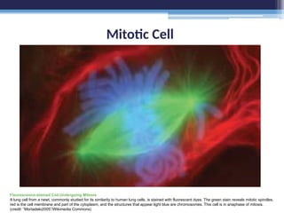
Mitotic Cell
Flourescence-stained CellUndergoing Mitosis
A lung cell from a newt, commonly studied for its similarity to human lung cells, is stained with fluorescent dyes. The green stain reveals mitotic spindles,
red is the cell membrane and part of the cytoplasm, and the structures that appear light blue are chromosomes. This cell is in anaphase of mitosis.
(credit: “Mortadelo2005”/Wikimedia Commons)
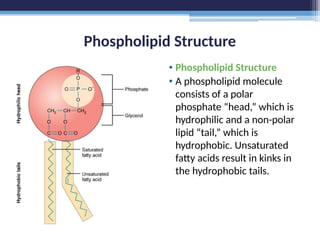
Phospholipid Structure
• PhospholipidStructure
• A phospholipid molecule
consists of a polar
phosphate “head,” which is
hydrophilic and a non-polar
lipid “tail,” which is
hydrophobic. Unsaturated
fatty acids result in kinks in
the hydrophobic tails.
6.
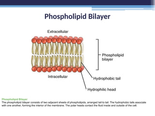
Phospholipid Bilayer
Phospholipid Bilayer
Thephospholipid bilayer consists of two adjacent sheets of phospholipids, arranged tail to tail. The hydrophobic tails associate
with one another, forming the interior of the membrane. The polar heads contact the fluid inside and outside of the cell.
7.
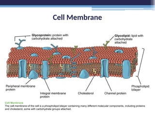
Cell Membrane
Cell Membrane
Thecell membrane of the cell is a phospholipid bilayer containing many different molecular components, including proteins
and cholesterol, some with carbohydrate groups attached.
8.
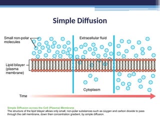
Simple Diffusion
Simple Diffusionacross the Cell (Plasma) Membrane
The structure of the lipid bilayer allows only small, non-polar substances such as oxygen and carbon dioxide to pass
through the cell membrane, down their concentration gradient, by simple diffusion.
9.
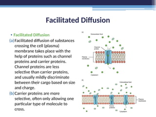
Facilitated Diffusion
• FacilitatedDiffusion
(a)Facilitated diffusion of substances
crossing the cell (plasma)
membrane takes place with the
help of proteins such as channel
proteins and carrier proteins.
Channel proteins are less
selective than carrier proteins,
and usually mildly discriminate
between their cargo based on size
and charge.
(b)Carrier proteins are more
selective, often only allowing one
particular type of molecule to
cross.
10.
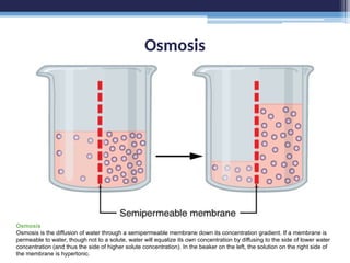
Osmosis
Osmosis
Osmosis is thediffusion of water through a semipermeable membrane down its concentration gradient. If a membrane is
permeable to water, though not to a solute, water will equalize its own concentration by diffusing to the side of lower water
concentration (and thus the side of higher solute concentration). In the beaker on the left, the solution on the right side of
the membrane is hypertonic.
11.
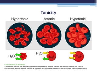
Tonicity
Concentration of Solutions
Ahypertonic solution has a solute concentration higher than another solution. An isotonic solution has a solute
concentration equal to another solution. A hypotonic solution has a solute concentration lower than another solution.
12.
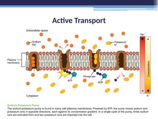
Active Transport
Sodium-Potassium Pump
Thesodium-potassium pump is found in many cell (plasma) membranes. Powered by ATP, the pump moves sodium and
potassium ions in opposite directions, each against its concentration gradient. In a single cycle of the pump, three sodium
ions are extruded from and two potassium ions are imported into the cell.
.
13.
Endocytosis
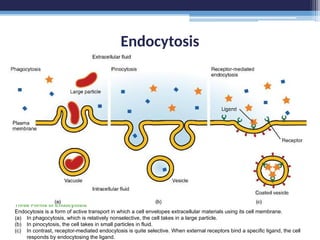
Three Forms ofEndocytosis
Endocytosis is a form of active transport in which a cell envelopes extracellular materials using its cell membrane.
(a) In phagocytosis, which is relatively nonselective, the cell takes in a large particle.
(b) In pinocytosis, the cell takes in small particles in fluid.
(c) In contrast, receptor-mediated endocytosis is quite selective. When external receptors bind a specific ligand, the cell
responds by endocytosing the ligand.
14.
Exocytosis
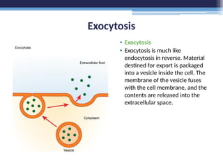
• Exocytosis
• Exocytosisis much like
endocytosis in reverse. Material
destined for export is packaged
into a vesicle inside the cell. The
membrane of the vesicle fuses
with the cell membrane, and the
contents are released into the
extracellular space.
Prototypical Human Cell
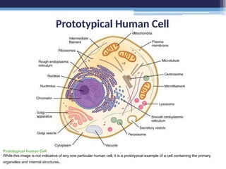
PrototypicalHuman Cell
While this image is not indicative of any one particular human cell, it is a prototypical example of a cell containing the primary
organelles and internal structures.
Golgi Apparatus
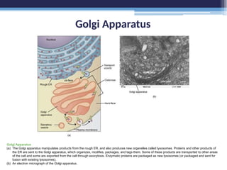
Golgi Apparatus
(a)The Golgi apparatus manipulates products from the rough ER, and also produces new organelles called lysosomes. Proteins and other products of
the ER are sent to the Golgi apparatus, which organizes, modifies, packages, and tags them. Some of these products are transported to other areas
of the cell and some are exported from the cell through exocytosis. Enzymatic proteins are packaged as new lysosomes (or packaged and sent for
fusion with existing lysosomes).
(b) An electron micrograph of the Golgi apparatus.
Cytoskeleton
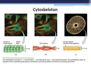
The Three Componentsof the Cytoskeleton
The cytoskeleton consists of (a) microtubules, (b) microfilaments, and (c) intermediate filaments. The cytoskeleton plays an
important role in maintaining cell shape and structure, promoting cellular movement, and aiding cell division.
Nucleus
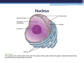
The Nucleus
The nucleusis the control center of the cell. The nucleus of living cells contains the genetic material that determines
the entire structure and function of that cell.
.
DNA Macrostructure
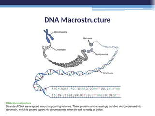
DNA Macrostructure
Strandsof DNA are wrapped around supporting histones. These proteins are increasingly bundled and condensed into
chromatin, which is packed tightly into chromosomes when the cell is ready to divide.
.
28.
Molecular Structure ofDNA
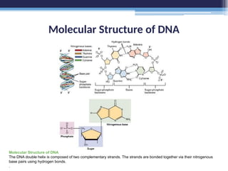
Molecular Structure of DNA
The DNA double helix is composed of two complementary strands. The strands are bonded together via their nitrogenous
base pairs using hydrogen bonds.
.
29.
DNA Replication
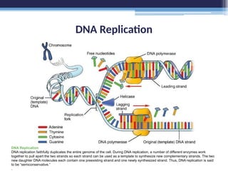
DNA Replication
DNAreplication faithfully duplicates the entire genome of the cell. During DNA replication, a number of different enzymes work
together to pull apart the two strands so each strand can be used as a template to synthesize new complementary strands. The two
new daughter DNA molecules each contain one preexisting strand and one newly synthesized strand. Thus, DNA replication is said
to be “semiconservative.”
.
Gene Expression: TheGenetic Code
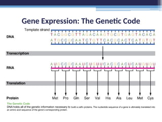
The Genetic Code
DNA holds all of the genetic information necessary to build a cell’s proteins. The nucleotide sequence of a gene is ultimately translated into
an amino acid sequence of the gene’s corresponding protein.
32.
Transcription
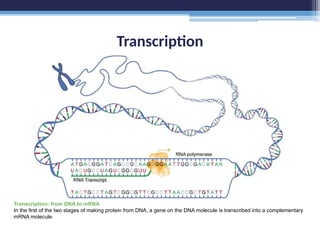
Transcription: from DNAto mRNA
In the first of the two stages of making protein from DNA, a gene on the DNA molecule is transcribed into a complementary
mRNA molecule.
33.
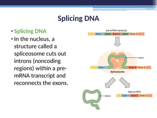
Splicing DNA
•Splicing DNA
•Inthe nucleus, a
structure called a
spliceosome cuts out
introns (noncoding
regions) within a pre-
mRNA transcript and
reconnects the exons.
34.
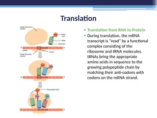
Translation
• Translation fromRNA to Protein
• During translation, the mRNA
transcript is “read” by a functional
complex consisting of the
ribosome and tRNA molecules.
tRNAs bring the appropriate
amino acids in sequence to the
growing polypeptide chain by
matching their anti-codons with
codons on the mRNA strand.
35.
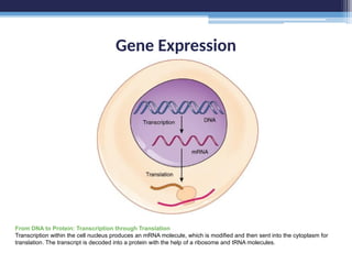
Gene Expression
From DNAto Protein: Transcription through Translation
Transcription within the cell nucleus produces an mRNA molecule, which is modified and then sent into the cytoplasm for
translation. The transcript is decoded into a protein with the help of a ribosome and tRNA molecules.
The Cell Cycle
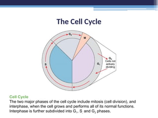
CellCycle
The two major phases of the cell cycle include mitosis (cell division), and
interphase, when the cell grows and performs all of its normal functions.
Interphase is further subdivided into G1, S, and G2 phases.
38.
Chromosomes
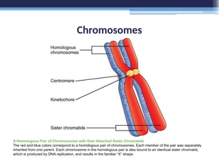
A Homologous Pairof Chromosomes with their Attached Sister Chromatids
The red and blue colors correspond to a homologous pair of chromosomes. Each member of the pair was separately
inherited from one parent. Each chromosome in the homologous pair is also bound to an identical sister chromatid,
which is produced by DNA replication, and results in the familiar “X” shape.
39.
Cell Division: MitosisFollowed by Cytokinesis
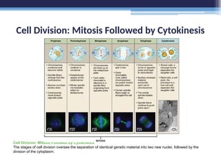
Cell Division: Mitosis Followed by Cytokinesis
The stages of cell division oversee the separation of identical genetic material into two new nuclei, followed by the
division of the cytoplasm.
40.
Control of theCell Cycle
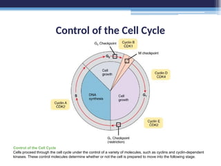
Control of the Cell Cycle
Cells proceed through the cell cycle under the control of a variety of molecules, such as cyclins and cyclin-dependent
kinases. These control molecules determine whether or not the cell is prepared to move into the following stage.
Hematopoiesis
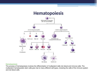
Hematopoiesis
The process ofhematopoiesis involves the differentiation of multipotent cells into blood and immune cells. The
multipotent hematopoietic stem cells give rise to many different cell types, including the cells of the immune system
and red blood cells.
43.
Hematopoiesis
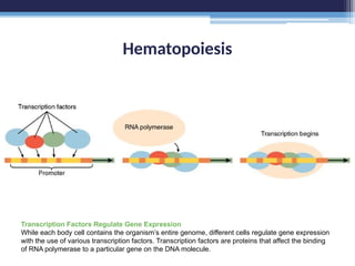
Transcription Factors RegulateGene Expression
While each body cell contains the organism’s entire genome, different cells regulate gene expression
with the use of various transcription factors. Transcription factors are proteins that affect the binding
of RNA polymerase to a particular gene on the DNA molecule.
44.
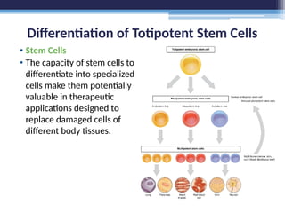
Differentiation of TotipotentStem Cells
• Stem Cells
• The capacity of stem cells to
differentiate into specialized
cells make them potentially
valuable in therapeutic
applications designed to
replace damaged cells of
different body tissues.
45.
Unless otherwise noted,the images and text used in this PowerPoint
are from:
Betts, J. G., Young, K. A., Wise, J. A., Johnson, E., Poe, B., Kruse, D.
H., Korol, O., Johnson, J. E., Womble, M., & DeSaix, P. (2022).
Chapter 9: Joints. In Anatomy and Physiology 2e. OpenStax.
https://openstax.org/books/anatomy-and-physiology-2e/pages/9-
introduction
Citation
Editor's Notes
#1 Unless otherwise noted, the images and text used in this PowerPoint are from:
Betts, J. G., Young, K. A., Wise, J. A., Johnson, E., Poe, B., Kruse, D. H., Korol, O., Johnson, J. E., Womble, M., & DeSaix, P. (2022). Chapter 9: Joints. In Anatomy and Physiology 2e. OpenStax. https://openstax.org/books/anatomy-and-physiology-2e/pages/9-introduction
#45 Unless otherwise noted, the images and text used in this PowerPoint are from:
Betts, J. G., Young, K. A., Wise, J. A., Johnson, E., Poe, B., Kruse, D. H., Korol, O., Johnson, J. E., Womble, M., & DeSaix, P. (2022). Chapter 9: Joints. In Anatomy and Physiology 2e. OpenStax. https://openstax.org/books/anatomy-and-physiology-2e/pages/9-introduction