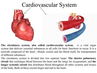
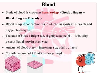
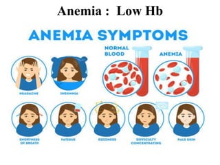
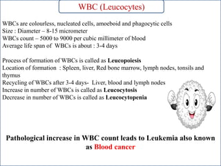
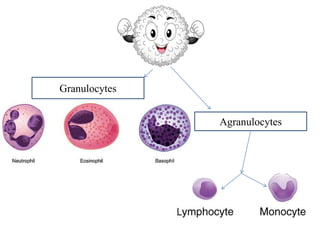
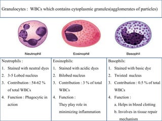
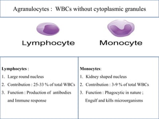
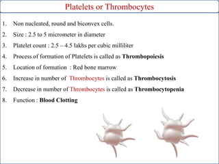
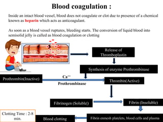
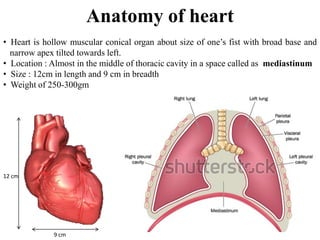
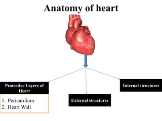
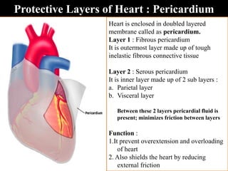
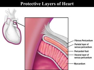
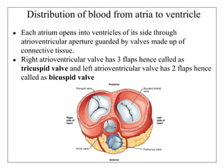
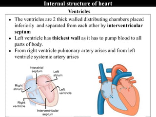
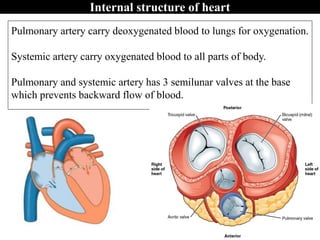
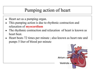
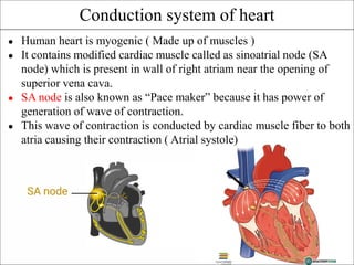
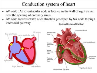
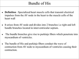
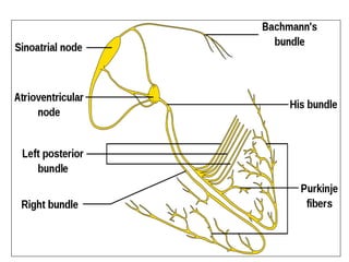
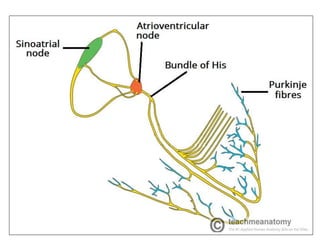
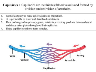
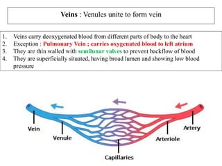
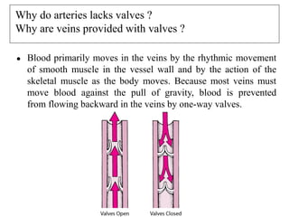
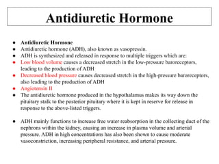
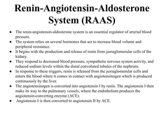
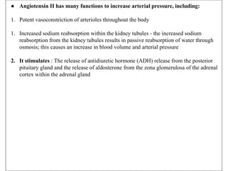
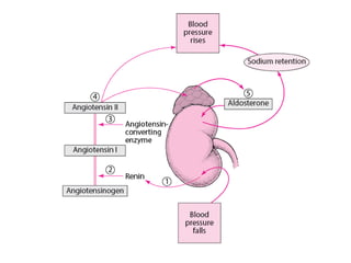
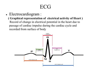
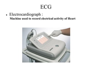
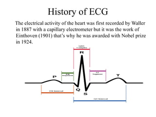
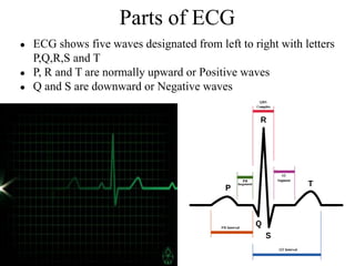
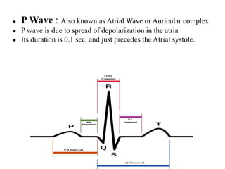
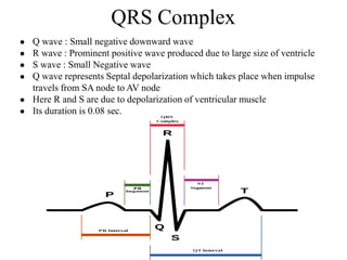
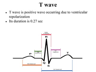
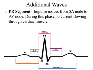
SlideShare a Scribd company logo
1 of 81
St. Wilfred’s Institute of Pharmacy, Panvel.
DTE CODE: 3485
TOPIC : Cardiovascular System
Presented by : Mr.Vijay Ikale(Ass.Professor Pharmacology)
Who discovered the circulation of blood ?
William Harvey (1 April 1578 – 3 June 1657) was an
English physician who made influential contributions
in anatomy and physiology.
He was the first known physician to describe completely,
and in detail, the systemic circulation and properties of
blood being pumped to the brain and the rest of the body
by the heart(1628).
Cardiovascular System
The circulatory system, also called cardiovascular system, is a vital organ
system that delivers essential substances to all cells for basic functions to occur. It is a
network composed of the heart , bloods vessels and the blood itself, for transportation
of different substances.
The circulatory system is divided into two separate loops: The shorter pulmonary
circuit that exchanges blood between the heart and the lungs for oxygenation; and the
longer systemic circuit that distributes blood throughout all other systems and tissues
of the body. Both of these circuits begin and end in the heart.
Blood
● Study of blood is known as heamatology (Greek : Haeme –
Blood , Logos – To study )
● Blood is liquid connective tissue which transports all nutrients and
oxygen to every cell
● Features of blood : Bright red, slightly alkaline(pH – 7.4), salty,
viscous liquid heavier than water
● Amount of blood present in average size adult : 5 liters
● Contributes around 8 % of total body weight
Blood Composition
45%
55%
Blood cells Plasma
Plasma
•Straw coloured, slightly alkaline and viscous fluid.
•It contains 90-92 % water and 8-10% solutes, most of which are
proteins(Plasma proteins are serum albumin, serum globulin,
heparin, fibrinogen and prothrombin)
• Other solutes are :
•Nutrients : such as glucose, amino acids, fatty acids, glycerols,
nitrogenous waste such as urea, uric acid ammonia and creatinine
•Gases : Oxygen, Carbon dioxide and Nitrogen
•Regulatory substances : Enzymes and Hormones
•Inorganic substances : Bicarbonates,Chlorides,Phosphates,Sulfates of s
Sodium, Potassium, Calcium, Magnesium
Blood cells
WBC (Leucocytes) RBC (Erythrocytes) Thrombocyte (Platelet)
RBC (Erythrocytes)
Erythros : Red , Kytos – Cell )
RBCs are red, circular, biconcave, non-nucleated cells
Size : Diameter - 7 micrometer and thickness - 2.5 micrometer
Adult Male RBCs count : 5.1 – 5.8 million RBCs per cubic millimeter
Female RBCs count : 4.3 to 5.2 million RBCs per cubic millimeter
Average life span of RBCs is about : 120 days
Process of formation of RBCs is called as erythropoiesis
Location of formation : Foetus – Spleen and liver , Adults – Red bone marrow
Recycling of RBCS after 120 days - Liver and spleen
Increase in number of RBCs is called as Polycythemia
Decrease in number of RBCs is called as Erythrocytopenia
Cytoplasm of RBCs contain a respiratory pigment called as haemoglobin which
helps in transport of oxygen and carbon dioxide
Normal haemoglobin content in male : 13-18 gm/100 ml of blood
Normal haemoglobin content in female : 11.-18 gm/100 ml of blood
Less haemoglobin leads to anemia
RBC (Erythrocytes)
Anemia : Low Hb
WBCs are colourless, nucleated cells, amoeboid and phagocytic cells
Size : Diameter – 8-15 micrometer
WBCs count – 5000 to 9000 per cubic millimeter of blood
Average life span of WBCs is about : 3-4 days
Process of formation of WBCs is called as Leucopoiesis
Location of formation : Spleen, liver, Red bone marrow, lymph nodes, tonsils and
thymus
Recycling of WBCs after 3-4 days- Liver, blood and lymph nodes
Increase in number of WBCs is called as Leucocytosis
Decrease in number of WBCs is called as Leucocytopenia
Pathological increase in WBC count leads to Leukemia also known
as Blood cancer
WBC (Leucocytes)
Granulocytes
Agranulocytes
Granulocytes : WBCs which contains cytoplasmic granules(agglomerates of particles)
Neutrophils :
1. Stained with neutral dyes
2. 3-5 Lobed nucleus
3. Contribution : 54-62 %
of total WBCs
4. Function : Phagocytic in
action
Eosinophils:
1. Stained with acidic dyes
2. Bilobed nucleus
3. Contribution : 3 % of total
WBCs
4. Function :
They play role in
minimizing inflammation
Basophils:
1. Stained with basic dye
2. Twisted nucleus
3. Contribution : 0.5 % of total
WBCs
4. Function :
a. Helps in blood clotting
b. Involves in tissue repair
. .mechanism
Agranulocytes : WBCs without cytoplasmic granules
Lymphocytes :
1. Large round nucleus
2. Contribution : 25-33 % of total WBCs
3. Function : Production of antibodies
and Immune response
Monocytes:
1. Kidney shaped nucleus
2. Contribution : 3-9 % of total WBCs
3. Function : Phagocytic in nature ;
Engulf and kills microorganisms
Platelets or Thrombocytes
1. Non nucleated, round and biconvex cells.
2. Size : 2.5 to 5 micrometer in diameter
3. Platelet count : 2.5 – 4.5 lakhs per cubic milliliter
4. Process of formation of Platelets is called as Thrombopoiesis
5. Location of formation : Red bone marrow
6. Increase in number of Thrombocytes is called as Thrombocytosis
7. Decrease in number of Thrombocytes is called as Thrombocytopenia
8. Function : Blood Clotting
Blood coagulation :
Inside an intact blood vessel, blood does not coagulate or clot due to presence of a chemical
known as heparin which acts as anticoagulant.
As soon as a blood vessel ruptures, bleeding starts. The conversion of liquid blood into
semisolid jelly is called as blood coagulation or clotting
Release of
Thromboplastin
Synthesis of enzyme Prothrombinase
Prothrombin(Inactive)
Thrombin(Active)
Fibrinogen (Soluble|) Fibrin (Insoluble)
Fibrin enmesh platelets, blood cells and plasma
Blood clotting
Ca++
Prothrombinase
Clotting Time : 2-8
min.
Heart
Anatomy of heart
• Heart is hollow muscular conical organ about size of one’s fist with broad base and
. narrow apex tilted towards left.
• Location : Almost in the middle of thoracic cavity in a space called as mediastinum
• Size : 12cm in length and 9 cm in breadth
• Weight of 250-300gm
12 cm
9 cm
Anatomy of heart
Protective Layers of
Heart
Internal structures
External structures
1. Pericardium
2. Heart Wall
Protective Layers of Heart : Pericardium
Heart is enclosed in doubled layered
membrane called as pericardium.
Layer 1 : Fibrous pericardium
It is outermost layer made up of tough
inelastic fibrous connective tissue
Layer 2 : Serous pericardium
It is inner layer made up of 2 sub layers :
a. Parietal layer
b. Visceral layer
Between these 2 layers pericardial fluid is
present; minimizes friction between layers
Function :
1.It prevent overextension and overloading
of heart
2. Also shields the heart by reducing
external friction
Protective Layers of Heart
Protective Layers of Heart : Heart Wall
Wall of heart made up of 3 layers namely ; outer Epicardium, middle
Myocardium and inner Endocardium
1.Epicardium : Consist of single layer of flat epithelial cells .
The epicardium functions to protect the inner heart layers and also assists in the
production of pericardial fluid. This fluid fills the pericardial cavity and helps to
reduce friction between pericardial membranes. Also found in this heart layer are
the coronary blood vessels, which supply the heart wall with blood.
2.Myocardium : Composed of cardiac muscle fibers responsible for contraction
and relaxation of heart. The myocardium is the thickest layer of the heart wall,
with its thickness varying in different parts of the heart. The myocardium of the
left ventricle is the thickest, as this ventricle is responsible for generating the
power needed to pump oxygenated blood from the heart to the rest of the body.
3.Endocardium : Consist of single layer of flat epithelial cells. This layer lines
the inner heart chambers, covers heart valves, and is continuous with the
endothelium of large blood vessels
External structure of heart
1. Heart is made up of 4 chambers : Two superior, small, thin walled receiving
chambers called as atria and two inferior, large, thick walled, distributing chambers
called as ventricles.
2. Externally wall of heart situated with grooves in which coronary artery and veins are
located .
a. Coronary artery supplies oxygenated blood to heart
b. Coronary vein collects deoxygenated blood from heart
3. From right ventricle pulmonary artery arises and from left ventricle systemic artery
arises
● Venae cavae : In humans there are the superior vena cava and
the inferior vena cava, and both empty into the right atrium.
● Superior vena cava : The superior vena cava is the large vein
which returns deoxygenated blood to the heart from the head, neck
and both upper limbs
● Inferior vena cava : The inferior vena cava returns deoxygenated
blood to the heart from the lower limbs and middle body
(Abdomen)
External structure of heart
Superior vena cava
Inferior vena cava
● Opening of inferior vena cava is guarded by Eustachian valve
and opening of coronary sinus is guarded by Thebesian valve.
● What are valves ?
● Valves are flap-like structures present in heart that allow blood
to flow in one direction.
Internal structure of heart
Internally heart is made up of 4 chambers with 2 atria and 2
ventricles
Atria : Two thin walled receiving chambers placed superiorly and
separated from each other by interatrial septum
The right atrium receives deoxygenated blood from all over the body
through superior vena cava, inferior vena cava and coronary sinus
● Left atrium receives oxygenated blood from the lungs through 4
openings of pulmonary veins
Internal structure of heart
RA : Deoxygenated
blood
LA: Oxygenated blood
Distribution of blood from atria to ventricle
● Each atrium opens into ventricles of its side through
atrioventricular aperture guarded by valves made up of
connective tissue.
● Right atrioventricular valve has 3 flaps hence called as
tricuspid valve and left atrioventricular valve has 2 flaps hence
called as bicuspid valve
Ventricles
● The ventricles are 2 thick walled distributing chambers placed
inferiorly and separated from each other by interventricular
septum
● Left ventricle has thickest wall as it has to pump blood to all
parts of body.
● From right ventricle pulmonary artery arises and from left
ventricle systemic artery arises
Internal structure of heart
Pulmonary artery carry deoxygenated blood to lungs for oxygenation.
Systemic artery carry oxygenated blood to all parts of body.
Pulmonary and systemic artery has 3 semilunar valves at the base
which prevents backward flow of blood.
Internal structure of heart
Pumping action of heart
● Heart act as a pumping organ.
● This pumping action is due to rhythmic contraction and
relaxation of myocardium
● The rhythmic contraction and relaxation of heart is known as
heart beat.
● Heart beats 72 times per minute ; also known as heart rate and
pumps 5 liter of blood per minute
Conduction system of heart
● Human heart is myogenic ( Made up of muscles )
● It contains modified cardiac muscle called as sinoatrial node (SA
node) which is present in wall of right atriam near the opening of
superior vena cava.
● SA node is also known as “Pace maker” because it has power of
generation of wave of contraction.
● This wave of contraction is conducted by cardiac muscle fiber to both
atria causing their contraction ( Atrial systole)
● AV node : Atrioventricular node is located in the wall of right atriam
near the opening of coronary sinus.
● AV node receives wave of contraction generated by SA node through
internodal pathway
Conduction system of heart
● Definition : Specialized heart muscle cells that transmit electrical
impulses from the AV node in the heart to the muscle cells of the
heart wall
● It arises from AV node and divides into 2 branches i.e right and left
bundle branches located in interventricular septum.
● The bundle branches give rise to purkinje fibers which penetrate into
myocardium of ventricles.
● The bundle of His and purkinje fibers conduct the wave of
contraction from AV node to myocardium of ventricles causing their
contraction.
Bundle of His
Working of Heart ( Cardiac Cycle )
● The events associated with one cardiac heart
beat is known as cardiac cycle
● It lasts for 0.8 seconds.
● Each heart beat includes :
1) Atrial systole
2) Ventricular systole
3) Joint diastole
● Atrial Systole : 0.1 Second.
During Atrial systole, the deoxygenated blood
from the right atrium enters the right ventricle
through atrioventricular aperture, the back
flow prevented by tricuspid valve AND
oxygenated blood from left atrium enters left
ventricle through atrioventricular aperture ,
the backflow is prevented by bicuspid valve
Working of Heart ( Cardiac Cycle )
● Ventricular Systole : 0.3 Seconds.
During Ventricular systole, the deoxygenated
blood from the right Ventricle enters the
pulmonary artery, which carry blood to lungs
for oxygenation .
Oxygenated blood from left Ventricle enters
systemic artery which is supplied all over the
body, the backflow is prevented by
semilunar valve .
Working of Heart ( Cardiac Cycle )
● Joint diastole : 0.4 seconds
During joint diastole both atria and ventricles
undergo relaxation
The right atrium receives deoxygenated blood
from all over the body through superior vena
cava, inferior vena cava and coronary sinus.
The left atrium receives oxygenated blood
from the lungs through two pairs of
pulmonary veins.
Regulation of cardiac activity
● Normal activities of heart are regulated by specialized muscles i.e.
cardiac muscle
● Its activity is auto regulated
● Then why heart rate increases or decreases ?
● Who controls heart rate ?
Medulla oblongata
Brain – Heart Connection
Nervous control of heart
The ANS is responsible for controlling many
physiological functions: inducing the force of contraction
of the heart, peripheral resistance of blood vessels and
the heart rate. The ANS has both sympathetic and
parasympathetic divisions that work together to maintain
balance.
The parasympathetic nervous system sends input into the
heart is via the vagus nerve (X). The vagus nerve forms
synapses with postganglionic cells in SA node and AV
node. When stimulated, acetylcholine which binds on to
M₂ receptors, which acts to decrease the heart rate
The sympathetic sends input into the heart is via the
postganglionic fibres from the sympathetic trunk which
innervate the SA node and AV node. The post ganglionic
fibres release noradrenaline, which acts on B₁
adrenoreceptors to increase heart rate as well as
increasing the force of contraction
Blood Vessels
The study of blood vessels is called as angiology.
Blood vessels are of three types : Arteries, Veins, Capillaries
Blood Vessels
Arteries :
Arteries carry blood away from the heart to different parts of body.
They carry oxygenated blood except pulmonary artery
Arteries :
They are thick walled, muscular, elastic and without valves inside
They are deeply situated in body, having narrow lumen and showing
high blood pressure for efficient circulation of blood. Histologically the
wall of artery is made up of three layers namely Outer tunica externa,
middle tunica media and inner tunica interna
Arteries divide and redivide into arterioles which further divide into
capillaries.
Capillaries : Capillaries are the thinnest blood vessels and formed by
division and redivision of arterioles.
1. Wall of capillary is made up of squamous epithelium.
2. It is permeable to water and dissolved substances.
3. Thus exchange of respiratory gases, nutrients, excretory products between blood
and tissue takes place through wall of capillaries.
4. These capillaries unite to form venules.
Veins : Venules unite to form vein
1. Veins carry deoxygenated blood from different parts of body to the heart
2. Exception : Pulmonary Vein ; carries oxygenated blood to left atrium
3. They are thin walled with semilunar valves to prevent backflow of blood
4. They are superficially situated, having broad lumen and showing low blood
pressure
Why do arteries lacks valves ?
Why are veins provided with valves ?
● Blood primarily moves in the veins by the rhythmic movement
of smooth muscle in the vessel wall and by the action of the
skeletal muscle as the body moves. Because most veins must
move blood against the pull of gravity, blood is prevented
from flowing backward in the veins by one-way valves.
Regulation of Blood Pressure
Blood Pressure
Blood Pressure : Pressure exerted by circulating blood on wall of arteries
Normal Blood Pressure : 120/80 mmHg
1. Systolic : 120mmHg When Ventricles Contracts
2. Diastolic : 80 mmHg When Ventricles Relaxes
How blood pressure is
controlled?
Control of Blood Pressure
There are several mechanisms through which the body regulates arterial pressure :
1. Baroreceptor Reflex
In response to small (acute) changes in blood pressure, the body responds through the
baroreceptors located within blood vessels.
Baroreceptors are a form of mechanoreceptor that become activated by the stretching of the
vessel. This sensory information is transferred to the central nervous system(Brain and Spinal
Cord) and used to influence peripheral vascular resistance and cardiac output.
Control of Blood Pressure
There are two forms of baroreceptors.
 High-Pressure Baroreceptors
Two baroreceptors are located within the high-pressure arterial
system.
1. The carotid baroreceptor responds to both increases and
decreases in blood pressure
2. The aortic arch baroreceptor responds only to increases in
blood pressure.
These both send signals in response to the physical distortion
of the vessel. The stretch of the vessel leads to an increase in
action potential relayed from the sensory endings located in the
tunica interna of the artery. These action potentials get
transmitted to the autonomic neurons. They secrete
neurotransmitters to affect the cardiovascular system.
● Activation of the aortic baroreceptor increases during high blood pressure and it
effectively inhibits the sympathetic nerve response.
● On the other hand, if an individual’s blood pressure were to fall such as in
hypovolemic shock, the rate of action potential from the baroreceptor would be
decreased due to reduced depolarization; this would lead to reduced inhibition of
sympathetic activity, resulting in a reflex to increase pressure.
● Low-Pressure Baroreceptors
● These baroreceptors are present within the low-pressure venous system. They exist
within large veins, pulmonary vessels, and within the walls of the right atrium and
ventricle. Changes in volume largely influence the baroreceptors in the venous
system. Decreased frequency in stimulation in low-pressure scenarios leads to the
secretion of antidiuretic hormone, renin, and aldosterone. These lead to a downstream
effect to regulate arterial pressure.
Control of Blood Pressure
Antidiuretic Hormone
● Antidiuretic Hormone
● Antidiuretic hormone (ADH), also known as vasopressin.
● ADH is synthesized and released in response to multiple triggers which are:
● Low blood volume causes a decreased stretch in the low-pressure baroreceptors,
leading to the production of ADH
● Decreased blood pressure causes decreased stretch in the high-pressure baroreceptors,
also leading to the production of ADH
● Angiotensin II
● The antidiuretic hormone produced in the hypothalamus makes its way down the
pituitary stalk to the posterior pituitary where it is kept in reserve for release in
response to the above-listed triggers.
● ADH mainly functions to increase free water reabsorption in the collecting duct of the
nephrons within the kidney, causing an increase in plasma volume and arterial
pressure. ADH in high concentrations has also been shown to cause moderate
vasoconstriction, increasing peripheral resistance, and arterial pressure.
Renin-Angiotensin-Aldosterone
System (RAAS)
● The renin-angiotensin-aldosterone system is an essential regulator of arterial blood
pressure.
● The system relies on several hormones that act to increase blood volume and
peripheral resistance.
● It begins with the production and release of renin from juxtaglomerular cells of the
kidney.
● They respond to decreased blood pressure, sympathetic nervous system activity, and
reduced sodium levels within the distal convoluted tubules of the nephrons.
● In response to these triggers, renin is released from the juxtaglomerular cells and
enters the blood where it comes in contact with angiotensinogen which is produced
continuously by the liver.
● The angiotensinogen is converted into angiotensin I by renin. The angiotensin I then
make its way to the pulmonary vessels, where the endothelium produces the
angiotensin-converting enzyme (ACE).
● Angiotensin I is then converted to angiotensin II by ACE.
● Angiotensin II has many functions to increase arterial pressure, including:
1. Potent vasoconstriction of arterioles throughout the body
1. Increased sodium reabsorption within the kidney tubules - the increased sodium
reabsorption from the kidney tubules results in passive reabsorption of water through
osmosis; this causes an increase in blood volume and arterial pressure
2. It stimulates : The release of antidiuretic hormone (ADH) release from the posterior
pituitary gland and the release of aldosterone from the zona glomerulosa of the adrenal
cortex within the adrenal gland
ECG
● Electrocardiogram :
( Graphical representation of electrical activity of Heart )
Record of change in electrical potential in the heart due to
passage of cardiac impulse during the cardiac cycle and
recorded from surface of body
ECG
● Electrocardiograph :
Machine used to record electrical activity of Heart
History of ECG
The electrical activity of the heart was first recorded by Waller
in 1887 with a capillary electrometer but it was the work of
Einthoven (1901) that’s why he was awarded with Nobel prize
in 1924.
Parts of ECG
● ECG shows five waves designated from left to right with letters
P,Q,R,S and T
● P, R and T are normally upward or Positive waves
● Q and S are downward or Negative waves
● P Wave : Also known as Atrial Wave or Auricular complex
● P wave is due to spread of depolarization in the atria
● Its duration is 0.1 sec. and just precedes the Atrial systole.
QRS Complex
● Q wave : Small negative downward wave
● R wave : Prominent positive wave produced due to large size of ventricle
● S wave : Small Negative wave
● Q wave represents Septal depolarization which takes place when impulse
travels from SA node to AV node
● Here R and S are due to depolarization of ventricular muscle
● Its duration is 0.08 sec.
T wave
● T wave is positive wave occurring due to ventricular
repolarization
● Its duration is 0.27 sec
Additional Waves
● PR Segment : Impulse moves from SA node to
AV node. During this phase no current flowing
through cardiac muscle.
Additional Waves
● ST Segment : During this phase no current
flowing through cardiac muscle.
Procedure for ECG

More Related Content

What's hot

Transport in humans
Transport in humansTransport in humans
Transport in humansXu Jia Xian
 
1E1N transport in humans 2012
1E1N transport in humans 20121E1N transport in humans 2012
1E1N transport in humans 2012Jacklyn Kong
 
Chapter 8 transport in humans
Chapter 8 transport in humansChapter 8 transport in humans
Chapter 8 transport in humanschenghong03
 
Circulatory system -The blood
Circulatory system -The bloodCirculatory system -The blood
Circulatory system -The bloodLivson Thomas
 
Human circulatory system
Human circulatory systemHuman circulatory system
Human circulatory systemMohdMafuz1
 
Transportation in humans (part 1) grade 7 notes
Transportation in humans (part 1) grade 7 notesTransportation in humans (part 1) grade 7 notes
Transportation in humans (part 1) grade 7 notesmeera chavda
 
The circulatory system
The circulatory systemThe circulatory system
The circulatory systemRuba Salah
 
Biochemical composition and functions of blood and lymph
Biochemical composition and functions of blood and lymphBiochemical composition and functions of blood and lymph
Biochemical composition and functions of blood and lymphJinnah University For Women
 
Heart & Circulation
Heart & CirculationHeart & Circulation
Heart & Circulationraj kumar
 
Form 3 Science Chapter 2
Form 3 Science Chapter 2Form 3 Science Chapter 2
Form 3 Science Chapter 2ft02nLqt
 
ppt on human circulatory system
ppt on human circulatory system ppt on human circulatory system
ppt on human circulatory system bhavik7888
 
Chapter 2 Blood Circulation and Transportation
Chapter 2 Blood Circulation and TransportationChapter 2 Blood Circulation and Transportation
Chapter 2 Blood Circulation and TransportationBrandon Loo
 

What's hot (19)

Transport in humans
Transport in humansTransport in humans
Transport in humans
 
1E1N transport in humans 2012
1E1N transport in humans 20121E1N transport in humans 2012
1E1N transport in humans 2012
 
Chapter 8 transport in humans
Chapter 8 transport in humansChapter 8 transport in humans
Chapter 8 transport in humans
 
Circulatory system -The blood
Circulatory system -The bloodCirculatory system -The blood
Circulatory system -The blood
 
Cardiovascular system
Cardiovascular systemCardiovascular system
Cardiovascular system
 
Chapter 12
Chapter 12Chapter 12
Chapter 12
 
Human circulatory system
Human circulatory systemHuman circulatory system
Human circulatory system
 
Cardiovascular system
Cardiovascular system Cardiovascular system
Cardiovascular system
 
Transportation in humans (part 1) grade 7 notes
Transportation in humans (part 1) grade 7 notesTransportation in humans (part 1) grade 7 notes
Transportation in humans (part 1) grade 7 notes
 
The circulatory system
The circulatory systemThe circulatory system
The circulatory system
 
Biochemical composition and functions of blood and lymph
Biochemical composition and functions of blood and lymphBiochemical composition and functions of blood and lymph
Biochemical composition and functions of blood and lymph
 
Heart & Circulation
Heart & CirculationHeart & Circulation
Heart & Circulation
 
Form 3 Science Chapter 2
Form 3 Science Chapter 2Form 3 Science Chapter 2
Form 3 Science Chapter 2
 
The Anatomy & Physiology of Blood.
The Anatomy & Physiology of Blood.The Anatomy & Physiology of Blood.
The Anatomy & Physiology of Blood.
 
Unit 30 Lymphatic System
Unit 30 Lymphatic SystemUnit 30 Lymphatic System
Unit 30 Lymphatic System
 
4. body fluid & blood
4. body fluid & blood4. body fluid & blood
4. body fluid & blood
 
ppt on human circulatory system
ppt on human circulatory system ppt on human circulatory system
ppt on human circulatory system
 
Chapter 2 Blood Circulation and Transportation
Chapter 2 Blood Circulation and TransportationChapter 2 Blood Circulation and Transportation
Chapter 2 Blood Circulation and Transportation
 
Circulation
CirculationCirculation
Circulation
 

Similar to Circulatory system

Cardiovascular system presentation.bert
Cardiovascular system presentation.bertCardiovascular system presentation.bert
Cardiovascular system presentation.bert2184692
 
Phsiyological structure and life process Suresh Chaudhary.docx
Phsiyological structure and life process Suresh Chaudhary.docxPhsiyological structure and life process Suresh Chaudhary.docx
Phsiyological structure and life process Suresh Chaudhary.docxsureshchaudhary72
 
Circulatory system grade 9
Circulatory system grade 9Circulatory system grade 9
Circulatory system grade 9MariaZafar30
 
Blood and Blood Vessels
Blood and Blood VesselsBlood and Blood Vessels
Blood and Blood VesselsJoyce Peh
 
Power point the cardiovascular system - anatomy and physiology
Power point   the cardiovascular system - anatomy and physiologyPower point   the cardiovascular system - anatomy and physiology
Power point the cardiovascular system - anatomy and physiologyStephen Collins
 
biology 1 the circulatory system
biology 1 the circulatory systembiology 1 the circulatory system
biology 1 the circulatory systemhafsah hafy
 
powerpoint-thecardiovascularsystem-anatomyandphysiology-141126132329-conversi...
powerpoint-thecardiovascularsystem-anatomyandphysiology-141126132329-conversi...powerpoint-thecardiovascularsystem-anatomyandphysiology-141126132329-conversi...
powerpoint-thecardiovascularsystem-anatomyandphysiology-141126132329-conversi...Jay5441
 
Cardiovascular system-
Cardiovascular system-Cardiovascular system-
Cardiovascular system-RitikKumar140
 
Cardiovascular system
Cardiovascular systemCardiovascular system
Cardiovascular systemsom allul
 
circulatory_system.ppt
circulatory_system.pptcirculatory_system.ppt
circulatory_system.pptAzeAmasolJosol
 
Circulatory system
Circulatory systemCirculatory system
Circulatory systemCanisBlaire
 

Similar to Circulatory system (20)

Chapter 12
Chapter 12Chapter 12
Chapter 12
 
Circulatory system
Circulatory systemCirculatory system
Circulatory system
 
Circulatory System
Circulatory  SystemCirculatory  System
Circulatory System
 
Circulatory system
Circulatory   system Circulatory   system
Circulatory system
 
Cardiovascular system presentation.bert
Cardiovascular system presentation.bertCardiovascular system presentation.bert
Cardiovascular system presentation.bert
 
Phsiyological structure and life process Suresh Chaudhary.docx
Phsiyological structure and life process Suresh Chaudhary.docxPhsiyological structure and life process Suresh Chaudhary.docx
Phsiyological structure and life process Suresh Chaudhary.docx
 
Circulatory system grade 9
Circulatory system grade 9Circulatory system grade 9
Circulatory system grade 9
 
Blood and Blood Vessels
Blood and Blood VesselsBlood and Blood Vessels
Blood and Blood Vessels
 
Power point the cardiovascular system - anatomy and physiology
Power point   the cardiovascular system - anatomy and physiologyPower point   the cardiovascular system - anatomy and physiology
Power point the cardiovascular system - anatomy and physiology
 
biology 1 the circulatory system
biology 1 the circulatory systembiology 1 the circulatory system
biology 1 the circulatory system
 
powerpoint-thecardiovascularsystem-anatomyandphysiology-141126132329-conversi...
powerpoint-thecardiovascularsystem-anatomyandphysiology-141126132329-conversi...powerpoint-thecardiovascularsystem-anatomyandphysiology-141126132329-conversi...
powerpoint-thecardiovascularsystem-anatomyandphysiology-141126132329-conversi...
 
Circulation
CirculationCirculation
Circulation
 
Cardiovascular system
Cardiovascular systemCardiovascular system
Cardiovascular system
 
Cardiovascular system-
Cardiovascular system-Cardiovascular system-
Cardiovascular system-
 
Cardiovascular system
Cardiovascular systemCardiovascular system
Cardiovascular system
 
circulatory_system.ppt
circulatory_system.pptcirculatory_system.ppt
circulatory_system.ppt
 
circulatory_system.ppt
circulatory_system.pptcirculatory_system.ppt
circulatory_system.ppt
 
Circulatory system
Circulatory systemCirculatory system
Circulatory system
 
Cardio vascular system
Cardio vascular systemCardio vascular system
Cardio vascular system
 
Circulatory system
Circulatory systemCirculatory system
Circulatory system
 

More from VIJAY IKALE

Signal transduction
Signal transductionSignal transduction
Signal transductionVIJAY IKALE
 
Peripheral nervous system
Peripheral nervous system Peripheral nervous system
Peripheral nervous system VIJAY IKALE
 
Digestive system
Digestive  systemDigestive  system
Digestive systemVIJAY IKALE
 

More from VIJAY IKALE (6)

Chemotherapy
ChemotherapyChemotherapy
Chemotherapy
 
Antacids
AntacidsAntacids
Antacids
 
Signal transduction
Signal transductionSignal transduction
Signal transduction
 
Peripheral nervous system
Peripheral nervous system Peripheral nervous system
Peripheral nervous system
 
Skeletal system
Skeletal systemSkeletal system
Skeletal system
 
Digestive system
Digestive  systemDigestive  system
Digestive system
 

Recently uploaded

POINT- BIOCHEMISTRY SEM 2 ENZYMES UNIT 5.pptx
POINT- BIOCHEMISTRY SEM 2 ENZYMES UNIT 5.pptxPOINT- BIOCHEMISTRY SEM 2 ENZYMES UNIT 5.pptx
POINT- BIOCHEMISTRY SEM 2 ENZYMES UNIT 5.pptxSayali Powar
 
Pharmacognosy Flower 3. Compositae 2023.pdf
Pharmacognosy Flower 3. Compositae 2023.pdfPharmacognosy Flower 3. Compositae 2023.pdf
Pharmacognosy Flower 3. Compositae 2023.pdfMahmoud M. Sallam
 
“Oh GOSH! Reflecting on Hackteria's Collaborative Practices in a Global Do-It...
“Oh GOSH! Reflecting on Hackteria's Collaborative Practices in a Global Do-It...“Oh GOSH! Reflecting on Hackteria's Collaborative Practices in a Global Do-It...
“Oh GOSH! Reflecting on Hackteria's Collaborative Practices in a Global Do-It...Marc Dusseiller Dusjagr
 
Types of Journalistic Writing Grade 8.pptx
Types of Journalistic Writing Grade 8.pptxTypes of Journalistic Writing Grade 8.pptx
Types of Journalistic Writing Grade 8.pptxEyham Joco
 
Full Stack Web Development Course for Beginners
Full Stack Web Development Course  for BeginnersFull Stack Web Development Course  for Beginners
Full Stack Web Development Course for BeginnersSabitha Banu
 
How to Configure Email Server in Odoo 17
How to Configure Email Server in Odoo 17How to Configure Email Server in Odoo 17
How to Configure Email Server in Odoo 17Celine George
 
KSHARA STURA .pptx---KSHARA KARMA THERAPY (CAUSTIC THERAPY)————IMP.OF KSHARA ...
KSHARA STURA .pptx---KSHARA KARMA THERAPY (CAUSTIC THERAPY)————IMP.OF KSHARA ...KSHARA STURA .pptx---KSHARA KARMA THERAPY (CAUSTIC THERAPY)————IMP.OF KSHARA ...
KSHARA STURA .pptx---KSHARA KARMA THERAPY (CAUSTIC THERAPY)————IMP.OF KSHARA ...M56BOOKSTORE PRODUCT/SERVICE
 
Earth Day Presentation wow hello nice great
Earth Day Presentation wow hello nice greatEarth Day Presentation wow hello nice great
Earth Day Presentation wow hello nice greatYousafMalik24
 
Historical philosophical, theoretical, and legal foundations of special and i...
Historical philosophical, theoretical, and legal foundations of special and i...Historical philosophical, theoretical, and legal foundations of special and i...
Historical philosophical, theoretical, and legal foundations of special and i...jaredbarbolino94
 
Framing an Appropriate Research Question 6b9b26d93da94caf993c038d9efcdedb.pdf
Framing an Appropriate Research Question 6b9b26d93da94caf993c038d9efcdedb.pdfFraming an Appropriate Research Question 6b9b26d93da94caf993c038d9efcdedb.pdf
Framing an Appropriate Research Question 6b9b26d93da94caf993c038d9efcdedb.pdfUjwalaBharambe
 
EPANDING THE CONTENT OF AN OUTLINE using notes.pptx
EPANDING THE CONTENT OF AN OUTLINE using notes.pptxEPANDING THE CONTENT OF AN OUTLINE using notes.pptx
EPANDING THE CONTENT OF AN OUTLINE using notes.pptxRaymartEstabillo3
 
Computed Fields and api Depends in the Odoo 17
Computed Fields and api Depends in the Odoo 17Computed Fields and api Depends in the Odoo 17
Computed Fields and api Depends in the Odoo 17Celine George
 
Introduction to AI in Higher Education_draft.pptx
Introduction to AI in Higher Education_draft.pptxIntroduction to AI in Higher Education_draft.pptx
Introduction to AI in Higher Education_draft.pptxpboyjonauth
 
Proudly South Africa powerpoint Thorisha.pptx
Proudly South Africa powerpoint Thorisha.pptxProudly South Africa powerpoint Thorisha.pptx
Proudly South Africa powerpoint Thorisha.pptxthorishapillay1
 
Roles & Responsibilities in Pharmacovigilance
Roles & Responsibilities in PharmacovigilanceRoles & Responsibilities in Pharmacovigilance
Roles & Responsibilities in PharmacovigilanceSamikshaHamane
 
Incoming and Outgoing Shipments in 1 STEP Using Odoo 17
Incoming and Outgoing Shipments in 1 STEP Using Odoo 17Incoming and Outgoing Shipments in 1 STEP Using Odoo 17
Incoming and Outgoing Shipments in 1 STEP Using Odoo 17Celine George
 
Presiding Officer Training module 2024 lok sabha elections
Presiding Officer Training module 2024 lok sabha electionsPresiding Officer Training module 2024 lok sabha elections
Presiding Officer Training module 2024 lok sabha electionsanshu789521
 

Recently uploaded (20)

POINT- BIOCHEMISTRY SEM 2 ENZYMES UNIT 5.pptx
POINT- BIOCHEMISTRY SEM 2 ENZYMES UNIT 5.pptxPOINT- BIOCHEMISTRY SEM 2 ENZYMES UNIT 5.pptx
POINT- BIOCHEMISTRY SEM 2 ENZYMES UNIT 5.pptx
 
Pharmacognosy Flower 3. Compositae 2023.pdf
Pharmacognosy Flower 3. Compositae 2023.pdfPharmacognosy Flower 3. Compositae 2023.pdf
Pharmacognosy Flower 3. Compositae 2023.pdf
 
“Oh GOSH! Reflecting on Hackteria's Collaborative Practices in a Global Do-It...
“Oh GOSH! Reflecting on Hackteria's Collaborative Practices in a Global Do-It...“Oh GOSH! Reflecting on Hackteria's Collaborative Practices in a Global Do-It...
“Oh GOSH! Reflecting on Hackteria's Collaborative Practices in a Global Do-It...
 
Types of Journalistic Writing Grade 8.pptx
Types of Journalistic Writing Grade 8.pptxTypes of Journalistic Writing Grade 8.pptx
Types of Journalistic Writing Grade 8.pptx
 
Full Stack Web Development Course for Beginners
Full Stack Web Development Course  for BeginnersFull Stack Web Development Course  for Beginners
Full Stack Web Development Course for Beginners
 
How to Configure Email Server in Odoo 17
How to Configure Email Server in Odoo 17How to Configure Email Server in Odoo 17
How to Configure Email Server in Odoo 17
 
KSHARA STURA .pptx---KSHARA KARMA THERAPY (CAUSTIC THERAPY)————IMP.OF KSHARA ...
KSHARA STURA .pptx---KSHARA KARMA THERAPY (CAUSTIC THERAPY)————IMP.OF KSHARA ...KSHARA STURA .pptx---KSHARA KARMA THERAPY (CAUSTIC THERAPY)————IMP.OF KSHARA ...
KSHARA STURA .pptx---KSHARA KARMA THERAPY (CAUSTIC THERAPY)————IMP.OF KSHARA ...
 
Earth Day Presentation wow hello nice great
Earth Day Presentation wow hello nice greatEarth Day Presentation wow hello nice great
Earth Day Presentation wow hello nice great
 
Historical philosophical, theoretical, and legal foundations of special and i...
Historical philosophical, theoretical, and legal foundations of special and i...Historical philosophical, theoretical, and legal foundations of special and i...
Historical philosophical, theoretical, and legal foundations of special and i...
 
Framing an Appropriate Research Question 6b9b26d93da94caf993c038d9efcdedb.pdf
Framing an Appropriate Research Question 6b9b26d93da94caf993c038d9efcdedb.pdfFraming an Appropriate Research Question 6b9b26d93da94caf993c038d9efcdedb.pdf
Framing an Appropriate Research Question 6b9b26d93da94caf993c038d9efcdedb.pdf
 
Model Call Girl in Bikash Puri Delhi reach out to us at 🔝9953056974🔝
Model Call Girl in Bikash Puri  Delhi reach out to us at 🔝9953056974🔝Model Call Girl in Bikash Puri  Delhi reach out to us at 🔝9953056974🔝
Model Call Girl in Bikash Puri Delhi reach out to us at 🔝9953056974🔝
 
EPANDING THE CONTENT OF AN OUTLINE using notes.pptx
EPANDING THE CONTENT OF AN OUTLINE using notes.pptxEPANDING THE CONTENT OF AN OUTLINE using notes.pptx
EPANDING THE CONTENT OF AN OUTLINE using notes.pptx
 
Computed Fields and api Depends in the Odoo 17
Computed Fields and api Depends in the Odoo 17Computed Fields and api Depends in the Odoo 17
Computed Fields and api Depends in the Odoo 17
 
Introduction to AI in Higher Education_draft.pptx
Introduction to AI in Higher Education_draft.pptxIntroduction to AI in Higher Education_draft.pptx
Introduction to AI in Higher Education_draft.pptx
 
ESSENTIAL of (CS/IT/IS) class 06 (database)
ESSENTIAL of (CS/IT/IS) class 06 (database)ESSENTIAL of (CS/IT/IS) class 06 (database)
ESSENTIAL of (CS/IT/IS) class 06 (database)
 
9953330565 Low Rate Call Girls In Rohini Delhi NCR
9953330565 Low Rate Call Girls In Rohini  Delhi NCR9953330565 Low Rate Call Girls In Rohini  Delhi NCR
9953330565 Low Rate Call Girls In Rohini Delhi NCR
 
Proudly South Africa powerpoint Thorisha.pptx
Proudly South Africa powerpoint Thorisha.pptxProudly South Africa powerpoint Thorisha.pptx
Proudly South Africa powerpoint Thorisha.pptx
 
Roles & Responsibilities in Pharmacovigilance
Roles & Responsibilities in PharmacovigilanceRoles & Responsibilities in Pharmacovigilance
Roles & Responsibilities in Pharmacovigilance
 
Incoming and Outgoing Shipments in 1 STEP Using Odoo 17
Incoming and Outgoing Shipments in 1 STEP Using Odoo 17Incoming and Outgoing Shipments in 1 STEP Using Odoo 17
Incoming and Outgoing Shipments in 1 STEP Using Odoo 17
 
Presiding Officer Training module 2024 lok sabha elections
Presiding Officer Training module 2024 lok sabha electionsPresiding Officer Training module 2024 lok sabha elections
Presiding Officer Training module 2024 lok sabha elections
 

Circulatory system

  • 1. St. Wilfred’s Institute of Pharmacy, Panvel. DTE CODE: 3485 TOPIC : Cardiovascular System Presented by : Mr.Vijay Ikale(Ass.Professor Pharmacology)
  • 2. Who discovered the circulation of blood ? William Harvey (1 April 1578 – 3 June 1657) was an English physician who made influential contributions in anatomy and physiology. He was the first known physician to describe completely, and in detail, the systemic circulation and properties of blood being pumped to the brain and the rest of the body by the heart(1628).
  • 3. Cardiovascular System The circulatory system, also called cardiovascular system, is a vital organ system that delivers essential substances to all cells for basic functions to occur. It is a network composed of the heart , bloods vessels and the blood itself, for transportation of different substances. The circulatory system is divided into two separate loops: The shorter pulmonary circuit that exchanges blood between the heart and the lungs for oxygenation; and the longer systemic circuit that distributes blood throughout all other systems and tissues of the body. Both of these circuits begin and end in the heart.
  • 4. Blood ● Study of blood is known as heamatology (Greek : Haeme – Blood , Logos – To study ) ● Blood is liquid connective tissue which transports all nutrients and oxygen to every cell ● Features of blood : Bright red, slightly alkaline(pH – 7.4), salty, viscous liquid heavier than water ● Amount of blood present in average size adult : 5 liters ● Contributes around 8 % of total body weight
  • 6. Plasma •Straw coloured, slightly alkaline and viscous fluid. •It contains 90-92 % water and 8-10% solutes, most of which are proteins(Plasma proteins are serum albumin, serum globulin, heparin, fibrinogen and prothrombin) • Other solutes are : •Nutrients : such as glucose, amino acids, fatty acids, glycerols, nitrogenous waste such as urea, uric acid ammonia and creatinine •Gases : Oxygen, Carbon dioxide and Nitrogen •Regulatory substances : Enzymes and Hormones •Inorganic substances : Bicarbonates,Chlorides,Phosphates,Sulfates of s Sodium, Potassium, Calcium, Magnesium
  • 7. Blood cells WBC (Leucocytes) RBC (Erythrocytes) Thrombocyte (Platelet) RBC (Erythrocytes) Erythros : Red , Kytos – Cell )
  • 8. RBCs are red, circular, biconcave, non-nucleated cells Size : Diameter - 7 micrometer and thickness - 2.5 micrometer Adult Male RBCs count : 5.1 – 5.8 million RBCs per cubic millimeter Female RBCs count : 4.3 to 5.2 million RBCs per cubic millimeter Average life span of RBCs is about : 120 days Process of formation of RBCs is called as erythropoiesis Location of formation : Foetus – Spleen and liver , Adults – Red bone marrow Recycling of RBCS after 120 days - Liver and spleen Increase in number of RBCs is called as Polycythemia Decrease in number of RBCs is called as Erythrocytopenia Cytoplasm of RBCs contain a respiratory pigment called as haemoglobin which helps in transport of oxygen and carbon dioxide Normal haemoglobin content in male : 13-18 gm/100 ml of blood Normal haemoglobin content in female : 11.-18 gm/100 ml of blood Less haemoglobin leads to anemia RBC (Erythrocytes)
  • 9.
  • 11. WBCs are colourless, nucleated cells, amoeboid and phagocytic cells Size : Diameter – 8-15 micrometer WBCs count – 5000 to 9000 per cubic millimeter of blood Average life span of WBCs is about : 3-4 days Process of formation of WBCs is called as Leucopoiesis Location of formation : Spleen, liver, Red bone marrow, lymph nodes, tonsils and thymus Recycling of WBCs after 3-4 days- Liver, blood and lymph nodes Increase in number of WBCs is called as Leucocytosis Decrease in number of WBCs is called as Leucocytopenia Pathological increase in WBC count leads to Leukemia also known as Blood cancer WBC (Leucocytes)
  • 12.
  • 13.
  • 15. Granulocytes : WBCs which contains cytoplasmic granules(agglomerates of particles) Neutrophils : 1. Stained with neutral dyes 2. 3-5 Lobed nucleus 3. Contribution : 54-62 % of total WBCs 4. Function : Phagocytic in action Eosinophils: 1. Stained with acidic dyes 2. Bilobed nucleus 3. Contribution : 3 % of total WBCs 4. Function : They play role in minimizing inflammation Basophils: 1. Stained with basic dye 2. Twisted nucleus 3. Contribution : 0.5 % of total WBCs 4. Function : a. Helps in blood clotting b. Involves in tissue repair . .mechanism
  • 16.
  • 17. Agranulocytes : WBCs without cytoplasmic granules Lymphocytes : 1. Large round nucleus 2. Contribution : 25-33 % of total WBCs 3. Function : Production of antibodies and Immune response Monocytes: 1. Kidney shaped nucleus 2. Contribution : 3-9 % of total WBCs 3. Function : Phagocytic in nature ; Engulf and kills microorganisms
  • 18.
  • 19. Platelets or Thrombocytes 1. Non nucleated, round and biconvex cells. 2. Size : 2.5 to 5 micrometer in diameter 3. Platelet count : 2.5 – 4.5 lakhs per cubic milliliter 4. Process of formation of Platelets is called as Thrombopoiesis 5. Location of formation : Red bone marrow 6. Increase in number of Thrombocytes is called as Thrombocytosis 7. Decrease in number of Thrombocytes is called as Thrombocytopenia 8. Function : Blood Clotting
  • 20. Blood coagulation : Inside an intact blood vessel, blood does not coagulate or clot due to presence of a chemical known as heparin which acts as anticoagulant. As soon as a blood vessel ruptures, bleeding starts. The conversion of liquid blood into semisolid jelly is called as blood coagulation or clotting Release of Thromboplastin Synthesis of enzyme Prothrombinase Prothrombin(Inactive) Thrombin(Active) Fibrinogen (Soluble|) Fibrin (Insoluble) Fibrin enmesh platelets, blood cells and plasma Blood clotting Ca++ Prothrombinase Clotting Time : 2-8 min.
  • 21.
  • 22. Heart
  • 23. Anatomy of heart • Heart is hollow muscular conical organ about size of one’s fist with broad base and . narrow apex tilted towards left. • Location : Almost in the middle of thoracic cavity in a space called as mediastinum • Size : 12cm in length and 9 cm in breadth • Weight of 250-300gm 12 cm 9 cm
  • 24. Anatomy of heart Protective Layers of Heart Internal structures External structures 1. Pericardium 2. Heart Wall
  • 25. Protective Layers of Heart : Pericardium Heart is enclosed in doubled layered membrane called as pericardium. Layer 1 : Fibrous pericardium It is outermost layer made up of tough inelastic fibrous connective tissue Layer 2 : Serous pericardium It is inner layer made up of 2 sub layers : a. Parietal layer b. Visceral layer Between these 2 layers pericardial fluid is present; minimizes friction between layers Function : 1.It prevent overextension and overloading of heart 2. Also shields the heart by reducing external friction
  • 27. Protective Layers of Heart : Heart Wall
  • 28.
  • 29. Protective Layers of Heart : Heart Wall Wall of heart made up of 3 layers namely ; outer Epicardium, middle Myocardium and inner Endocardium 1.Epicardium : Consist of single layer of flat epithelial cells . The epicardium functions to protect the inner heart layers and also assists in the production of pericardial fluid. This fluid fills the pericardial cavity and helps to reduce friction between pericardial membranes. Also found in this heart layer are the coronary blood vessels, which supply the heart wall with blood. 2.Myocardium : Composed of cardiac muscle fibers responsible for contraction and relaxation of heart. The myocardium is the thickest layer of the heart wall, with its thickness varying in different parts of the heart. The myocardium of the left ventricle is the thickest, as this ventricle is responsible for generating the power needed to pump oxygenated blood from the heart to the rest of the body. 3.Endocardium : Consist of single layer of flat epithelial cells. This layer lines the inner heart chambers, covers heart valves, and is continuous with the endothelium of large blood vessels
  • 30. External structure of heart 1. Heart is made up of 4 chambers : Two superior, small, thin walled receiving chambers called as atria and two inferior, large, thick walled, distributing chambers called as ventricles. 2. Externally wall of heart situated with grooves in which coronary artery and veins are located . a. Coronary artery supplies oxygenated blood to heart b. Coronary vein collects deoxygenated blood from heart 3. From right ventricle pulmonary artery arises and from left ventricle systemic artery arises
  • 31.
  • 32. ● Venae cavae : In humans there are the superior vena cava and the inferior vena cava, and both empty into the right atrium. ● Superior vena cava : The superior vena cava is the large vein which returns deoxygenated blood to the heart from the head, neck and both upper limbs ● Inferior vena cava : The inferior vena cava returns deoxygenated blood to the heart from the lower limbs and middle body (Abdomen) External structure of heart
  • 34. ● Opening of inferior vena cava is guarded by Eustachian valve and opening of coronary sinus is guarded by Thebesian valve. ● What are valves ? ● Valves are flap-like structures present in heart that allow blood to flow in one direction.
  • 35. Internal structure of heart Internally heart is made up of 4 chambers with 2 atria and 2 ventricles Atria : Two thin walled receiving chambers placed superiorly and separated from each other by interatrial septum The right atrium receives deoxygenated blood from all over the body through superior vena cava, inferior vena cava and coronary sinus
  • 36. ● Left atrium receives oxygenated blood from the lungs through 4 openings of pulmonary veins Internal structure of heart RA : Deoxygenated blood LA: Oxygenated blood
  • 37. Distribution of blood from atria to ventricle ● Each atrium opens into ventricles of its side through atrioventricular aperture guarded by valves made up of connective tissue. ● Right atrioventricular valve has 3 flaps hence called as tricuspid valve and left atrioventricular valve has 2 flaps hence called as bicuspid valve
  • 38.
  • 39. Ventricles ● The ventricles are 2 thick walled distributing chambers placed inferiorly and separated from each other by interventricular septum ● Left ventricle has thickest wall as it has to pump blood to all parts of body. ● From right ventricle pulmonary artery arises and from left ventricle systemic artery arises Internal structure of heart
  • 40. Pulmonary artery carry deoxygenated blood to lungs for oxygenation. Systemic artery carry oxygenated blood to all parts of body. Pulmonary and systemic artery has 3 semilunar valves at the base which prevents backward flow of blood. Internal structure of heart
  • 41. Pumping action of heart ● Heart act as a pumping organ. ● This pumping action is due to rhythmic contraction and relaxation of myocardium ● The rhythmic contraction and relaxation of heart is known as heart beat. ● Heart beats 72 times per minute ; also known as heart rate and pumps 5 liter of blood per minute
  • 42. Conduction system of heart ● Human heart is myogenic ( Made up of muscles ) ● It contains modified cardiac muscle called as sinoatrial node (SA node) which is present in wall of right atriam near the opening of superior vena cava. ● SA node is also known as “Pace maker” because it has power of generation of wave of contraction. ● This wave of contraction is conducted by cardiac muscle fiber to both atria causing their contraction ( Atrial systole)
  • 43. ● AV node : Atrioventricular node is located in the wall of right atriam near the opening of coronary sinus. ● AV node receives wave of contraction generated by SA node through internodal pathway Conduction system of heart
  • 44. ● Definition : Specialized heart muscle cells that transmit electrical impulses from the AV node in the heart to the muscle cells of the heart wall ● It arises from AV node and divides into 2 branches i.e right and left bundle branches located in interventricular septum. ● The bundle branches give rise to purkinje fibers which penetrate into myocardium of ventricles. ● The bundle of His and purkinje fibers conduct the wave of contraction from AV node to myocardium of ventricles causing their contraction. Bundle of His
  • 45.
  • 46.
  • 47.
  • 48. Working of Heart ( Cardiac Cycle ) ● The events associated with one cardiac heart beat is known as cardiac cycle ● It lasts for 0.8 seconds. ● Each heart beat includes : 1) Atrial systole 2) Ventricular systole 3) Joint diastole ● Atrial Systole : 0.1 Second. During Atrial systole, the deoxygenated blood from the right atrium enters the right ventricle through atrioventricular aperture, the back flow prevented by tricuspid valve AND oxygenated blood from left atrium enters left ventricle through atrioventricular aperture , the backflow is prevented by bicuspid valve
  • 49. Working of Heart ( Cardiac Cycle ) ● Ventricular Systole : 0.3 Seconds. During Ventricular systole, the deoxygenated blood from the right Ventricle enters the pulmonary artery, which carry blood to lungs for oxygenation . Oxygenated blood from left Ventricle enters systemic artery which is supplied all over the body, the backflow is prevented by semilunar valve .
  • 50. Working of Heart ( Cardiac Cycle ) ● Joint diastole : 0.4 seconds During joint diastole both atria and ventricles undergo relaxation The right atrium receives deoxygenated blood from all over the body through superior vena cava, inferior vena cava and coronary sinus. The left atrium receives oxygenated blood from the lungs through two pairs of pulmonary veins.
  • 51. Regulation of cardiac activity ● Normal activities of heart are regulated by specialized muscles i.e. cardiac muscle ● Its activity is auto regulated ● Then why heart rate increases or decreases ? ● Who controls heart rate ?
  • 53. Brain – Heart Connection
  • 54. Nervous control of heart The ANS is responsible for controlling many physiological functions: inducing the force of contraction of the heart, peripheral resistance of blood vessels and the heart rate. The ANS has both sympathetic and parasympathetic divisions that work together to maintain balance. The parasympathetic nervous system sends input into the heart is via the vagus nerve (X). The vagus nerve forms synapses with postganglionic cells in SA node and AV node. When stimulated, acetylcholine which binds on to M₂ receptors, which acts to decrease the heart rate The sympathetic sends input into the heart is via the postganglionic fibres from the sympathetic trunk which innervate the SA node and AV node. The post ganglionic fibres release noradrenaline, which acts on B₁ adrenoreceptors to increase heart rate as well as increasing the force of contraction
  • 55. Blood Vessels The study of blood vessels is called as angiology. Blood vessels are of three types : Arteries, Veins, Capillaries
  • 56. Blood Vessels Arteries : Arteries carry blood away from the heart to different parts of body. They carry oxygenated blood except pulmonary artery
  • 57. Arteries : They are thick walled, muscular, elastic and without valves inside They are deeply situated in body, having narrow lumen and showing high blood pressure for efficient circulation of blood. Histologically the wall of artery is made up of three layers namely Outer tunica externa, middle tunica media and inner tunica interna
  • 58. Arteries divide and redivide into arterioles which further divide into capillaries.
  • 59. Capillaries : Capillaries are the thinnest blood vessels and formed by division and redivision of arterioles. 1. Wall of capillary is made up of squamous epithelium. 2. It is permeable to water and dissolved substances. 3. Thus exchange of respiratory gases, nutrients, excretory products between blood and tissue takes place through wall of capillaries. 4. These capillaries unite to form venules.
  • 60. Veins : Venules unite to form vein 1. Veins carry deoxygenated blood from different parts of body to the heart 2. Exception : Pulmonary Vein ; carries oxygenated blood to left atrium 3. They are thin walled with semilunar valves to prevent backflow of blood 4. They are superficially situated, having broad lumen and showing low blood pressure
  • 61. Why do arteries lacks valves ? Why are veins provided with valves ? ● Blood primarily moves in the veins by the rhythmic movement of smooth muscle in the vessel wall and by the action of the skeletal muscle as the body moves. Because most veins must move blood against the pull of gravity, blood is prevented from flowing backward in the veins by one-way valves.
  • 62.
  • 64. Blood Pressure Blood Pressure : Pressure exerted by circulating blood on wall of arteries Normal Blood Pressure : 120/80 mmHg 1. Systolic : 120mmHg When Ventricles Contracts 2. Diastolic : 80 mmHg When Ventricles Relaxes How blood pressure is controlled?
  • 65. Control of Blood Pressure There are several mechanisms through which the body regulates arterial pressure : 1. Baroreceptor Reflex In response to small (acute) changes in blood pressure, the body responds through the baroreceptors located within blood vessels. Baroreceptors are a form of mechanoreceptor that become activated by the stretching of the vessel. This sensory information is transferred to the central nervous system(Brain and Spinal Cord) and used to influence peripheral vascular resistance and cardiac output.
  • 66. Control of Blood Pressure There are two forms of baroreceptors.  High-Pressure Baroreceptors Two baroreceptors are located within the high-pressure arterial system. 1. The carotid baroreceptor responds to both increases and decreases in blood pressure 2. The aortic arch baroreceptor responds only to increases in blood pressure. These both send signals in response to the physical distortion of the vessel. The stretch of the vessel leads to an increase in action potential relayed from the sensory endings located in the tunica interna of the artery. These action potentials get transmitted to the autonomic neurons. They secrete neurotransmitters to affect the cardiovascular system.
  • 67. ● Activation of the aortic baroreceptor increases during high blood pressure and it effectively inhibits the sympathetic nerve response. ● On the other hand, if an individual’s blood pressure were to fall such as in hypovolemic shock, the rate of action potential from the baroreceptor would be decreased due to reduced depolarization; this would lead to reduced inhibition of sympathetic activity, resulting in a reflex to increase pressure. ● Low-Pressure Baroreceptors ● These baroreceptors are present within the low-pressure venous system. They exist within large veins, pulmonary vessels, and within the walls of the right atrium and ventricle. Changes in volume largely influence the baroreceptors in the venous system. Decreased frequency in stimulation in low-pressure scenarios leads to the secretion of antidiuretic hormone, renin, and aldosterone. These lead to a downstream effect to regulate arterial pressure. Control of Blood Pressure
  • 68. Antidiuretic Hormone ● Antidiuretic Hormone ● Antidiuretic hormone (ADH), also known as vasopressin. ● ADH is synthesized and released in response to multiple triggers which are: ● Low blood volume causes a decreased stretch in the low-pressure baroreceptors, leading to the production of ADH ● Decreased blood pressure causes decreased stretch in the high-pressure baroreceptors, also leading to the production of ADH ● Angiotensin II ● The antidiuretic hormone produced in the hypothalamus makes its way down the pituitary stalk to the posterior pituitary where it is kept in reserve for release in response to the above-listed triggers. ● ADH mainly functions to increase free water reabsorption in the collecting duct of the nephrons within the kidney, causing an increase in plasma volume and arterial pressure. ADH in high concentrations has also been shown to cause moderate vasoconstriction, increasing peripheral resistance, and arterial pressure.
  • 69. Renin-Angiotensin-Aldosterone System (RAAS) ● The renin-angiotensin-aldosterone system is an essential regulator of arterial blood pressure. ● The system relies on several hormones that act to increase blood volume and peripheral resistance. ● It begins with the production and release of renin from juxtaglomerular cells of the kidney. ● They respond to decreased blood pressure, sympathetic nervous system activity, and reduced sodium levels within the distal convoluted tubules of the nephrons. ● In response to these triggers, renin is released from the juxtaglomerular cells and enters the blood where it comes in contact with angiotensinogen which is produced continuously by the liver. ● The angiotensinogen is converted into angiotensin I by renin. The angiotensin I then make its way to the pulmonary vessels, where the endothelium produces the angiotensin-converting enzyme (ACE). ● Angiotensin I is then converted to angiotensin II by ACE.
  • 70. ● Angiotensin II has many functions to increase arterial pressure, including: 1. Potent vasoconstriction of arterioles throughout the body 1. Increased sodium reabsorption within the kidney tubules - the increased sodium reabsorption from the kidney tubules results in passive reabsorption of water through osmosis; this causes an increase in blood volume and arterial pressure 2. It stimulates : The release of antidiuretic hormone (ADH) release from the posterior pituitary gland and the release of aldosterone from the zona glomerulosa of the adrenal cortex within the adrenal gland
  • 71.
  • 72. ECG ● Electrocardiogram : ( Graphical representation of electrical activity of Heart ) Record of change in electrical potential in the heart due to passage of cardiac impulse during the cardiac cycle and recorded from surface of body
  • 73. ECG ● Electrocardiograph : Machine used to record electrical activity of Heart
  • 74. History of ECG The electrical activity of the heart was first recorded by Waller in 1887 with a capillary electrometer but it was the work of Einthoven (1901) that’s why he was awarded with Nobel prize in 1924.
  • 75. Parts of ECG ● ECG shows five waves designated from left to right with letters P,Q,R,S and T ● P, R and T are normally upward or Positive waves ● Q and S are downward or Negative waves
  • 76. ● P Wave : Also known as Atrial Wave or Auricular complex ● P wave is due to spread of depolarization in the atria ● Its duration is 0.1 sec. and just precedes the Atrial systole.
  • 77. QRS Complex ● Q wave : Small negative downward wave ● R wave : Prominent positive wave produced due to large size of ventricle ● S wave : Small Negative wave ● Q wave represents Septal depolarization which takes place when impulse travels from SA node to AV node ● Here R and S are due to depolarization of ventricular muscle ● Its duration is 0.08 sec.
  • 78. T wave ● T wave is positive wave occurring due to ventricular repolarization ● Its duration is 0.27 sec
  • 79. Additional Waves ● PR Segment : Impulse moves from SA node to AV node. During this phase no current flowing through cardiac muscle.
  • 80. Additional Waves ● ST Segment : During this phase no current flowing through cardiac muscle.