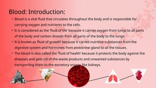
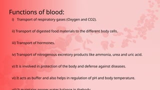
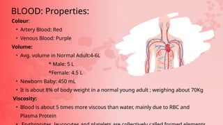
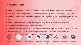
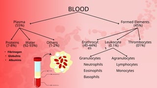
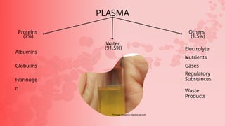
Blood and it’s
Composition
• Blood is a vital fluid that circulates throughout the body and is responsible for
carrying oxygen and nutrients to the cells.
• It is considered as the ‘fluid of life’ because it carries oxygen from lungs to all parts
of the body and carbon dioxide from all parts of the body to the lungs.
• It is known as ‘fluid of growth’ because it carries nutritive substances from the
digestive system and hormones from endocrine gland to all the tissues.
• The blood is also called the ‘fluid of health’ because it protects the body against the
diseases and gets rid of the waste products and unwanted substances by
transporting them to the excretory organs like kidneys.
Blood: Introduction:
i) Transport of respiratory gases (Oxygen and CO2).
ii) Transport of digested food materials to the different body cells.
iii) Transport of hormones.
iv) Transport of nitrogenous excretory products like ammonia, urea and uric acid.
v) It is involved in protection of the body and defense against diseases.
vi) It acts as buffer and also helps in regulation of pH and body temperature.
Functions of blood:
Colour:
• Artery Blood: Red
• Venous Blood: Purple
Volume:
• Avg. volume in Normal Adult:4-6L
* Male: 5 L
*Female: 4.5 L
• Newborn Baby: 450 mL
• It is about 8% of body weight in a normal young adult ; weighing about 70Kg
Viscosity:
• Blood is about 5 times more viscous than water, mainly due to RBC and
Plasma Protein
BLOOD: Properties:
• Blood is composed of plasma, red blood cells, white blood cells, and platelets.
• Plasma is the liquid portion of blood that makes up about 55% of blood volume.
• Red blood cells, also called erythrocytes, are responsible for carrying oxygen to the
cells.
• White blood cells, also called leukocytes, play a role in the immune system.
• Platelets are important for blood clotting.
• The blood is also called the ‘fluid of health’ because it protects the body against the
diseases and gets rid of the waste products and unwanted substances by
transporting them to the excretory organs like kidneys.
Composition:
Platelets
RBC Macrophage Monocyte Lymphocy
te
Neutrophi
l
Eosinophil Basophil
BLOOD
Formed Elements
(45%)
Plasma
(55%)
Proteins Water Others Erythrocyt
es
Thrombocytes
Leukocyte
s
Agranulocytes
Lymphocytes
Monocytes
Granulocytes
Neutrophils
Eosinophils
Basophils
(7-8%) (92-93%) (1-2%) (40-44%) (0.1%) (01%)
• Fibrinogen
• Globulins
• Albumins
• Plasma is a yellowish liquid component of blood that makes up about 55% of
total blood volume.
• It is composed of mostly water, electrolytes, and proteins, including albumin,
globulins, and fibrinogen.
• Plasma serves several important functions in the body, including maintaining
blood volume and pressure, transporting nutrients and waste products, and
playing a critical role in the immune response.
• Plasma can be separated from other blood components and used for
transfusions, such as in cases of severe bleeding or blood loss.
• Plasma also contains clotting factors, which can be used to treat certain
bleeding disorders.
• Certain medical conditions, such as liver disease or kidney disease, can affect
PLASMA:
•Fibrinogen, globulins and albumins are the major proteins.
•Fibrinogens are needed for clotting or coagulation of blood.
•Globulins primarily are involved in defense mechanisms of the body
•Albumins help in osmotic balance.
•Plasma also contains small amounts of minerals like Na+, Ca++, Mg++,
HCO3-, Cl-, etc. Glucose, amino acids, lipids, etc., are also present in the
plasma as they are always in transit in the body.
•Factors for coagulation or clotting of blood are also present in the plasma
in an inactive form. Plasma without the clotting factors is called serum.
PLASMA:
PLASMA
Others
(1.5%)
Proteins
(7%)
Water
(91.5%)
Albumins
Globulins
Fibrinoge
n
Electrolyte
s
Nutrients
Gases
Regulatory
Substances
Waste
Products
*image showing plasma serum
• Red blood cells (RBCs) are the non-nucleated formed elements in the blood.
• RBC’s are also known as erythrocytes (erythros = red). Red color of the red blood
cell is due to the presence of the coloring pigment called hemoglobin. RBCs play
a vital role in transport of respiratory gases. RBCs are larger in number
compared to the other two blood cells, namely white blood cells and platelets. 5
million-5.5million /cubic mm of blood
• RBC’s are non-nucleated, except the only mammal that has Nucleated RBC is
Camel 🐪
Erythrocytes: RBC
• In Adult Male, RBC count is 5 million/cu mm and in Female RBC count is 4.5
million/cu mm
• RBC’s are generally disc and biconcave (dumbbell ) shaped.
• RBC’s are larger in number compared to WBC and Platelets.
• Old RBC’s are destroyed in Reticuloendothelial system.
• RBCs have an average life span of 120 days after which they are destroyed in the
spleen (graveyard of RBCs).
• Haem’s solution is a specific type of dilution used in manual red blood cell (RBC)
counting
Erythrocytes: RBC
• Transport of Oxygen from Lungs to tissue and CO2 from tissue to Lungs
• Buffering action in Blood (Maintains acid-base balance)
• Helps in determination of blood group (RBC carries the blood group antigen)
• RBCs contain enzymes that help protect the body from oxidative damage
and maintain the integrity of cell membranes.
• RBCs play a role in the immune system by interacting with white blood cells
to help fight infections.
• RBCs are produced in the bone marrow and have a lifespan of about 120
days. After this time, they are removed from circulation and broken down by
the liver and spleen.
• Spleen = blood bank of body & Graveyard of RBC
Function: RBC
Destruction: RBC
• White blood cells (WBCs) are also known as leukocytes and are a type of blood cell that
helps the body fight infection and disease.
• There are several types of WBCs, including neutrophils, lymphocytes, monocytes,
eosinophils, and basophils, each with a specific role in the immune system.
• WBCs are produced in the bone marrow and circulate in the blood and lymphatic system.
• Abnormal WBC counts can be indicative of certain medical conditions, such as infections,
autoimmune disorders, and some types of cancer.
• WBC counts can be measured through a simple blood test called a complete blood count
(CBC).
• Certain medications and medical treatments, such as chemotherapy, can affect WBC
counts and increase the risk of infections.
• Maintaining a healthy immune system through proper diet, exercise, and hygiene can
help prevent some WBC-related health problems.
Leukocytes: WBC
• WBC's are colourless. They do not have haemoglobin and are nucleated cells. It is found
in the bone marrow, spleen, thymus and lymph nodes. They are capable of amoeboid
movement
• Lifespan = 3 to 4 weeks
• 8000-1000 wbc/cubic mm of blood
• Turk’s solution is a specific type of dilution used in manual White blood cell (WBC)
counting
• The white blood corpuscles can be grouped into two categories:
1. Granulocytes
2. Agranulocytes.
Leukocytes: WBC
Granulocytes
They contain granules in their cytoplasm.Their nucleus is irregular or lobed. The
granulocytes are of three types
(i) Neutrophils
(ii) Eosinophils
(iii) Basophils
i) Neutrophils
• They are large in size and have a 2 - 7 lobed nucleus. These corpuscles form 60% - 65% of
the total leucocytes. Their numbers are increased during infection and inflammation.
ii) Eosinophils
• It has a bilobed nucleus and constitute 2% - 3% of the total leucocytes. Their number
increases during conditions of allergy and parasitic infections. It brings about
detoxification of toxins.
iii) Basophils
• Basophils have lobed nucleus. They form0.5-1.0% of the total leucocytes. They release
Agranulocytes
Granules are not found in the cytoplasm of
these cells. The agranulocytes are of two types:
(i) Lymphocytes (ii) Monocytes
i) Lymphocytes
These are about 20-25% of the total leucocytes.They produce antibodies during bacterial
and
viral infections.
ii) Monocytes
They are the largest of the leucocytes and are amoeboid in shape. These cells form 5 - 6 %
of
• Platelets, also known as thrombocytes, are small, disc-shaped cell
fragments found in the blood of vertebrates.
• They are formed by fragmentation of megakaryocytes, which are found in
the bone marrow, and destroyed in Liver & spleen
• Platelets lack a nucleus and have a lifespan of about 8-10 days.
• They are essential for blood clotting and wound healing.
• Platelets play a crucial role in hemostasis, the process by which bleeding
is stopped.
• They are responsible for forming blood clots at the site of injury, which
Thrombocytes: Platelets
• Platelets also release growth factors that stimulate the repair and regeneration of
damaged tissues.
• Additionally, platelets are involved in the immune response and play a role in the
formation of new blood vessels.
• There are about 2,50,000 – 4,00,000 platelets / cubic mm of blood.
• The solution commonly used in platelet count tests is called Platelet Diluting Fluid. This
fluid typically contains formaldehyde and ammonium oxalate
Thrombocytes: Platelets
od Groups:
oncept of blood grouping wasdeveloped by Karl Landsteiner (1900). He identified blood groups A, B
O.
ood group was recognized by Decastello and Sturli (1902).
od Donation:
n an individual receives a mismatched blood group from the donor agglutination (clumping) of
d occurs in
ody which leads to death.
grouping
other antigen, the Rh antigen similar to one present in Rhesus monkeys (hence Rh), is also observed
the surface of RBCs of majority (nearly 80 per cent) of humans. Discovered by Karl lannister
ch individuals are called Rh positive (Rh+ve) and those in whom this antigen is absent are called Rh
gative (Rh-ve)
throblastosis foetalis.
special case of Rh incompatibility (mismatching) has been observed between the Rh-ve blood of a
egnant mother with Rh+ve blood of the foetus.
antigens of the foetus do not get exposed to the Rh-ve blood of the mother in the first pregnancy as
e two bloods are well separated by the placenta.
wever, during the delivery of the first child, there is a possibility of exposure of the maternal blood to
mall amounts of the Rh+ve blood from the foetus.
such cases, the mother starts preparing antibodies against Rh antigen in her blood.
case of her subsequent pregnancies, the Rh antibodies from the mother (Rh-ve) can leak into
e blood of the foetus (Rh+ve) and destroy the foetal RBCs.
is could be fatal to the foetus or could cause severe anaemia and jaundice to the baby. This
ndition is called erythroblastosis foetalis.
is can be avoided by administering anti-Rh antibodies to the mother immediately after the
livery of the first child.(anti-d injection)
Blood and it’s composition; blood composition and function

Blood and it’s composition; blood composition and function

  • 1.
  • 2.
    • Blood isa vital fluid that circulates throughout the body and is responsible for carrying oxygen and nutrients to the cells. • It is considered as the ‘fluid of life’ because it carries oxygen from lungs to all parts of the body and carbon dioxide from all parts of the body to the lungs. • It is known as ‘fluid of growth’ because it carries nutritive substances from the digestive system and hormones from endocrine gland to all the tissues. • The blood is also called the ‘fluid of health’ because it protects the body against the diseases and gets rid of the waste products and unwanted substances by transporting them to the excretory organs like kidneys. Blood: Introduction:
  • 3.
    i) Transport ofrespiratory gases (Oxygen and CO2). ii) Transport of digested food materials to the different body cells. iii) Transport of hormones. iv) Transport of nitrogenous excretory products like ammonia, urea and uric acid. v) It is involved in protection of the body and defense against diseases. vi) It acts as buffer and also helps in regulation of pH and body temperature. Functions of blood:
  • 4.
    Colour: • Artery Blood:Red • Venous Blood: Purple Volume: • Avg. volume in Normal Adult:4-6L * Male: 5 L *Female: 4.5 L • Newborn Baby: 450 mL • It is about 8% of body weight in a normal young adult ; weighing about 70Kg Viscosity: • Blood is about 5 times more viscous than water, mainly due to RBC and Plasma Protein BLOOD: Properties:
  • 5.
    • Blood iscomposed of plasma, red blood cells, white blood cells, and platelets. • Plasma is the liquid portion of blood that makes up about 55% of blood volume. • Red blood cells, also called erythrocytes, are responsible for carrying oxygen to the cells. • White blood cells, also called leukocytes, play a role in the immune system. • Platelets are important for blood clotting. • The blood is also called the ‘fluid of health’ because it protects the body against the diseases and gets rid of the waste products and unwanted substances by transporting them to the excretory organs like kidneys. Composition: Platelets RBC Macrophage Monocyte Lymphocy te Neutrophi l Eosinophil Basophil
  • 6.
    BLOOD Formed Elements (45%) Plasma (55%) Proteins WaterOthers Erythrocyt es Thrombocytes Leukocyte s Agranulocytes Lymphocytes Monocytes Granulocytes Neutrophils Eosinophils Basophils (7-8%) (92-93%) (1-2%) (40-44%) (0.1%) (01%) • Fibrinogen • Globulins • Albumins
  • 7.
    • Plasma isa yellowish liquid component of blood that makes up about 55% of total blood volume. • It is composed of mostly water, electrolytes, and proteins, including albumin, globulins, and fibrinogen. • Plasma serves several important functions in the body, including maintaining blood volume and pressure, transporting nutrients and waste products, and playing a critical role in the immune response. • Plasma can be separated from other blood components and used for transfusions, such as in cases of severe bleeding or blood loss. • Plasma also contains clotting factors, which can be used to treat certain bleeding disorders. • Certain medical conditions, such as liver disease or kidney disease, can affect PLASMA:
  • 8.
    •Fibrinogen, globulins andalbumins are the major proteins. •Fibrinogens are needed for clotting or coagulation of blood. •Globulins primarily are involved in defense mechanisms of the body •Albumins help in osmotic balance. •Plasma also contains small amounts of minerals like Na+, Ca++, Mg++, HCO3-, Cl-, etc. Glucose, amino acids, lipids, etc., are also present in the plasma as they are always in transit in the body. •Factors for coagulation or clotting of blood are also present in the plasma in an inactive form. Plasma without the clotting factors is called serum. PLASMA:
  • 9.
  • 10.
    • Red bloodcells (RBCs) are the non-nucleated formed elements in the blood. • RBC’s are also known as erythrocytes (erythros = red). Red color of the red blood cell is due to the presence of the coloring pigment called hemoglobin. RBCs play a vital role in transport of respiratory gases. RBCs are larger in number compared to the other two blood cells, namely white blood cells and platelets. 5 million-5.5million /cubic mm of blood • RBC’s are non-nucleated, except the only mammal that has Nucleated RBC is Camel 🐪 Erythrocytes: RBC
  • 11.
    • In AdultMale, RBC count is 5 million/cu mm and in Female RBC count is 4.5 million/cu mm • RBC’s are generally disc and biconcave (dumbbell ) shaped. • RBC’s are larger in number compared to WBC and Platelets. • Old RBC’s are destroyed in Reticuloendothelial system. • RBCs have an average life span of 120 days after which they are destroyed in the spleen (graveyard of RBCs). • Haem’s solution is a specific type of dilution used in manual red blood cell (RBC) counting Erythrocytes: RBC
  • 12.
    • Transport ofOxygen from Lungs to tissue and CO2 from tissue to Lungs • Buffering action in Blood (Maintains acid-base balance) • Helps in determination of blood group (RBC carries the blood group antigen) • RBCs contain enzymes that help protect the body from oxidative damage and maintain the integrity of cell membranes. • RBCs play a role in the immune system by interacting with white blood cells to help fight infections. • RBCs are produced in the bone marrow and have a lifespan of about 120 days. After this time, they are removed from circulation and broken down by the liver and spleen. • Spleen = blood bank of body & Graveyard of RBC Function: RBC
  • 13.
  • 14.
    • White bloodcells (WBCs) are also known as leukocytes and are a type of blood cell that helps the body fight infection and disease. • There are several types of WBCs, including neutrophils, lymphocytes, monocytes, eosinophils, and basophils, each with a specific role in the immune system. • WBCs are produced in the bone marrow and circulate in the blood and lymphatic system. • Abnormal WBC counts can be indicative of certain medical conditions, such as infections, autoimmune disorders, and some types of cancer. • WBC counts can be measured through a simple blood test called a complete blood count (CBC). • Certain medications and medical treatments, such as chemotherapy, can affect WBC counts and increase the risk of infections. • Maintaining a healthy immune system through proper diet, exercise, and hygiene can help prevent some WBC-related health problems. Leukocytes: WBC
  • 15.
    • WBC's arecolourless. They do not have haemoglobin and are nucleated cells. It is found in the bone marrow, spleen, thymus and lymph nodes. They are capable of amoeboid movement • Lifespan = 3 to 4 weeks • 8000-1000 wbc/cubic mm of blood • Turk’s solution is a specific type of dilution used in manual White blood cell (WBC) counting • The white blood corpuscles can be grouped into two categories: 1. Granulocytes 2. Agranulocytes. Leukocytes: WBC
  • 16.
    Granulocytes They contain granulesin their cytoplasm.Their nucleus is irregular or lobed. The granulocytes are of three types (i) Neutrophils (ii) Eosinophils (iii) Basophils i) Neutrophils • They are large in size and have a 2 - 7 lobed nucleus. These corpuscles form 60% - 65% of the total leucocytes. Their numbers are increased during infection and inflammation. ii) Eosinophils • It has a bilobed nucleus and constitute 2% - 3% of the total leucocytes. Their number increases during conditions of allergy and parasitic infections. It brings about detoxification of toxins. iii) Basophils • Basophils have lobed nucleus. They form0.5-1.0% of the total leucocytes. They release
  • 17.
    Agranulocytes Granules are notfound in the cytoplasm of these cells. The agranulocytes are of two types: (i) Lymphocytes (ii) Monocytes i) Lymphocytes These are about 20-25% of the total leucocytes.They produce antibodies during bacterial and viral infections. ii) Monocytes They are the largest of the leucocytes and are amoeboid in shape. These cells form 5 - 6 % of
  • 18.
    • Platelets, alsoknown as thrombocytes, are small, disc-shaped cell fragments found in the blood of vertebrates. • They are formed by fragmentation of megakaryocytes, which are found in the bone marrow, and destroyed in Liver & spleen • Platelets lack a nucleus and have a lifespan of about 8-10 days. • They are essential for blood clotting and wound healing. • Platelets play a crucial role in hemostasis, the process by which bleeding is stopped. • They are responsible for forming blood clots at the site of injury, which Thrombocytes: Platelets
  • 19.
    • Platelets alsorelease growth factors that stimulate the repair and regeneration of damaged tissues. • Additionally, platelets are involved in the immune response and play a role in the formation of new blood vessels. • There are about 2,50,000 – 4,00,000 platelets / cubic mm of blood. • The solution commonly used in platelet count tests is called Platelet Diluting Fluid. This fluid typically contains formaldehyde and ammonium oxalate Thrombocytes: Platelets
  • 21.
    od Groups: oncept ofblood grouping wasdeveloped by Karl Landsteiner (1900). He identified blood groups A, B O. ood group was recognized by Decastello and Sturli (1902).
  • 22.
    od Donation: n anindividual receives a mismatched blood group from the donor agglutination (clumping) of d occurs in ody which leads to death.
  • 23.
    grouping other antigen, theRh antigen similar to one present in Rhesus monkeys (hence Rh), is also observed the surface of RBCs of majority (nearly 80 per cent) of humans. Discovered by Karl lannister ch individuals are called Rh positive (Rh+ve) and those in whom this antigen is absent are called Rh gative (Rh-ve) throblastosis foetalis. special case of Rh incompatibility (mismatching) has been observed between the Rh-ve blood of a egnant mother with Rh+ve blood of the foetus. antigens of the foetus do not get exposed to the Rh-ve blood of the mother in the first pregnancy as e two bloods are well separated by the placenta. wever, during the delivery of the first child, there is a possibility of exposure of the maternal blood to mall amounts of the Rh+ve blood from the foetus.
  • 24.
    such cases, themother starts preparing antibodies against Rh antigen in her blood. case of her subsequent pregnancies, the Rh antibodies from the mother (Rh-ve) can leak into e blood of the foetus (Rh+ve) and destroy the foetal RBCs. is could be fatal to the foetus or could cause severe anaemia and jaundice to the baby. This ndition is called erythroblastosis foetalis. is can be avoided by administering anti-Rh antibodies to the mother immediately after the livery of the first child.(anti-d injection)