


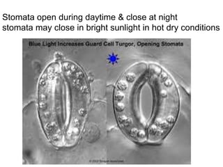
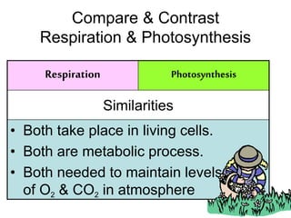
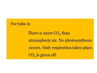
![Breathing
is an involuntary function of the CNS
a respiratory / breathing center in the
medulla oblongata [part of brain stem] :
establishes basic breathing pattern
Brain stem Spinal cord](https://image.slidesharecdn.com/chapter7-respirationpart2-141111210504-conversion-gate02/85/BIOLOGY-FORM-4-CHAPTER-7-RESPIRATION-PART-2-39-320.jpg)
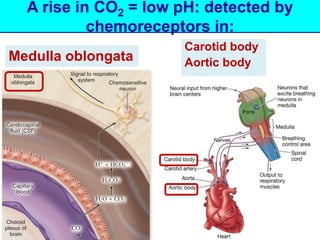
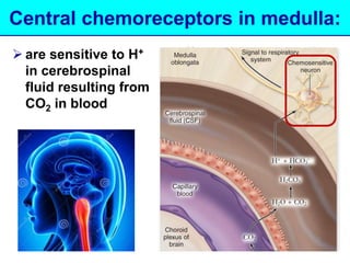
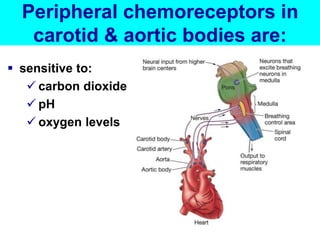

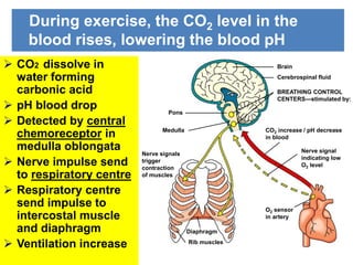
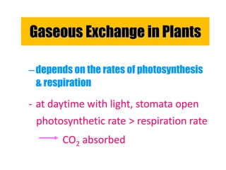
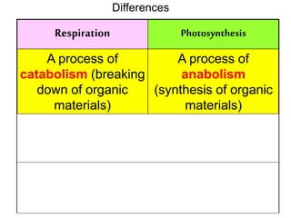
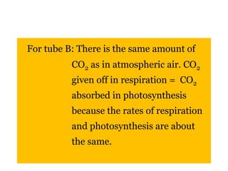
![CO2
Water
Carbonic acid pH
Central chemoreceptor
[medulla oblongata]
Impulse
Respiratory centre
Detected by
Impulse
Intercostals muscle diaphragm
Ventilation faster CO2 eliminate faster](https://image.slidesharecdn.com/chapter7-respirationpart2-141111210504-conversion-gate02/85/BIOLOGY-FORM-4-CHAPTER-7-RESPIRATION-PART-2-59-320.jpg)


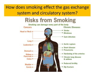


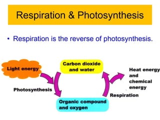


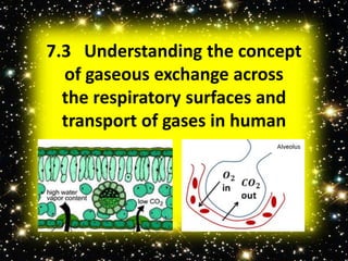
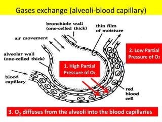
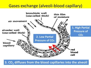
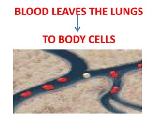
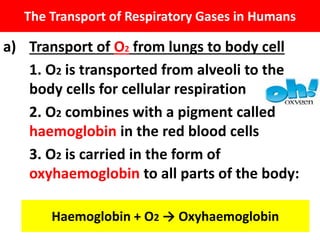
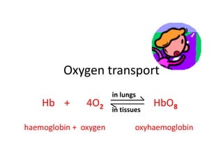
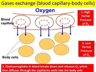
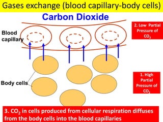
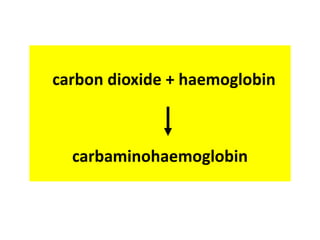
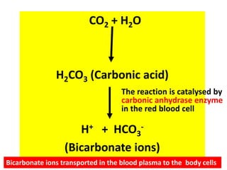
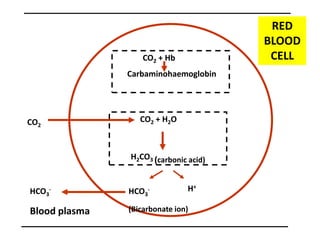
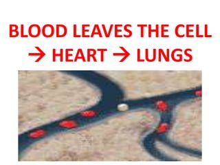
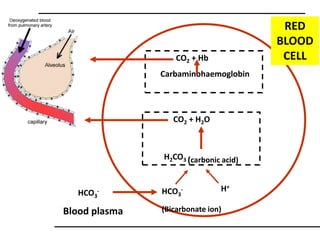
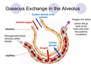
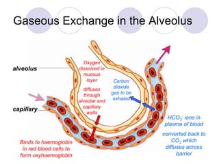
The document discusses respiration and the transport of gases in the human body. It explains: 1) How gases are exchanged between the alveoli and blood in the lungs, and between the blood and body cells, via diffusion. 2) How oxygen is transported from the lungs to body cells by binding to hemoglobin in red blood cells and being carried to tissues. 3) How carbon dioxide is transported from tissues to the lungs, carried in three forms: as carbonic acid, carbaminohaemoglobin, and bicarbonate ions.


![breathing and exchange of gases (1).pptx [Repaired].pptx](https://cdn.slidesharecdn.com/ss_thumbnails/breathingandexchangeofgases1-250919170828-d5147614-thumbnail.jpg?width=640&height=640&fit=bounds)


