SlideShare a Scribd company logo
Biology | Form 4 | Chapter 3: Movement of Substances across the Plasma Membrane | Experiment
Experiment 3.1

Aim                 To study the movement of substances across the Visking tubing
Problem statement   What are the factors affecting the movement of substances across a semi-permeable membrane?
Hypothesis          The movement of the molecules across a semi-permeable membrane depends on the sizes of the molecules.
Variables           (a) Manipulated variable: Size of solute molecules
                    (b) Responding variable: Colour of the solution in Visking tubing
                    (c) Fixed variable: Volume of solutions, time, surrounding temperature
Materials           5% starch suspension, 20% glucose solution, distilled water, Benedict’s solution, iodine solution, Visking tubing and thread
Apparatus           Boiling tube, test tubes, pipette, test tube holder and Bunsen burner
Procedure


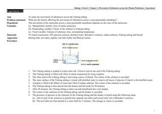
                    1.   The Visking tubing is soaked in water until soft. A knot is tied at one end of the Visking tubing.
                    2.   The Visking tubing is filled with 10ml of starch suspension by using a pipette.
                    3.   The other end of the Visking tubing is tied using a piece of thread. The colour of the solution is recorded.
                    4.   The outer surface of the Visking tubing is rinsed with distilled water to remove all traces of glucose of starch in the distilled water.
                    5.   A beaker is filled with 300ml of water and 10ml of iodine solution. The colour of the solution is recorded.
                    6.   The Visking tubing is then placed into the beaker and left aside for 30 minutes.
                    7.   After 30 minutes, the Visking tubing is taken out and transferred into a dry beaker.
                    8.   The colour of the solutions in the Visking tubing and the beaker is recorded.
                    9.   The presence of glucose in the solutions of the Visking tubing and the beaker is tested using the following steps.
                         (a) 2ml of each of the solutions is poured into separate test tubes and mixed with 1ml of Benedict’s solotion.
                         (b) The test tubes are then heated in a water bath for 3 minutes. The change in colour is recorded.


                                                                                                                                                                    1
Biology | Form 4 | Chapter 3: Movement of Substances across the Plasma Membrane | Experiment


Observation                               Content of Visking tubing                         Contents of beaker
                     Test
                                     Initial colour        Final colour            Initial colour        Final colour
               Iondine test               Clear             Blue black                 Brown                Brown
               Benedict’s test            Blue                 Blue                     Blue           Orange precipitate

Discussion    1.  At the end of the experiment, the solution in the Visking tubing turns blue-black. This shows the both iodine and starch are presence
                  in the Visking tubing.
              2. Iodine molecules in the beaker can diffuse throught the semi-permeable membrane of the Visking tubing.
              3. Starch molecules cannot diffuse through the Visking tubing. As the result, the colour of the solution in the beaker remains yellow.
              4. The brick red precipitate formed shows that glucose is found in the beaker at the end of the experiment.
              5. This shows that the Visking tubing has pores big enough for the glucose and iodine molecules to diffuse through but too small for
                  the starch molecules to diffuse through.
Conclusion    The Visking tubing is a semi-permeable membrane that only allows substances with small molecules to diffuse through it. The move of
              molecules across a semi-permeable membrane depends on their sizes. The hypothesis is acceptable.


                                                                                                                                                          2
Biology | Form 4 | Chapter 3: Movement of Substances across the Plasma Membrane | Experiment
Experiment 3.2

Aim                 To demonstrate the process of osmosis using a simple osmometer.
Problem Statement   How does osmosis take place?
Hypothesis          Osmosis takes place when water moves from a region of low solute concentration to a region of high solute concentration across a
                    semi-permeable membrane.
Variables           (a) Manipulated variable: Concentration of solution
                    (b) Responding variable: The level of sucrose solution in the capillary tube
                    (c) Fixed variable: Surrounding temperature
Materials           Visking tubing, 30% sucrose solution, distilled water and thread
Apparatus           Beaker, retord stand and clamp, capillary tube, pipette and marker pen
Procedure


                    1.   The Visking tubing is soaked in water until soft. A knot is tied at one end of the Visking tubing.
                    2.   The Visking tubing is filled with 20 ml of sucrose solution using pipette.
                    3.   The other end of Visking tubing is then fitted onto one end of the capillary tube and is tied with a piece of thread.
                    4.   The outer surface of the Visking tubing is rinsed with distilled water.
                    5.   The Visking tubing is immersed into a beaker of distilled water and the capillary tube is clamped to a retord stand.
                    6.   The initial level of the sucrose solution in the capillary tube is marked with a marker pen.
                    7.   The level of sucrose solution in the capillary tube is marked again after 30 minutes.
                    8.   Steps 1 to 7 are repeated to set up a control experiment. Distilled water is used instead of sucrose solution in Visking tubing.
Observation         1.   The final level of sucrose solution in the capillary tube is higher than the initial level.
                    2.   The water level in the control experiment remains unchanged.


                                                                                                                                                                 3
Biology | Form 4 | Chapter 3: Movement of Substances across the Plasma Membrane | Experiment
Discussion   1.  The Visking tubing is a semi-permeable membrane. It has pores that are small enough for water molecules to diffuse through it but
                 not big enough for sucrose molecules to diffuse out of it.
             2. As the concentration of the sucrose is higher than that of water in the beaker, water molecules will diffuse into the Visking tubing
                 from the beaker. This causes the level of sucrose solution in the capillary tube to move higher.
             3. In the control experiment where the concentration of distilled water in the Visking tubing and the beaker are the same, the water
                 level remains unchanged because the rate of water molecules moving into the Visking tubing is the same as the rate of water moving
                 out of it.
Conclusion   The hypothesis is accepted that osmosis is a process by which water moves from a region of low solute concentration to a region of high
             solute concentration across a selectively-permeable membrane.


                                                                                                                                                        4
Biology | Form 4 | Chapter 3: Movement of Substances across the Plasma Membrane | Experiment
Activity 3.1

Aim            To study the wffects of hypotonic, hypertonic and isotonic solutions on plant cells.
Materials      Onion, 0.5 M sucrose solution, 1.0 M sucrose solution and distilled water
Apparatus      Light microscope, glass slides, cover slips, scalpel, forceps, dropper, white tile and petri dish
Procedure      1. A thin layer of epidermis is peeled off from the inside of an onion scale leaf.
               2. The epidermis is placed in a drop of distilled water on a glass silde lebelled A. the glass slide is covered with a cover slip.
               3. Silde A is observed under the microscope and the epidermal cells are drawn.
               4. Another piece of epidermis is obtained and placed in a drop of 0.5 M sucrose solution on another galss slide lebelled B. the glass
                   slide is covered with a cover slip.
               5. Slide B is observed under the microscope and the epidermal cells are drawn.
               6. Steps 4 to 5 are repeated using 1.0 M sucrose solution and another glass slide, C to replace the sucrose solution.
               7. After observing under the microscope, the epidermis in the 1.0 M sucrose solution is transferred into a petri dish filled with distilled
                   water for a few minutes. The epidermis is again placed in a drop of distilled water on a glass slide labeled D. similar to the glass
                   slides before, glass slide D is covered using a cover slip.
               8. Slide D is observed again under the microscope and the epidermal cells are drawn.
Observation                  Slide                           Observation                    Shape or condition of the cell
                A (distilled water)                                                     Turgid, very large vacuole


                B (0.5 M sucrose solution)                                             Turgid, vacuole is in the center of the
                                                                                       cell


                C (1.0 M sucrose solution)                                             Flaccid, small vacuole, cytoplasm
                                                                                       shrinks, plasma membrane separates
                                                                                       from the cell wall


                                                                                                                                                           5
Biology | Form 4 | Chapter 3: Movement of Substances across the Plasma Membrane | Experiment


              D (from 1.0 M sucrose                                                   Turgid, very large vacuole
              solution into distilled water)


Discussiom   1.   The distilled water is less concentrated than or hypotonic to the cell sap of onion cells. When the onion cells are placed in it, water
                  diffuses into the vacuoles of the cells by osmosis. This causes the vacuoles to expand and press against the plasma membrane and
                  the cell wall. The cells are said to be turgid.
             2.   0.5 M sucrose solution is isotonic to the cell sap of onion cells. The cells neither gain nor lose water by osmosis.
             3.   1.0 M sucrose solution is more concentrated or hypertonic to the cell sap of onion cells. Then the onion cells are placed in it, water
                  diffuses out from the vacuoles of the cells by osmosis. This causes the vacuoles to shrink and the plasma membrane to detach from
                  the cell wall. The cells are filled with water and said to be plasmolysed by the solution and have become flaccid.
             4.   When the onion cells in the 1.0 M sucrose solution are transferred into distilled water which is hypotonic to water diffuses into the
                  onion cells by osmosis. The cells are filled with water and are said to have undergone deplasmolysis. The cells are turgid.
Conclusion   1.   When a plant cell is placed in a hypotonic solution, the cell becomes turgid as water diffuses into it by osmosis.
             2.   When a plant cell is placed in an isotonic solution, the cell neither gain nor lose water by osmosis.
             3.   When a plant cell is placed in a hypertonic solution, the cell becomes flaccid as water diffuses out of it by osmosis. The cell
                  undergoes plasmolysis.
             4.   When a plant cell placed in a hypertonic solution is transferred back into a hypotonic solution, the cell becomes turgid again as
                  water diffuses into it by osmosis.


                                                                                                                                                          6
Biology | Form 4 | Chapter 3: Movement of Substances across the Plasma Membrane | Experiment
Activity 3.2

Aim            To study the effects of hypotonic, hypertonic and isotonic solutions on animal cells such as red blood cells.
Materials      Chicken blood, 0.85% sodium chloride solution and distilled water
Apparatus      Light microscope, glass slides, cover slips, test tubes, pipette and beaker
Procedure      1. Four slides are prepared with labels: A, B, C and D.
               2. A drop of chicken blood is dropped onto slide A and covered with a cover slip.
               3. The slide is observed under a microscope and the shape of the red blood cells is drawn.
               4. A drop of distilled water is dropped onto slide B and covered with a cover slip. A drop of blood is put at one slide of the cover slip.
               5. When the blood is drawn into the water, the slide is observed under the microscope. The shape of the red blood cells is drawn.
               6. Steps 4 to 5 are repeated using 0.85% sodium chloride solution on slide C and 5% sodium chloride solution on slide D to replace
                   distilled water.
Observation
                Slide                               A                          B                           C                            D
                Observation


                shape or condition Biconcave dics shape             Fragments of plasma Biconcave dics shape               Crinkling of plasma
                of red blood cell                                   membrane haemolysis                                    membrane cell(crenation)

Discussion     1.   When the red blood cells are placed in the distilled water which is hypotonic, water diffuses into the red blood cells. This causes the
                    cells to swell and burst. As a result, the haemoglobin spills out of the red blood cells. Under the microscope, no red blood cells are
                    observed. There are only broken cell membranes. The red blood cells are said to have undergone haemolysis.
               2.   The shape of the red blood cells in the 0.85% sodium chloride solution remains the same when observed under the microscope. The
                    red blood cells remain the same as it is placed in an isotonic solution,
               3.   When the red blood cells are placed in 5% sodium chloride solution which is hypertonic, the water diffuses out from the red blood
                    cells. This causes the cells to shrink. This process is called crenation.
Conclusion     1.   In an isotonic solution, red blood cells remain the same.
               2.   In a hypotonic solution, red blood cells burst and undergo haemolysis.
               3.   In a hypertonic solution, red blood cells shrink and crenate.


                                                                                                                                                            7
Biology | Form 4 | Chapter 3: Movement of Substances across the Plasma Membrane | Experiment
Experiment 3.3

Aim                 To determine the concentration of an external solution which is isotonic to the cell sap of plants.
Problem statement   Which concentration of the sucrose solution is isotonic to the cell sap of plants?
Hypothesis          0.36 M sucrose solution is isotonic to the cell sap of plants.
Variables           (a) Manipulated variable: Concentration of sucrose solution
                    (b) Responding variable: Length of potato strips
                    (c) Fixed variable: Temperature, volume of sucrose solution used, time
Materials           A potato, distilled water, filter paper, sucrose solutions with concentrations of 0.1 M, 0.2 M, 0.3 M, 0.4 M, 0.5 M and 0.6 M
Apparatus           Seven tested tubes, cork borer, scalpel, forceps, beaker, ruler, glass rod and white tile
Procedure           1. Seven cylindrical strips of potato are obtained using a cork borer. Each strip is measured and cut into a length of 5 cm.
                    2. Seven test tubes were labeled A, B, C, D, E, F and G.
                    3. The test tubes are then filled with the different concentrations of solutions as listed below and placed upright on a test tube rack.
                         Test tube A: 10 ml of distilled water                   Test tube E: 10 ml of 0.4 M sucrose solution
                         Test tube B: 10 ml of 0.1 M sucrose solution            Test tube F: 10 ml of 0.5 M sucrose solution
                         Test tube C: 10 ml of 0.2 M sucrose solution            Test tube G: 10 ml of 0.6 M sucrose solution
                         Test tube D: 10 ml of 0.3 M sucrose solution
                    4. One strip of potato is then added into each of the test tubes A to G, and left to stand for an hour.
                    5. After an hour, each potato strip is removed from the test tube using the forceps and wiped dry with filter paper. The texture of the
                        potato strip is observed and the length is measured and recorded in a table.
                    6. From the results obtained, a graph of the change in length of the potato strips aginst the concentration of sucrose solution is plotted.
                        The concentration at which the change in length is zero is the concentration of the potato cell sap.
Observation
                                                                   Length of potato strip (cm)
                      Test tube              solution                                                Change in length        Texture
                                                                     Initial          Final
                          A         distilled water                   5.00             5.95                 +0.95            Turgid
                          B         0.1 M sucrose solution            5.00             5.60                 +0.60            Turgid
                          C         0.2 M sucrose solution            5.00             5.35                 +0.35            Turgid
                          D         0.3 M sucrose solution            5.00             5.10                 +0.10            Turgid
                          E         0.4 M sucrose solution            5.00             4.95                 -0.05            Flaccid
                          F         0.5 M sucrose solution            5.00             4.80                 -0.20            Flaccid
                          G         0.6 M sucrose solution            5.00             4.70                 -0.30            Flaccid

                                                                                                                                                                8
Biology | Form 4 | Chapter 3: Movement of Substances across the Plasma Membrane | Experiment


Discussion   1.  The distilled water an sucrose solutions from 0.1 M to 0.3 M are hypotonic to potato cell sap. Water diffuses into the cells by
                 osmosis and the potato strips becomes turgid and longer.
             2. The 0.4 M to 0.6 M sucrose solutions are hypertonic to the potato cell sap. This causes water to leave the potato cell sap by osmosis.
                 The potato strips become mere and more falccid and shorter as the concentration of the sucrose solution increases.
             3. The concentration of sucrose solution that is isotonic to the potato cell sap can be obtained by reading from the graph. The
                 concentration at which there is no change in the length in the potato strip is the concentration of the potato cell sap. This is because
                 in an isotonic solution, there is no net movement of water into the cells and out of the cells.
Conclusion   The concentration of the potato cell sap is equivalent to 0.36 M sucrose solution. The hypothesis is accepted.


                                                                                                                                                         9

More Related Content

What's hot

F4 Experiments
F4 ExperimentsF4 Experiments
F4 Experimentsmarjerin
 
28.prinsip archimedes
28.prinsip archimedes28.prinsip archimedes
28.prinsip archimedes
Atiqah Azmi
 
Jadual ion kimia
Jadual ion kimia Jadual ion kimia
Jadual ion kimia
alhalfeast
 
Teknik menjawab soalan sains
Teknik menjawab soalan sainsTeknik menjawab soalan sains
Teknik menjawab soalan sainssazaser
 
Langkah membina negara maju dan sejahtera
Langkah membina negara maju dan sejahteraLangkah membina negara maju dan sejahtera
Langkah membina negara maju dan sejahtera
JANGAN TINGGAL DAKU
 
Peribahasa dan cogan kata dalam karangan
Peribahasa dan cogan kata dalam karanganPeribahasa dan cogan kata dalam karangan
Peribahasa dan cogan kata dalam karangan
nooraini kasturi
 
5.1 Mitosis tingkatan 4
5.1 Mitosis tingkatan 45.1 Mitosis tingkatan 4
5.1 Mitosis tingkatan 4
Jummy Masindah Jm
 
Nota hukum gas
Nota hukum gasNota hukum gas
Nota hukum gas
Lokman Hakim
 
Biologi Ting 4 (Bab 3 - Pergerakan Bahan Merentas Membran Plasma
Biologi Ting 4 (Bab 3 - Pergerakan Bahan Merentas Membran PlasmaBiologi Ting 4 (Bab 3 - Pergerakan Bahan Merentas Membran Plasma
Biologi Ting 4 (Bab 3 - Pergerakan Bahan Merentas Membran Plasma
Fawwaz Rahim
 
6. Polimer Semula Jadi
6. Polimer Semula Jadi6. Polimer Semula Jadi
6. Polimer Semula Jadi
ROSNANI MOHAMED JAMALLUDIN
 
Eksotermik & endotermik
Eksotermik & endotermikEksotermik & endotermik
Eksotermik & endotermik
Gee3145
 
FIZIK TG 5 KANTA
FIZIK TG 5 KANTAFIZIK TG 5 KANTA
FIZIK TG 5 KANTA
Ramli Rem
 
MM Tingkatan 5, 3.1.2 mengira premium
MM Tingkatan 5, 3.1.2 mengira premiumMM Tingkatan 5, 3.1.2 mengira premium
MM Tingkatan 5, 3.1.2 mengira premium
Noor Syamila Mohd
 
4.0 haba
4.0 haba4.0 haba
4.0 haba
MrHan Physics
 
Sejarah Paper3 SPM Soalan KBKK
Sejarah Paper3 SPM  Soalan KBKKSejarah Paper3 SPM  Soalan KBKK
Sejarah Paper3 SPM Soalan KBKK
Zhang Ewe
 
Formula fizik f4
Formula fizik f4Formula fizik f4
Formula fizik f4
Sal Aw
 
Susunan ayat biasa dan susunan ayat songsang
Susunan ayat biasa dan susunan ayat songsangSusunan ayat biasa dan susunan ayat songsang
Susunan ayat biasa dan susunan ayat songsangHT Hong
 
Bab 2 Struktur sel dan organisasi sel
Bab 2 Struktur sel dan organisasi selBab 2 Struktur sel dan organisasi sel
Bab 2 Struktur sel dan organisasi sel
lina jahari
 
SAINS TINGKATAN 4 BAB 8
SAINS TINGKATAN 4 BAB 8SAINS TINGKATAN 4 BAB 8
SAINS TINGKATAN 4 BAB 8
sumayy fa
 
Organisasi sel amoeba (sarah)
Organisasi sel amoeba (sarah)Organisasi sel amoeba (sarah)
Organisasi sel amoeba (sarah)
Nurul Azuwin
 

What's hot (20)

F4 Experiments
F4 ExperimentsF4 Experiments
F4 Experiments
 
28.prinsip archimedes
28.prinsip archimedes28.prinsip archimedes
28.prinsip archimedes
 
Jadual ion kimia
Jadual ion kimia Jadual ion kimia
Jadual ion kimia
 
Teknik menjawab soalan sains
Teknik menjawab soalan sainsTeknik menjawab soalan sains
Teknik menjawab soalan sains
 
Langkah membina negara maju dan sejahtera
Langkah membina negara maju dan sejahteraLangkah membina negara maju dan sejahtera
Langkah membina negara maju dan sejahtera
 
Peribahasa dan cogan kata dalam karangan
Peribahasa dan cogan kata dalam karanganPeribahasa dan cogan kata dalam karangan
Peribahasa dan cogan kata dalam karangan
 
5.1 Mitosis tingkatan 4
5.1 Mitosis tingkatan 45.1 Mitosis tingkatan 4
5.1 Mitosis tingkatan 4
 
Nota hukum gas
Nota hukum gasNota hukum gas
Nota hukum gas
 
Biologi Ting 4 (Bab 3 - Pergerakan Bahan Merentas Membran Plasma
Biologi Ting 4 (Bab 3 - Pergerakan Bahan Merentas Membran PlasmaBiologi Ting 4 (Bab 3 - Pergerakan Bahan Merentas Membran Plasma
Biologi Ting 4 (Bab 3 - Pergerakan Bahan Merentas Membran Plasma
 
6. Polimer Semula Jadi
6. Polimer Semula Jadi6. Polimer Semula Jadi
6. Polimer Semula Jadi
 
Eksotermik & endotermik
Eksotermik & endotermikEksotermik & endotermik
Eksotermik & endotermik
 
FIZIK TG 5 KANTA
FIZIK TG 5 KANTAFIZIK TG 5 KANTA
FIZIK TG 5 KANTA
 
MM Tingkatan 5, 3.1.2 mengira premium
MM Tingkatan 5, 3.1.2 mengira premiumMM Tingkatan 5, 3.1.2 mengira premium
MM Tingkatan 5, 3.1.2 mengira premium
 
4.0 haba
4.0 haba4.0 haba
4.0 haba
 
Sejarah Paper3 SPM Soalan KBKK
Sejarah Paper3 SPM  Soalan KBKKSejarah Paper3 SPM  Soalan KBKK
Sejarah Paper3 SPM Soalan KBKK
 
Formula fizik f4
Formula fizik f4Formula fizik f4
Formula fizik f4
 
Susunan ayat biasa dan susunan ayat songsang
Susunan ayat biasa dan susunan ayat songsangSusunan ayat biasa dan susunan ayat songsang
Susunan ayat biasa dan susunan ayat songsang
 
Bab 2 Struktur sel dan organisasi sel
Bab 2 Struktur sel dan organisasi selBab 2 Struktur sel dan organisasi sel
Bab 2 Struktur sel dan organisasi sel
 
SAINS TINGKATAN 4 BAB 8
SAINS TINGKATAN 4 BAB 8SAINS TINGKATAN 4 BAB 8
SAINS TINGKATAN 4 BAB 8
 
Organisasi sel amoeba (sarah)
Organisasi sel amoeba (sarah)Organisasi sel amoeba (sarah)
Organisasi sel amoeba (sarah)
 

Viewers also liked

Onion and cheek cell lab
Onion and cheek cell labOnion and cheek cell lab
Onion and cheek cell labD Sanders
 
Laporan Praktikum Difusi Osmosis dan Plasmolisis
Laporan Praktikum Difusi Osmosis dan PlasmolisisLaporan Praktikum Difusi Osmosis dan Plasmolisis
Laporan Praktikum Difusi Osmosis dan Plasmolisis
Nur Meili Zakiyah
 
ReverseOsmosisLabReport
ReverseOsmosisLabReportReverseOsmosisLabReport
ReverseOsmosisLabReportJanet Mok
 
Aprevised osmosis lab
Aprevised osmosis labAprevised osmosis lab
Aprevised osmosis labsbarkanic
 
Osmosis report powerpoint
Osmosis report powerpointOsmosis report powerpoint
Osmosis report powerpointclairebloom
 
Osmosis, diffusion, active transport
Osmosis, diffusion, active transportOsmosis, diffusion, active transport
Osmosis, diffusion, active transportMarthese Azzopardi
 
Diffusion and osmosis student handout
Diffusion and osmosis   student handout Diffusion and osmosis   student handout
Diffusion and osmosis student handout ilanasaxe
 
Bio22 Lab - Ex 4 (Cell Transport - Osmosis in Human RBC)
Bio22 Lab - Ex 4 (Cell Transport - Osmosis in Human RBC)Bio22 Lab - Ex 4 (Cell Transport - Osmosis in Human RBC)
Bio22 Lab - Ex 4 (Cell Transport - Osmosis in Human RBC)
Josh Protacio
 
1 p9 onion cells 291110
1 p9 onion cells 2911101 p9 onion cells 291110
1 p9 onion cells 291110drgav2005
 
Ib biology lab_report_sample
Ib biology lab_report_sampleIb biology lab_report_sample
Ib biology lab_report_sample
npopova
 
Biologi f4 sbp akhir tahun 2008
Biologi f4 sbp akhir tahun 2008Biologi f4 sbp akhir tahun 2008
Biologi f4 sbp akhir tahun 2008
Stanley Lee
 
Experiment 2 transport of materials across cell membranes and plant cell wate...
Experiment 2 transport of materials across cell membranes and plant cell wate...Experiment 2 transport of materials across cell membranes and plant cell wate...
Experiment 2 transport of materials across cell membranes and plant cell wate...
Nadine Uy
 
Classificazione software
Classificazione softwareClassificazione software
Classificazione software
Salvatore Cianciabella
 
Igcse2010 -osmosis & diffusion
Igcse2010 -osmosis & diffusionIgcse2010 -osmosis & diffusion
Igcse2010 -osmosis & diffusion
biologia, geologia , poesía , vida,
 
Osmosis project of chemistry
Osmosis project of chemistryOsmosis project of chemistry
Osmosis project of chemistry
Ayushi Sharma
 
Tor the onion router
Tor  the onion routerTor  the onion router
Tor the onion routerAshly Liza
 
Diffusion lab
Diffusion labDiffusion lab
Diffusion lab14771
 

Viewers also liked (20)

Cells Osmosis
Cells OsmosisCells Osmosis
Cells Osmosis
 
Onion and cheek cell lab
Onion and cheek cell labOnion and cheek cell lab
Onion and cheek cell lab
 
Laporan Praktikum Difusi Osmosis dan Plasmolisis
Laporan Praktikum Difusi Osmosis dan PlasmolisisLaporan Praktikum Difusi Osmosis dan Plasmolisis
Laporan Praktikum Difusi Osmosis dan Plasmolisis
 
ReverseOsmosisLabReport
ReverseOsmosisLabReportReverseOsmosisLabReport
ReverseOsmosisLabReport
 
Aprevised osmosis lab
Aprevised osmosis labAprevised osmosis lab
Aprevised osmosis lab
 
Osmosis report powerpoint
Osmosis report powerpointOsmosis report powerpoint
Osmosis report powerpoint
 
Osmosis, diffusion, active transport
Osmosis, diffusion, active transportOsmosis, diffusion, active transport
Osmosis, diffusion, active transport
 
Diffusion and osmosis student handout
Diffusion and osmosis   student handout Diffusion and osmosis   student handout
Diffusion and osmosis student handout
 
Igcse2010 fernando enzymes
Igcse2010 fernando enzymesIgcse2010 fernando enzymes
Igcse2010 fernando enzymes
 
Bio22 Lab - Ex 4 (Cell Transport - Osmosis in Human RBC)
Bio22 Lab - Ex 4 (Cell Transport - Osmosis in Human RBC)Bio22 Lab - Ex 4 (Cell Transport - Osmosis in Human RBC)
Bio22 Lab - Ex 4 (Cell Transport - Osmosis in Human RBC)
 
1 p9 onion cells 291110
1 p9 onion cells 2911101 p9 onion cells 291110
1 p9 onion cells 291110
 
Ib biology lab_report_sample
Ib biology lab_report_sampleIb biology lab_report_sample
Ib biology lab_report_sample
 
Biologi f4 sbp akhir tahun 2008
Biologi f4 sbp akhir tahun 2008Biologi f4 sbp akhir tahun 2008
Biologi f4 sbp akhir tahun 2008
 
Experiment 2 transport of materials across cell membranes and plant cell wate...
Experiment 2 transport of materials across cell membranes and plant cell wate...Experiment 2 transport of materials across cell membranes and plant cell wate...
Experiment 2 transport of materials across cell membranes and plant cell wate...
 
Classificazione software
Classificazione softwareClassificazione software
Classificazione software
 
Igcse2010 -osmosis & diffusion
Igcse2010 -osmosis & diffusionIgcse2010 -osmosis & diffusion
Igcse2010 -osmosis & diffusion
 
Plasmolysis
PlasmolysisPlasmolysis
Plasmolysis
 
Osmosis project of chemistry
Osmosis project of chemistryOsmosis project of chemistry
Osmosis project of chemistry
 
Tor the onion router
Tor  the onion routerTor  the onion router
Tor the onion router
 
Diffusion lab
Diffusion labDiffusion lab
Diffusion lab
 

Similar to 132441402 chapter-3-movement-of-substances-across-the-plasma-membrane

The movement of substances in the plasma membrane - Osmosis
The movement of substances in the plasma membrane - OsmosisThe movement of substances in the plasma membrane - Osmosis
The movement of substances in the plasma membrane - Osmosis
delcampotangaan
 
4 biological membranes
4 biological membranes4 biological membranes
4 biological membranes
Jaden Francis
 
Movement of substances 2013
Movement of substances 2013Movement of substances 2013
Movement of substances 2013Jacklyn Kong
 
C3 -.pptx
C3 -.pptxC3 -.pptx
C3 -.pptx
NingCheng12
 
Practical
PracticalPractical
Practical
pcjmanju
 
Movement in and out of a cell
Movement in and out of a cellMovement in and out of a cell
Movement in and out of a cell
Nathan Ssekamatte
 
Movement of substances
Movement of substancesMovement of substances
Movement of substances
chenghong03
 
Englishcell2 po poprawie
Englishcell2 po poprawieEnglishcell2 po poprawie
Englishcell2 po poprawiemastx
 
Dialysis
DialysisDialysis
Dialysis
Afra Fathima
 
Dialysis lecture 3
Dialysis lecture 3Dialysis lecture 3
Dialysis lecture 3
Sidra Shaffique
 
[M1.3] READ - Movement In & Out of Cells (PowerPoint).ppt
[M1.3] READ - Movement In & Out of Cells (PowerPoint).ppt[M1.3] READ - Movement In & Out of Cells (PowerPoint).ppt
[M1.3] READ - Movement In & Out of Cells (PowerPoint).ppt
GitaFaroka2
 
Diffustion osmosis 2014
  Diffustion osmosis 2014  Diffustion osmosis 2014
Diffustion osmosis 2014
Lorraine Stratton
 
4. cell membrane and transport 7-1-19.pptx
4. cell membrane and transport  7-1-19.pptx4. cell membrane and transport  7-1-19.pptx
4. cell membrane and transport 7-1-19.pptx
Selva Kumari
 
passive-transport.pptx
passive-transport.pptxpassive-transport.pptx
passive-transport.pptx
Sora519727
 
10. Transport System in Organisms E-learning - Diffusion & Osmosis
10. Transport System in Organisms E-learning - Diffusion & Osmosis10. Transport System in Organisms E-learning - Diffusion & Osmosis
10. Transport System in Organisms E-learning - Diffusion & Osmosis
harrywwh
 

Similar to 132441402 chapter-3-movement-of-substances-across-the-plasma-membrane (20)

The movement of substances in the plasma membrane - Osmosis
The movement of substances in the plasma membrane - OsmosisThe movement of substances in the plasma membrane - Osmosis
The movement of substances in the plasma membrane - Osmosis
 
4 biological membranes
4 biological membranes4 biological membranes
4 biological membranes
 
Movement of substances 2013
Movement of substances 2013Movement of substances 2013
Movement of substances 2013
 
C3 -.pptx
C3 -.pptxC3 -.pptx
C3 -.pptx
 
Diffusion prac
Diffusion pracDiffusion prac
Diffusion prac
 
Practical
PracticalPractical
Practical
 
Movement in and out of a cell
Movement in and out of a cellMovement in and out of a cell
Movement in and out of a cell
 
Lect 1 biochemiical
Lect 1 biochemiicalLect 1 biochemiical
Lect 1 biochemiical
 
Ppt diffusion and osmosis
Ppt diffusion and osmosisPpt diffusion and osmosis
Ppt diffusion and osmosis
 
Movement of substances
Movement of substancesMovement of substances
Movement of substances
 
Osmosis
OsmosisOsmosis
Osmosis
 
Englishcell2 po poprawie
Englishcell2 po poprawieEnglishcell2 po poprawie
Englishcell2 po poprawie
 
Dialysis
DialysisDialysis
Dialysis
 
Dialysis lecture 3
Dialysis lecture 3Dialysis lecture 3
Dialysis lecture 3
 
Micro lab 3
Micro lab 3Micro lab 3
Micro lab 3
 
[M1.3] READ - Movement In & Out of Cells (PowerPoint).ppt
[M1.3] READ - Movement In & Out of Cells (PowerPoint).ppt[M1.3] READ - Movement In & Out of Cells (PowerPoint).ppt
[M1.3] READ - Movement In & Out of Cells (PowerPoint).ppt
 
Diffustion osmosis 2014
  Diffustion osmosis 2014  Diffustion osmosis 2014
Diffustion osmosis 2014
 
4. cell membrane and transport 7-1-19.pptx
4. cell membrane and transport  7-1-19.pptx4. cell membrane and transport  7-1-19.pptx
4. cell membrane and transport 7-1-19.pptx
 
passive-transport.pptx
passive-transport.pptxpassive-transport.pptx
passive-transport.pptx
 
10. Transport System in Organisms E-learning - Diffusion & Osmosis
10. Transport System in Organisms E-learning - Diffusion & Osmosis10. Transport System in Organisms E-learning - Diffusion & Osmosis
10. Transport System in Organisms E-learning - Diffusion & Osmosis
 

132441402 chapter-3-movement-of-substances-across-the-plasma-membrane

  • 1. Biology | Form 4 | Chapter 3: Movement of Substances across the Plasma Membrane | Experiment Experiment 3.1 Aim To study the movement of substances across the Visking tubing Problem statement What are the factors affecting the movement of substances across a semi-permeable membrane? Hypothesis The movement of the molecules across a semi-permeable membrane depends on the sizes of the molecules. Variables (a) Manipulated variable: Size of solute molecules (b) Responding variable: Colour of the solution in Visking tubing (c) Fixed variable: Volume of solutions, time, surrounding temperature Materials 5% starch suspension, 20% glucose solution, distilled water, Benedict’s solution, iodine solution, Visking tubing and thread Apparatus Boiling tube, test tubes, pipette, test tube holder and Bunsen burner Procedure 1. The Visking tubing is soaked in water until soft. A knot is tied at one end of the Visking tubing. 2. The Visking tubing is filled with 10ml of starch suspension by using a pipette. 3. The other end of the Visking tubing is tied using a piece of thread. The colour of the solution is recorded. 4. The outer surface of the Visking tubing is rinsed with distilled water to remove all traces of glucose of starch in the distilled water. 5. A beaker is filled with 300ml of water and 10ml of iodine solution. The colour of the solution is recorded. 6. The Visking tubing is then placed into the beaker and left aside for 30 minutes. 7. After 30 minutes, the Visking tubing is taken out and transferred into a dry beaker. 8. The colour of the solutions in the Visking tubing and the beaker is recorded. 9. The presence of glucose in the solutions of the Visking tubing and the beaker is tested using the following steps. (a) 2ml of each of the solutions is poured into separate test tubes and mixed with 1ml of Benedict’s solotion. (b) The test tubes are then heated in a water bath for 3 minutes. The change in colour is recorded. 1
  • 2. Biology | Form 4 | Chapter 3: Movement of Substances across the Plasma Membrane | Experiment Observation Content of Visking tubing Contents of beaker Test Initial colour Final colour Initial colour Final colour Iondine test Clear Blue black Brown Brown Benedict’s test Blue Blue Blue Orange precipitate Discussion 1. At the end of the experiment, the solution in the Visking tubing turns blue-black. This shows the both iodine and starch are presence in the Visking tubing. 2. Iodine molecules in the beaker can diffuse throught the semi-permeable membrane of the Visking tubing. 3. Starch molecules cannot diffuse through the Visking tubing. As the result, the colour of the solution in the beaker remains yellow. 4. The brick red precipitate formed shows that glucose is found in the beaker at the end of the experiment. 5. This shows that the Visking tubing has pores big enough for the glucose and iodine molecules to diffuse through but too small for the starch molecules to diffuse through. Conclusion The Visking tubing is a semi-permeable membrane that only allows substances with small molecules to diffuse through it. The move of molecules across a semi-permeable membrane depends on their sizes. The hypothesis is acceptable. 2
  • 3. Biology | Form 4 | Chapter 3: Movement of Substances across the Plasma Membrane | Experiment Experiment 3.2 Aim To demonstrate the process of osmosis using a simple osmometer. Problem Statement How does osmosis take place? Hypothesis Osmosis takes place when water moves from a region of low solute concentration to a region of high solute concentration across a semi-permeable membrane. Variables (a) Manipulated variable: Concentration of solution (b) Responding variable: The level of sucrose solution in the capillary tube (c) Fixed variable: Surrounding temperature Materials Visking tubing, 30% sucrose solution, distilled water and thread Apparatus Beaker, retord stand and clamp, capillary tube, pipette and marker pen Procedure 1. The Visking tubing is soaked in water until soft. A knot is tied at one end of the Visking tubing. 2. The Visking tubing is filled with 20 ml of sucrose solution using pipette. 3. The other end of Visking tubing is then fitted onto one end of the capillary tube and is tied with a piece of thread. 4. The outer surface of the Visking tubing is rinsed with distilled water. 5. The Visking tubing is immersed into a beaker of distilled water and the capillary tube is clamped to a retord stand. 6. The initial level of the sucrose solution in the capillary tube is marked with a marker pen. 7. The level of sucrose solution in the capillary tube is marked again after 30 minutes. 8. Steps 1 to 7 are repeated to set up a control experiment. Distilled water is used instead of sucrose solution in Visking tubing. Observation 1. The final level of sucrose solution in the capillary tube is higher than the initial level. 2. The water level in the control experiment remains unchanged. 3
  • 4. Biology | Form 4 | Chapter 3: Movement of Substances across the Plasma Membrane | Experiment Discussion 1. The Visking tubing is a semi-permeable membrane. It has pores that are small enough for water molecules to diffuse through it but not big enough for sucrose molecules to diffuse out of it. 2. As the concentration of the sucrose is higher than that of water in the beaker, water molecules will diffuse into the Visking tubing from the beaker. This causes the level of sucrose solution in the capillary tube to move higher. 3. In the control experiment where the concentration of distilled water in the Visking tubing and the beaker are the same, the water level remains unchanged because the rate of water molecules moving into the Visking tubing is the same as the rate of water moving out of it. Conclusion The hypothesis is accepted that osmosis is a process by which water moves from a region of low solute concentration to a region of high solute concentration across a selectively-permeable membrane. 4
  • 5. Biology | Form 4 | Chapter 3: Movement of Substances across the Plasma Membrane | Experiment Activity 3.1 Aim To study the wffects of hypotonic, hypertonic and isotonic solutions on plant cells. Materials Onion, 0.5 M sucrose solution, 1.0 M sucrose solution and distilled water Apparatus Light microscope, glass slides, cover slips, scalpel, forceps, dropper, white tile and petri dish Procedure 1. A thin layer of epidermis is peeled off from the inside of an onion scale leaf. 2. The epidermis is placed in a drop of distilled water on a glass silde lebelled A. the glass slide is covered with a cover slip. 3. Silde A is observed under the microscope and the epidermal cells are drawn. 4. Another piece of epidermis is obtained and placed in a drop of 0.5 M sucrose solution on another galss slide lebelled B. the glass slide is covered with a cover slip. 5. Slide B is observed under the microscope and the epidermal cells are drawn. 6. Steps 4 to 5 are repeated using 1.0 M sucrose solution and another glass slide, C to replace the sucrose solution. 7. After observing under the microscope, the epidermis in the 1.0 M sucrose solution is transferred into a petri dish filled with distilled water for a few minutes. The epidermis is again placed in a drop of distilled water on a glass slide labeled D. similar to the glass slides before, glass slide D is covered using a cover slip. 8. Slide D is observed again under the microscope and the epidermal cells are drawn. Observation Slide Observation Shape or condition of the cell A (distilled water) Turgid, very large vacuole B (0.5 M sucrose solution) Turgid, vacuole is in the center of the cell C (1.0 M sucrose solution) Flaccid, small vacuole, cytoplasm shrinks, plasma membrane separates from the cell wall 5
  • 6. Biology | Form 4 | Chapter 3: Movement of Substances across the Plasma Membrane | Experiment D (from 1.0 M sucrose Turgid, very large vacuole solution into distilled water) Discussiom 1. The distilled water is less concentrated than or hypotonic to the cell sap of onion cells. When the onion cells are placed in it, water diffuses into the vacuoles of the cells by osmosis. This causes the vacuoles to expand and press against the plasma membrane and the cell wall. The cells are said to be turgid. 2. 0.5 M sucrose solution is isotonic to the cell sap of onion cells. The cells neither gain nor lose water by osmosis. 3. 1.0 M sucrose solution is more concentrated or hypertonic to the cell sap of onion cells. Then the onion cells are placed in it, water diffuses out from the vacuoles of the cells by osmosis. This causes the vacuoles to shrink and the plasma membrane to detach from the cell wall. The cells are filled with water and said to be plasmolysed by the solution and have become flaccid. 4. When the onion cells in the 1.0 M sucrose solution are transferred into distilled water which is hypotonic to water diffuses into the onion cells by osmosis. The cells are filled with water and are said to have undergone deplasmolysis. The cells are turgid. Conclusion 1. When a plant cell is placed in a hypotonic solution, the cell becomes turgid as water diffuses into it by osmosis. 2. When a plant cell is placed in an isotonic solution, the cell neither gain nor lose water by osmosis. 3. When a plant cell is placed in a hypertonic solution, the cell becomes flaccid as water diffuses out of it by osmosis. The cell undergoes plasmolysis. 4. When a plant cell placed in a hypertonic solution is transferred back into a hypotonic solution, the cell becomes turgid again as water diffuses into it by osmosis. 6
  • 7. Biology | Form 4 | Chapter 3: Movement of Substances across the Plasma Membrane | Experiment Activity 3.2 Aim To study the effects of hypotonic, hypertonic and isotonic solutions on animal cells such as red blood cells. Materials Chicken blood, 0.85% sodium chloride solution and distilled water Apparatus Light microscope, glass slides, cover slips, test tubes, pipette and beaker Procedure 1. Four slides are prepared with labels: A, B, C and D. 2. A drop of chicken blood is dropped onto slide A and covered with a cover slip. 3. The slide is observed under a microscope and the shape of the red blood cells is drawn. 4. A drop of distilled water is dropped onto slide B and covered with a cover slip. A drop of blood is put at one slide of the cover slip. 5. When the blood is drawn into the water, the slide is observed under the microscope. The shape of the red blood cells is drawn. 6. Steps 4 to 5 are repeated using 0.85% sodium chloride solution on slide C and 5% sodium chloride solution on slide D to replace distilled water. Observation Slide A B C D Observation shape or condition Biconcave dics shape Fragments of plasma Biconcave dics shape Crinkling of plasma of red blood cell membrane haemolysis membrane cell(crenation) Discussion 1. When the red blood cells are placed in the distilled water which is hypotonic, water diffuses into the red blood cells. This causes the cells to swell and burst. As a result, the haemoglobin spills out of the red blood cells. Under the microscope, no red blood cells are observed. There are only broken cell membranes. The red blood cells are said to have undergone haemolysis. 2. The shape of the red blood cells in the 0.85% sodium chloride solution remains the same when observed under the microscope. The red blood cells remain the same as it is placed in an isotonic solution, 3. When the red blood cells are placed in 5% sodium chloride solution which is hypertonic, the water diffuses out from the red blood cells. This causes the cells to shrink. This process is called crenation. Conclusion 1. In an isotonic solution, red blood cells remain the same. 2. In a hypotonic solution, red blood cells burst and undergo haemolysis. 3. In a hypertonic solution, red blood cells shrink and crenate. 7
  • 8. Biology | Form 4 | Chapter 3: Movement of Substances across the Plasma Membrane | Experiment Experiment 3.3 Aim To determine the concentration of an external solution which is isotonic to the cell sap of plants. Problem statement Which concentration of the sucrose solution is isotonic to the cell sap of plants? Hypothesis 0.36 M sucrose solution is isotonic to the cell sap of plants. Variables (a) Manipulated variable: Concentration of sucrose solution (b) Responding variable: Length of potato strips (c) Fixed variable: Temperature, volume of sucrose solution used, time Materials A potato, distilled water, filter paper, sucrose solutions with concentrations of 0.1 M, 0.2 M, 0.3 M, 0.4 M, 0.5 M and 0.6 M Apparatus Seven tested tubes, cork borer, scalpel, forceps, beaker, ruler, glass rod and white tile Procedure 1. Seven cylindrical strips of potato are obtained using a cork borer. Each strip is measured and cut into a length of 5 cm. 2. Seven test tubes were labeled A, B, C, D, E, F and G. 3. The test tubes are then filled with the different concentrations of solutions as listed below and placed upright on a test tube rack. Test tube A: 10 ml of distilled water Test tube E: 10 ml of 0.4 M sucrose solution Test tube B: 10 ml of 0.1 M sucrose solution Test tube F: 10 ml of 0.5 M sucrose solution Test tube C: 10 ml of 0.2 M sucrose solution Test tube G: 10 ml of 0.6 M sucrose solution Test tube D: 10 ml of 0.3 M sucrose solution 4. One strip of potato is then added into each of the test tubes A to G, and left to stand for an hour. 5. After an hour, each potato strip is removed from the test tube using the forceps and wiped dry with filter paper. The texture of the potato strip is observed and the length is measured and recorded in a table. 6. From the results obtained, a graph of the change in length of the potato strips aginst the concentration of sucrose solution is plotted. The concentration at which the change in length is zero is the concentration of the potato cell sap. Observation Length of potato strip (cm) Test tube solution Change in length Texture Initial Final A distilled water 5.00 5.95 +0.95 Turgid B 0.1 M sucrose solution 5.00 5.60 +0.60 Turgid C 0.2 M sucrose solution 5.00 5.35 +0.35 Turgid D 0.3 M sucrose solution 5.00 5.10 +0.10 Turgid E 0.4 M sucrose solution 5.00 4.95 -0.05 Flaccid F 0.5 M sucrose solution 5.00 4.80 -0.20 Flaccid G 0.6 M sucrose solution 5.00 4.70 -0.30 Flaccid 8
  • 9. Biology | Form 4 | Chapter 3: Movement of Substances across the Plasma Membrane | Experiment Discussion 1. The distilled water an sucrose solutions from 0.1 M to 0.3 M are hypotonic to potato cell sap. Water diffuses into the cells by osmosis and the potato strips becomes turgid and longer. 2. The 0.4 M to 0.6 M sucrose solutions are hypertonic to the potato cell sap. This causes water to leave the potato cell sap by osmosis. The potato strips become mere and more falccid and shorter as the concentration of the sucrose solution increases. 3. The concentration of sucrose solution that is isotonic to the potato cell sap can be obtained by reading from the graph. The concentration at which there is no change in the length in the potato strip is the concentration of the potato cell sap. This is because in an isotonic solution, there is no net movement of water into the cells and out of the cells. Conclusion The concentration of the potato cell sap is equivalent to 0.36 M sucrose solution. The hypothesis is accepted. 9