1
• By the end of this sessions you will be able to:
– Identify different parts of anesthesia mashine
– Define anesthesia breathing circuit.
– Perform leak check of a machine
2
1. List the parts of anesthesia machine?
2. Anesthesia machine is not necessary for the cases done
under regional block. Write/wrong
3. It is possible to give two d/t volatile anesthetics at the
same time by one machine. Write/wrong
4. List medical gases you know
3
No piece of equipment is more intimately associated with
the practice of anesthesiology than the anesthesia
machine.
Anesthetists uses anesthesia machine to control the
patient’s ventilation, ensure oxygen delivery & administer
IAA
Misuse of anesthesia gas delivery systems is three times
more likely than failure of the device to cause equipment-
related adverse outcomes.
An operator’s lack of familiarity with the equipment or a
failure to check machine function, or both, are the most
frequent causes.
4
• In its most basic form, the anesthesia machine receives
medical gases from a gas supply, controls the flow and
reduces the pressure of desired gases to a safe level,
vaporizes volatile anesthetics into the final gas mixture, and
delivers the gases at the common gas outlet to the breathing
circuit connected to the patient’s airway
5
vaporizer
bellow
Corrugate
d tube
Soda lime
Flow
meter
ventilator
APL valve
Scavenging
system
6
7
8
• Convert supply gases from high pressure to low
pressure.
• Convert liquid agent to gas & Deliver in a controlled
manner.
• Provide positive pressure for ventilation.
• Alert the provider to malfunction.
• Prevent delivery of a hypoxic mixture.
9
• Anesthetic machines, regardless of their manufacturer,
consist of the same basic components.
• These include : -
1. Gas Supply
2. Pressure regulators
3. Fail-safe device
4. Flow meters (Rotameter)
5. Vaporizer
6. Common gas outlate
7. Oxygen flush valve
8. Breathing Systems / Limb
10
ØCylinder gas supply
ØCentral supply
ØConcentrator
11
• May supply O2, N2O, N2, Air or CO2
• Accidental connection of a wrong gas cylinder is
prevented by;
üHanger-yoke assemblies that utilize a pin index
safety system (PISS)
üUsing color coded cylinders
• In North America, O2 = green, nitrous oxide = blue, CO2
= gray, air = yellow, helium = brown, N2 = black.
• In the UK, white is used for O2 and black and white for air.
12
American Oxygen cylinder
13
• Cylinders are available in different sizes and
are filled to various pressures
• The content of the cylinder is depending on the
pressure and the original volume of the cylinder.
• Therefore, it is possible to calculate accurately
how long a given flow rate of oxygen can be
maintained before the cylinder is empty.
14
Calculation of cylinder contents:
Duration of flow (min) =
Current Cylinder Pressure × Conversion Factor)
Flow rate (L/min)
= (total cylinder pressure - remaining presser) × k
Flow rate (L/min)
where k =total cylinder volume/total cylinder pressure
Cylinder Type Max Pressure Max Volume k
D – Cylinder 2216 psi 350 L (gas) 0.16
E – Cylinder 2216 psi 625 L (gas) 0.28
H –cylinder 6000-8000
15
Oxygen
§ Medical grade oxygen (99% or 99.5% pure) is manufactured
by fractional distillation of liquefied air.
§ stored as a compressed gas at room temperature or
refrigerated as a liquid.
Nitrous Oxide
§ manufactured by heating ammonium nitrate
§ Stored in cylinders some part as liquid & some part as gaseous
state.
§ The volume remaining in a cylinder is not proportional to
cylinder pressure. So the only reliable way to determine residual
volume of N2O is to weigh the cylinder.
§ Nitrous oxide E cylinder can contain up to745 psig.
16
17
• Oxygen is stored in a large oxygen tank at some place in the
hospital and delivered to each room by a pipe line.
• The nominal pressure of gas in pipelines in UK is 4 bar (400
kPa).
• Correct pipe line tube to correct hose of anesthesia machine
- DISS (Diameter-index safety system)
- color coded tubes
18
• Concentrating atmospheric air;
– ( absorbing nitrogen by using zeolite granules &
releasing oxygen to the pt)
• Concentrating ability: 90 to 96%
• The product gas from the concentrator is thought to be
93% Oxygen.
• Limitations:
– dependent on electric power supply.
– a potential possibility of Ar accumulation in
rebreathing anaesthesia systems with reduced fresh
gas flow.
19
20
• The gas pressure from the cylinder to the patient is divided
in to different pressure systems:
–High pressure system
–Intermediate pressure system
–Low pressure system
21
22
Hanger yoak
• Receives gasses from the high pressure E cylinders
attached to the back of the anesthesia machine (2200
psig for O2, 745 psig for N2O)
• Consists of:
– Hanger Yolk (reserve gas cylinder holder)
– Pressure Reducing Device (Regulator)
– Check valve (prevent reverse flow of gas)
– Cylinder Pressure Indicator (Gauge)
• Usually not present when pipeline gas supply on.
23
Receives gasses from the regulator or the hospital pipeline at
pressures of 40-55 psig.
E.g. pipeline inlet connections, pipeline pressure indicators,
piping, oxygen pressure failure devices, the oxygen flush,
Master switch and the flow control valves.
24
• The low-pressure system is downstream of the flow control
devices.
• Include flowmeters, Vaporizers , hypoxia prevention safety
devices, unidirectional valves, pressure relief devices, and
the common gas outlet.
25
• The pressure in the cylinder is 137 Bar; too high for
the anesthetic machine.
• The pressure is reduced to 4 Bar to protect the
machine (This pressure would still harm or kill a
patient).
• After the rotameters (flowmwter), the pressure is
reduced to < 1/3 Bar to protect the patient.
• This requires pressure regulators or pressure release
valves Pressure regulators and pressure release
valves maintain a constant pressure
26
• One-stage pressure regulation (Draeger)
• two-stage pressure regulation (Datex-Ohmeda)
• cylinder gas pressure reduced to 45–47 psig1 before it
enters the flow valve.
• A high-pressure relief valve: opend when the supply
pressure exceeds the machine's maximum safety limit
(95–110 psig).
• After passing through pressure gauges and check
valves, the pipeline gases share a common pathway
with the cylinder gases.
27
28
• These devices sense O2 pressure that derived from
the gas inlet or secondary regulator.
• It allow other gases to flow only if there Is sufficient O2
pressure in the safety device .
• If the piloting pressure line falls below a threshold (eg,
20 psig),the shut-off valves close, preventing the
administration of any other gases
29
• Measure gases before mixing with other gases, entering
the active vaporizer, & exiting the mashin CGO.
• It comprise a flow control valve, a bobbin and a flow tube.
• Gas flow into the flow meter raises a float/bobbin
• Bobbin rotate constantly at the center to minimize friction
with the tube’s
• Calculated in Liters per minute {4-6 lit/min}
• The flow tubes and valve controls are color- coded
• The O2 flow meter is positioned to the right to prevent
hypoxia if there is leakage from a flow meter
30
31
§ Changes liquid agent IAA like halothane,.. to a vapor
and add a controlled amount of this vapor to the fresh
gas flow in a controlled manner
§ They must be located between the flow meters and the
common gas outlet
§ Unless the machine accepts only one vaporizer at a
time, all anesthesia machines should have an
interlocking device that prevents the concurrent use of
more than one vaporizer.
32
33
Classification
Based on method of regulating output concentration
1. Concentration calibrated (Variable bypass)
◦Vaporizer output is controlled by single knob / dial
calibrated in Volumes percent.
34
2. Measured flow/ flow meter controlled
• Use a measured flow of carrier gas–oxygen, to pick up
anesthetic vapor.
• No longer available for sale.
• Receives O2 as the carrier gas from a separate dedicated
flow meter
35
Based on method of vaporization
A. Flow –over
• a stream of carrier gas passes over the surface of
the liquid.
• Most commonly used.
B. Bubble through
• gas entering the vaporizer passes through the liquid
anesthetics and becomes saturated with vapor.
36
Based on Temperature compensated
A) mechanical thermo-compensation
– altering the splitting ratio of gases by metal strip
– When the temperature decreases (vaporizer cools ), the
metal contras & the strip to bend, allowing more gas to
pass through the vaporizing chamber.
– The opposite occurs if the vaporizer becomes too warm.
B) supplied heat
– An electric heater used to
maintain a constant temperature.
37
Depending on the specificity
1. Agent specific
• all modern vaporizers
• are flow- over vaporizer
• are temperature- compensated by heat - sensitive metallic
strip.
• Capable of delivering a constant concentration of agent
regardless of temperature changes or flow through the
vaporizer.
2. Multiple agents
• are bubble- through vaporizers.
• copper kettle and vernitrol
38
6. Common (Fresh) Gas Outlet :
– connects the machine to the breathing circuit.
– Anesthesia machine has only one common gas outlet(vs multiple
gas inlets)
– An anti disconnect retaining device is used to prevent accidental
39
7. Oxygen flush valve
– provides a high flow (35–75 L/min) of oxygen directly
to the CGO,
– bypassing the flow meters and vaporizers.
– Used for rapidly refill or flush the breathing circuit, but
may cause barotrauma to occur.
v Use cautiously whenever a patient is connected to
the breathing circuit
40
Definition:
qA breathing system: is an assembly of components which
connects the Pt airway to the anesthesia machine, creating
an artificial atmosphere from & to the Pt breathes.
provide the final conduit for the delivery of anesthetic
gases and oxygen to the patient.
They are designed to allow either spontaneous or
intermittent positive pressure ventilation (IPPV).
41
42
üA Fresh gas entry port
üoxygen analyzer
ügas sampling line,
üspirometer
üA reservoir for gases
üAPL(adjustable pressure limiting) valve
üCorrugated tubes pressure gauge
üConnection for scavenging system
ØA carbon dioxide absorber
ØOne way flow directing valves
Ømechanical ventilator,
43
Fresh gas enters the circle system through a connection
from the common gas outlet of the anesthesia machine.
Two unidirectional valves are situated in different limbs of
the corrugated tubing such that one functions for
inhalation and the other for exhalation.
These valves
Ø permit positive-pressure breathing and
Ø prevent the rebreathing of exhaled gases until they have
passed through the carbon dioxide absorbent canister and
have had their oxygen content replenished.
44
• Rebreathing and hypercapnia can occur if the unidirectional
valves stick in the open position, and total occlusion of the
circuit can occur if they are stuck in the closed position.
• If the expiratory valve is stuck in the closed position ,breath
stacking &barotraumas can occur.
• If the unidirectional valves are functioning properly, the only
dead space in the circle system is between the Y-piece and the
patient.
45
• Measures partial pressure of oxygen in %.
• The only machine safety device that evaluates the integrity
of the low-pressure circuit in an ongoing fashion.
• Placed into the inspiratory or expiratory limb of the circle
system’s breathing circuit—but not into the fresh gas line.
• Should have a low-level alarm that is automatically
activated
46
• Used to measure exhaled tidal volume in the breathing
circuit on all anesthesia machines, typically near the
exhalation valve.
• Modern anesthesia machines measure the actual delivered
and exhaled tidal volumes at the Y-connector.
• Changes in exhaled tidal volumes usually represent
changes in ventilator settings, circuit leaks, disconnections,
or ventilator malfunction.
• Spirometers are prone to errors caused by inertia, friction,
and water condensation.
47
• Usually it reflects air way pressure
• can be placed somewhere between the expiratory and
inspiratory valves but the exact location depends on the
model of anesthesia machine
• Exact value can be obtained from the Y connection.
• A rise in airway pressure may due to: decrease pulmonary
compliance, increase VT, obstruction in the breathing circuit ,
tracheal tube, or the patient’s airway
• A drop in pressure may indicate an increase compliance, a
decrease in VT, or a leak in the circuit.
48
A. Passive Humidifiers (heat and moisture exchanger
(HME) units)
contain a hygroscopic material that traps exhaled
humidification and heat, which is released upon
subsequent inhalation.
also act as effective filters from bacterial or viral cross-
contamination.
49
s/e
increase apparatus dead space (> 60 mL),
Increase breathing-circuit resistance and the work of
breathing during spontaneous respirations.
Risk of obstruction if excessive saturation of an HME
with water or secretions
50
B. Active Humidifiers
•add water to gas by passing the gas over a water chamber
(passover humidifier), (bubble-through humidifier)…
•are more effective than passive ones
•More valuable in children( prevent both hypothermia &
plugging trachea by dried secretions
Hazards
– thermal lung injury (T should not exceed 41°C),
– nosocomial infection,
– increased airway resistance
– interference with flow meter function, and
– an increased likelihood of circuit disconnection.
51
The inspiratory and expiratory corrugated tubes serve as
conduits for delivery of gases to and from the patient.
Their large bore provides minimal resistance, and the
corrugations provide flexibility, resist kinking, and promote
turbulent instead of laminar flow.
52
Also called as pressure relief or pop-off valve
Used to adjust the pressure in the breathing system.
Left fully open: during spontaneous ventilation
partially closed : during manual or assisted bag
ventilation,;;;; allows venting of excess gas from the
breathing system into the waste gas scavenging system
In mechanical Ventilation: The APL valve is excluded from
the circuit when the selector switch is changed from manual
to automatic ventilation
53
ü Widely used in OR and ICU, and incorporated in all
modern anesthesia machines.
ü Generate positive pressure and forces gas to flow in to
the upper airway
ü are powered by compressed gas, electricity, or both.
ü give the anesthetist a free hand because patients got
ventilation from the mechanical anesthesia.
ü When the “bag/vent” selector switch is set to “vent,” the
reservoir bag and APL valve are eliminated from the
circle system and the
54
Types:
1. Double circuit or bellows ventilators
◦ are most commonly used in modern anaesthesia workstations.
◦ Pneumatic force (driving gas) compresses a bellows, which
empties its contents (patient gas from flow meters and
vaporizer) into the patient.
◦ The driving gas circuit is located outside the bellows, and the
patient gas circuit is inside the bellows.
55
Classification of below:
Is based on the direction of bellows movement during
the expiratory phase.
- Ascending (standing) – safer & most electronic
ventilators have an ascending bellows design
- descending (hanging)
2. Single circuit or Piston Ventilators:
◦electrically driven piston substituted for the bellows,
and the ventilator requires either minimal or no
pneumatic (oxygen) power
◦Advat: deliver accurate tidal volumes
56
Ø Available in a range of sizes (e.g. 0.5 and 3 liters)
function
Ø as a reservoir of anesthetic gas
Ø method of generating PPV
ü are designed to increase in compliance as their volume increases
ü The bag also serves as a safety device because its distensibility
limits pressure in the breathing circuit to less than 60 cm H2O,
even when the APL valve is closed.
57
• Scavenging is the collection and subsequent removal
of vented waste gases from the breathing system and
prevent pollution of the operating theatre.
• The excess gas comes from either the APL valve if the
bag/vent selector switch is set to “bag” or from the
ventilator relief valve if the bag/vent selector switch is
set to “vent.”
• All excess gas from the patient exits the breathing
system through these valves.
58
Scavenging system can be connects to the breathing
system with a 30 mm conical connector
Pollution of the operating room environment with
anesthetic gases may pose a health hazard to surgical
personnel.
vThe anesthetist must be certain that the scavenging
system is operational and adjusted properly to ensure
adequate scavenging.
59
• Is controversial, confusing and even contradictory
• Traditionally classified as: Open, Semi open, Semi closed
and Closed, according to the presence or absence of
– A gas reservoir bag in the system
– Rebreathing of exhaled gases
– Means to chemically neutralized exhaled CO2 and
– unidirectional valves,
• The most commonly used anesthetic breathing systems are
the circle system, Mapleson F (Jackson-Rees system
and Bain circuit.
60
61
Mapleson Circuits
◦ Mapleson described 5 different arrangements of breathing
circuits by rearranging fresh gas inflow tubing, reservoir
tubing, facemask, reservoir bag, and an pop of valve
◦ Termed as Mapleson A-E.
◦ The Mapleson F system, which is a Jackson-Rees
modification of the Mapleson D system, was added later.
62
◦ Advantages
Used during transport of children
Minimal dead space, low resistance to breathing
lightweight, inexpensive, and simple.
◦ Disadvantages
Scavenging (variable ability, depending on FGF used)
High flows required (cools children, more costly)
Lack of humidification/heat (except Bain)
Unrecognized kink of inner hose in Bain
Pollution & higher cost
Difficult to assemble
63
64
• In Mapleson system Breathing-circuit efficiency is measured
by the fresh gas flow required to reduce CO2 rebreathing to
a negligible value.
65
Mapleson Systems Uses FGF SV FGF IPPV
A Magill
Lack
Spontaneous
Gen Anaesthesia
70-100 ml/kg/min Min 3 x MV
B Very uncommon,
not in use today
C Resuscitation
Bagging
Min 15 lpm
D Bain Spontaneous
IPPV, Gen. Anaes
150-200
ml/kg/min
70-100 ml/kg/min
E Ayres T Piece Very uncommon,
not in use today
F Jackson Rees Paediatric
<25 Kg
2.5 – 3 x MV
Min 4 lpm
Very efficient for spontaneous ventilation since a FGF = MV will
be enough to prevent re-breathing.
inefficient during controlled ventilation.
◦ Since No gas is vented during expiration, high unpredictable
FGF (> 3 times MV) needed to prevent re-breathing
66
• FGF forces alveolar gas away from pt toward APL valve.
• Efficient during Controlled Ventilation.
• Developed to facilitate scavenging of waste gas.
• Bain circuit is a modification of Mapleson D.
67
• Is a modification(coaxial version) of Mapleson D.
• Fresh gas enters through narrow inner tube near the reservoir
bag
• Exhaled gas exits through corrugated outer tube.
• Used for both spontaneous and controlled ventilation.
• FGF required to prevent re-breathing:
- 200-300ml/kg/min with spontaneous breathing .
- 70ml/kg/min with controlled ventilation.
68
 Advantage
 Warming of the fresh gas, Ease of scavenging
 Ease of scavenging waste gases.
 is lightweight, easily sterilized, reusable, and useful when access to the
patient is limited, such as during head and neck surgery
 Disadvantage
- Unrecognized disconnection
- Kinking (twist) of inner fresh gas flow tubing
- Requires high flows
- The outer expiratory tube should be transparent to allow inspection of the
inner tube.
69
• It is Jackson Rees modification of the Ayres T Piece
• is the most commonly used circuit in neonates, infants, and paediatric
patients less than 20 kg in weight or less than 5 years of age.
• Suitable for spontaneous and controlled ventilation
• the bag on expiratory limb used to monitoring or assisting the respiration
and to venting out excess gases.
70
Disadvantage
Scavenging is limited.
wastage of fresh gases and delaying induction by inhalation agents.
Summary
The relative efficiency of different Mapleson systems for
preventing rebreathing during spontaneous ventilation is A > DF>
C > B.
The relative efficiency of different Mapleson systems for
preventing rebreathing during controlled ventilation is DF > B > C
> A.
71
Ø It is the most popular anesthetic breathing system in world.
Ø It is so named because its essential components are arranged
in a circular manner.
Ø The circle system prevents rebreathing of carbon dioxide by
chemical neutralization of CO2 with CO2 absorbents.
Ø Rebreathing of exhaled gases result in:
- Some conservation of airway moisture & body heat and
- Decreased pollution of the surrounding atmosphere
72
It may be used as a closed or semi-closed system.
Closed System:
üThe APL is closed , No gas escapes from the system.
üThere is total rebreathing
üthe inflow gas exactly matches that being consumed by the patient.
üAlways contains 3 unidirectional valves (insp, exp, APL).
Semi-closed:
Øis the most commonly used breathing system.
ØThe APL is opened ,allowing excess gas to escape from the system.
ØThere is partial rebreathing.
ØFGF is less than minute ventilation.
ØExamples – The machine we use everyday!
73
1. A fresh gas inlet
2. Inspiratory and expiratory unidirectional check valves
3. Inspiratory and expiratory corrugated tubing
4. A Y-piece connector
5. An adjustable pressure-limiting (APL) valve, also referred
to as an overflow or “pop-off valve
6. A reservoir bag
7. A canister containing carbon dioxide absorbent
8. A bag/vent selector switch
9. A mechanical anesthesia ventilator.
74
Although there could be different arrangement, the ff is
preferred to decrease rebreathing of CO2
1. Unidirectional valve should be located between the
patient and the reservoir bag on both the inspiratory and
expiratory limbs of the circuit
2. the fresh gas inflow cannot enter the circuit between the
expiratory valve and the patient
3. the pop-off valve cannot be located between the patient
and the inspiratory valve.
75
76
• Chemical neutralization of CO2 is achieved by directing exhaled
gases through a container (canister) containing a carbon dioxide
absorbent such as soda lime , calcium hydroxide lime (Amsorb) or
bara lyme.
• Soda lime: contains calcium hydroxide (80%), along with sodium
hydroxide (4%), water (15%), and a small amount of potassium
hydroxide(1%) and silica(0.2%)
• Hydroxide act as an activators, water to assure optimal
activity and Silica is to give hardness so as to minimize
the formation of alkaline dust.
77
Ideal CO2 absorbent
- lack of reactivity with
common anesthetics
- lack of toxicity
- low resistance to air
flow
- low cost
- ease of handling
- efficient in CO2
absorption.
78
Absorption process of CO2
 is a series of chemical reactions; not a physical process
like soaking water into a sponge.
It begins with the reaction of CO2 with the water present
in soda lime granules
 CO2 + H2O → H2 CO3
H2 CO3 + NaOH → Na2 CO3 (rapid) + 2 H2O + Heat
H2 CO3 + Ca (OH)2 → CACO3, (slow) + 2 H2O + Heat
79
Reaction end products include
◦ heat (the heat of neutralization)
◦ Water &
◦ calcium carbonate.
The formed water is useful for humidifying the inhaled gases
Accumulation of this highly alkaline of water in the bottom of the
canister can warm the canister and produce burns sensation on
contact with the skin.
Failure of the canister to become warm to touch should alert the
anesthetist to the possibility that chemical neutralization of carbon
dioxide is not taking place.
80
Carbon dioxide neutralization:
Influenced by
◦ Size of granules( normal size = 4-8 mesh)
◦ Presence or absence of channeling in the canister (areas of loosely
packed granules).
The Color changed when the soda lime exhausted. so change it
when 50-70% of its color changed.
Toxic products
1. Carbon monoxide by dry absorbent (eg, sodium or potassium
hydroxide) causes carboxyhemoglobin specially at a higher
temperature (desflurane > sevoflurane
2. Compound A : due to degradation of sevoflurane
3. absorb and later release of medically active particles
- Delayed emergency
81
Advantages of Circle System:
1. maintenance of relatively stable inspired gas concentrations
2. conservation of respiratory moisture and heat
3. prevention of operating room pollution.
Disadvantage :
1. Complex design and multiple connection sites:
2. Bulkiness with less portability.
3. Unidirectional valves and CO2 absorbent…. -Increased
resistance to breathing
4. Bacterial Contamination
5. Toxic products
6. difficulty of predicting inspired gas concentrations during low
fresh gas flows
82
83
ü This checkout should be conducted before administration of anesthesia.
ü Users are encouraged to modify this guideline to accommodate differences
in equipment design and variations in local clinical practice.
ü Such local modifications should have appropriate peer review.
ü Users should refer to the appropriate operator manuals for specific
procedures and precautions.
1. Verify backup ventilation equipment is available and
functioning
High-Pressure System
2. Check O2 cylinder supply
A. Open O2 cylinder and verify at least half full (about 1000 psig).
b. Close cylinder
3. Check central pipeline supplies;
check that hoses are connected and pipeline gauges read about 50
psig
Low-Pressure System
4. Check initial status of low-pressure system
 a. Close flow control valves and turn vaporizers off.
 b. Check fill level and tighten vaporizers' filler caps.
84
5. Perform leak check of machine low-pressure system
a. Verify that the machine master switch and flow control valves
are off.
b. Attach suction bulb to common (fresh) gas outlet.
c. Squeeze bulb repeatedly until fully collapsed.
d. Verify bulb stays fully collapsed for at least 10 seconds.
e. Open one vaporizer at a time and repeat steps c & d.
f. Remove suction bulb, and reconnect fresh gas hose.
85
86
6. Turn on machine master switch and all other necessary
electrical equipment.
7. Test flowmeters
A. Adjust flow of all gases through their full range, checking
for smooth operation of floats and undamaged flow tubes.
B. Attempt to create a hypoxic O2/N2O mixture and verify
correct changes in flow and/or alarm.
Scavenging System
8. Adjust and check scavenging system
A. Ensure proper connections between the scavenging system and
both APL (pop-off) valve and ventilator relief valve
B. Adjust waste-gas vacuum (if possible).
C. Fully open APL valve and occlude Y-piece.
D. With minimum O2 flow, allow scavenger reservoir bag to
collapse completely and verify that absorber pressure gauge
reads about zero.
E. With the O2 flush activated, allow scavenger reservoir bag to
distend fully, and then verify that absorber pressure gauge reads
< 10 cm H2O.
87
Breathing System
9. Calibrate O2 monitor
a. Ensure monitor reads 21% in room air.
b. Verify low-O2 alarm is enabled and functioning.
c. Reinstall sensor in circuit and flush breathing system
with O2.
d. Verify that monitor now reads greater than 90%.
10. Check initial status breathing system
a. Set selector switch to Bag mode.
b. Check that breathing circuit is complete, undamaged and unobstructed.
c. Verify that CO2 absorbent is adequate.
d. Install breathing-circuit accessory equipment (eg, humidifier, PEEP valve)
to be used during the cas
88
11. Perform leak check of the breathing system
A. Set all gas flows to zero (or minimum).
B. Close APL (pop-off) valve and occlude Y-piece.
C. Pressurize breathing system to about 30 cm H2O with O2 flush
D. Ensure that pressure remains fixed for at least 10 seconds.
E. Open APL (pop-off) valve and ensure that pressure decreases
Manual and Automatic Ventilation Systems
12. Test ventilation systems and unidirectional valves
a. Place a second breathing bag on Y-piece
b. Set appropriate ventilator parameters for next patient
c. Switch to automatic-ventilation (ventilator) mode.
d. Turn ventilator on and fill bellows and breathing bag with O2 flush
89
90
E. Set O2 flow to minimum, other gas flows to zero.
F. Verify that during inspiration bellows deliver appropriate
tidal volume and that during expiration bellows fill
completely.
G. Set fresh gas flow to about 5 L min–1.
H. Verify that the ventilator bellows and simulated lungs fill
and empty appropriately without sustained pressure at
end expiration.
I. Check for proper action of unidirectional valves.
J. Exercise breathing circuit accessories to ensure proper
function.
91
K. Turn ventilator off and switch to manual ventilation
(bag/APL) mode.
L. Ventilate manually and ensure inflation and deflation of
artificial lungs and appropriate feel of system resistance
and compliance.
M. Remove second breathing bag from Y-piece.
Monitors
13. Check, calibrate, and/or set alarm limits of all monitors:
capnograph, pulse oximeter, O2 analyzer, respiratory-
volume monitor (spirometer), pressure monitor with high
and low airway-pressure alarms.
92
Final Position
14. Check final status of machine
A. Vaporizers off
B. APL valve open
C. Selector switch to Bag mode
D. All flow meters to zero (or minimum)
E. Patient suction level adequate
F. Breathing system ready to use
137 bar - cylinder
50 bar - pipline
4 bar - machine
1 bar - pt
1bar = 14.5psig = 760mmHg
93
So Take a Deep
Breathm and Go Home

basics on understanding Anesthesia Machine.pdf

  • 1.
  • 2.
    • By theend of this sessions you will be able to: – Identify different parts of anesthesia mashine – Define anesthesia breathing circuit. – Perform leak check of a machine 2
  • 3.
    1. List theparts of anesthesia machine? 2. Anesthesia machine is not necessary for the cases done under regional block. Write/wrong 3. It is possible to give two d/t volatile anesthetics at the same time by one machine. Write/wrong 4. List medical gases you know 3
  • 4.
    No piece ofequipment is more intimately associated with the practice of anesthesiology than the anesthesia machine. Anesthetists uses anesthesia machine to control the patient’s ventilation, ensure oxygen delivery & administer IAA Misuse of anesthesia gas delivery systems is three times more likely than failure of the device to cause equipment- related adverse outcomes. An operator’s lack of familiarity with the equipment or a failure to check machine function, or both, are the most frequent causes. 4
  • 5.
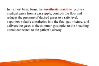
    • In itsmost basic form, the anesthesia machine receives medical gases from a gas supply, controls the flow and reduces the pressure of desired gases to a safe level, vaporizes volatile anesthetics into the final gas mixture, and delivers the gases at the common gas outlet to the breathing circuit connected to the patient’s airway 5
  • 6.
  • 7.
  • 8.
  • 9.
    • Convert supplygases from high pressure to low pressure. • Convert liquid agent to gas & Deliver in a controlled manner. • Provide positive pressure for ventilation. • Alert the provider to malfunction. • Prevent delivery of a hypoxic mixture. 9
  • 10.
    • Anesthetic machines,regardless of their manufacturer, consist of the same basic components. • These include : - 1. Gas Supply 2. Pressure regulators 3. Fail-safe device 4. Flow meters (Rotameter) 5. Vaporizer 6. Common gas outlate 7. Oxygen flush valve 8. Breathing Systems / Limb 10
  • 11.
    ØCylinder gas supply ØCentralsupply ØConcentrator 11
  • 12.
    • May supplyO2, N2O, N2, Air or CO2 • Accidental connection of a wrong gas cylinder is prevented by; üHanger-yoke assemblies that utilize a pin index safety system (PISS) üUsing color coded cylinders • In North America, O2 = green, nitrous oxide = blue, CO2 = gray, air = yellow, helium = brown, N2 = black. • In the UK, white is used for O2 and black and white for air. 12
  • 13.
  • 14.
    • Cylinders areavailable in different sizes and are filled to various pressures • The content of the cylinder is depending on the pressure and the original volume of the cylinder. • Therefore, it is possible to calculate accurately how long a given flow rate of oxygen can be maintained before the cylinder is empty. 14
  • 15.
    Calculation of cylindercontents: Duration of flow (min) = Current Cylinder Pressure × Conversion Factor) Flow rate (L/min) = (total cylinder pressure - remaining presser) × k Flow rate (L/min) where k =total cylinder volume/total cylinder pressure Cylinder Type Max Pressure Max Volume k D – Cylinder 2216 psi 350 L (gas) 0.16 E – Cylinder 2216 psi 625 L (gas) 0.28 H –cylinder 6000-8000 15
  • 16.
    Oxygen § Medical gradeoxygen (99% or 99.5% pure) is manufactured by fractional distillation of liquefied air. § stored as a compressed gas at room temperature or refrigerated as a liquid. Nitrous Oxide § manufactured by heating ammonium nitrate § Stored in cylinders some part as liquid & some part as gaseous state. § The volume remaining in a cylinder is not proportional to cylinder pressure. So the only reliable way to determine residual volume of N2O is to weigh the cylinder. § Nitrous oxide E cylinder can contain up to745 psig. 16
  • 17.
  • 18.
    • Oxygen isstored in a large oxygen tank at some place in the hospital and delivered to each room by a pipe line. • The nominal pressure of gas in pipelines in UK is 4 bar (400 kPa). • Correct pipe line tube to correct hose of anesthesia machine - DISS (Diameter-index safety system) - color coded tubes 18
  • 19.
    • Concentrating atmosphericair; – ( absorbing nitrogen by using zeolite granules & releasing oxygen to the pt) • Concentrating ability: 90 to 96% • The product gas from the concentrator is thought to be 93% Oxygen. • Limitations: – dependent on electric power supply. – a potential possibility of Ar accumulation in rebreathing anaesthesia systems with reduced fresh gas flow. 19
  • 20.
  • 21.
    • The gaspressure from the cylinder to the patient is divided in to different pressure systems: –High pressure system –Intermediate pressure system –Low pressure system 21
  • 22.
  • 23.
    • Receives gassesfrom the high pressure E cylinders attached to the back of the anesthesia machine (2200 psig for O2, 745 psig for N2O) • Consists of: – Hanger Yolk (reserve gas cylinder holder) – Pressure Reducing Device (Regulator) – Check valve (prevent reverse flow of gas) – Cylinder Pressure Indicator (Gauge) • Usually not present when pipeline gas supply on. 23
  • 24.
    Receives gasses fromthe regulator or the hospital pipeline at pressures of 40-55 psig. E.g. pipeline inlet connections, pipeline pressure indicators, piping, oxygen pressure failure devices, the oxygen flush, Master switch and the flow control valves. 24
  • 25.
    • The low-pressuresystem is downstream of the flow control devices. • Include flowmeters, Vaporizers , hypoxia prevention safety devices, unidirectional valves, pressure relief devices, and the common gas outlet. 25
  • 26.
    • The pressurein the cylinder is 137 Bar; too high for the anesthetic machine. • The pressure is reduced to 4 Bar to protect the machine (This pressure would still harm or kill a patient). • After the rotameters (flowmwter), the pressure is reduced to < 1/3 Bar to protect the patient. • This requires pressure regulators or pressure release valves Pressure regulators and pressure release valves maintain a constant pressure 26
  • 27.
    • One-stage pressureregulation (Draeger) • two-stage pressure regulation (Datex-Ohmeda) • cylinder gas pressure reduced to 45–47 psig1 before it enters the flow valve. • A high-pressure relief valve: opend when the supply pressure exceeds the machine's maximum safety limit (95–110 psig). • After passing through pressure gauges and check valves, the pipeline gases share a common pathway with the cylinder gases. 27
  • 28.
  • 29.
    • These devicessense O2 pressure that derived from the gas inlet or secondary regulator. • It allow other gases to flow only if there Is sufficient O2 pressure in the safety device . • If the piloting pressure line falls below a threshold (eg, 20 psig),the shut-off valves close, preventing the administration of any other gases 29
  • 30.
    • Measure gasesbefore mixing with other gases, entering the active vaporizer, & exiting the mashin CGO. • It comprise a flow control valve, a bobbin and a flow tube. • Gas flow into the flow meter raises a float/bobbin • Bobbin rotate constantly at the center to minimize friction with the tube’s • Calculated in Liters per minute {4-6 lit/min} • The flow tubes and valve controls are color- coded • The O2 flow meter is positioned to the right to prevent hypoxia if there is leakage from a flow meter 30
  • 31.
  • 32.
    § Changes liquidagent IAA like halothane,.. to a vapor and add a controlled amount of this vapor to the fresh gas flow in a controlled manner § They must be located between the flow meters and the common gas outlet § Unless the machine accepts only one vaporizer at a time, all anesthesia machines should have an interlocking device that prevents the concurrent use of more than one vaporizer. 32
  • 33.
  • 34.
    Classification Based on methodof regulating output concentration 1. Concentration calibrated (Variable bypass) ◦Vaporizer output is controlled by single knob / dial calibrated in Volumes percent. 34
  • 35.
    2. Measured flow/flow meter controlled • Use a measured flow of carrier gas–oxygen, to pick up anesthetic vapor. • No longer available for sale. • Receives O2 as the carrier gas from a separate dedicated flow meter 35
  • 36.
    Based on methodof vaporization A. Flow –over • a stream of carrier gas passes over the surface of the liquid. • Most commonly used. B. Bubble through • gas entering the vaporizer passes through the liquid anesthetics and becomes saturated with vapor. 36
  • 37.
    Based on Temperaturecompensated A) mechanical thermo-compensation – altering the splitting ratio of gases by metal strip – When the temperature decreases (vaporizer cools ), the metal contras & the strip to bend, allowing more gas to pass through the vaporizing chamber. – The opposite occurs if the vaporizer becomes too warm. B) supplied heat – An electric heater used to maintain a constant temperature. 37
  • 38.
    Depending on thespecificity 1. Agent specific • all modern vaporizers • are flow- over vaporizer • are temperature- compensated by heat - sensitive metallic strip. • Capable of delivering a constant concentration of agent regardless of temperature changes or flow through the vaporizer. 2. Multiple agents • are bubble- through vaporizers. • copper kettle and vernitrol 38
  • 39.
    6. Common (Fresh)Gas Outlet : – connects the machine to the breathing circuit. – Anesthesia machine has only one common gas outlet(vs multiple gas inlets) – An anti disconnect retaining device is used to prevent accidental 39
  • 40.
    7. Oxygen flushvalve – provides a high flow (35–75 L/min) of oxygen directly to the CGO, – bypassing the flow meters and vaporizers. – Used for rapidly refill or flush the breathing circuit, but may cause barotrauma to occur. v Use cautiously whenever a patient is connected to the breathing circuit 40
  • 41.
    Definition: qA breathing system:is an assembly of components which connects the Pt airway to the anesthesia machine, creating an artificial atmosphere from & to the Pt breathes. provide the final conduit for the delivery of anesthetic gases and oxygen to the patient. They are designed to allow either spontaneous or intermittent positive pressure ventilation (IPPV). 41
  • 42.
  • 43.
    üA Fresh gasentry port üoxygen analyzer ügas sampling line, üspirometer üA reservoir for gases üAPL(adjustable pressure limiting) valve üCorrugated tubes pressure gauge üConnection for scavenging system ØA carbon dioxide absorber ØOne way flow directing valves Ømechanical ventilator, 43
  • 44.
    Fresh gas entersthe circle system through a connection from the common gas outlet of the anesthesia machine. Two unidirectional valves are situated in different limbs of the corrugated tubing such that one functions for inhalation and the other for exhalation. These valves Ø permit positive-pressure breathing and Ø prevent the rebreathing of exhaled gases until they have passed through the carbon dioxide absorbent canister and have had their oxygen content replenished. 44
  • 45.
    • Rebreathing andhypercapnia can occur if the unidirectional valves stick in the open position, and total occlusion of the circuit can occur if they are stuck in the closed position. • If the expiratory valve is stuck in the closed position ,breath stacking &barotraumas can occur. • If the unidirectional valves are functioning properly, the only dead space in the circle system is between the Y-piece and the patient. 45
  • 46.
    • Measures partialpressure of oxygen in %. • The only machine safety device that evaluates the integrity of the low-pressure circuit in an ongoing fashion. • Placed into the inspiratory or expiratory limb of the circle system’s breathing circuit—but not into the fresh gas line. • Should have a low-level alarm that is automatically activated 46
  • 47.
    • Used tomeasure exhaled tidal volume in the breathing circuit on all anesthesia machines, typically near the exhalation valve. • Modern anesthesia machines measure the actual delivered and exhaled tidal volumes at the Y-connector. • Changes in exhaled tidal volumes usually represent changes in ventilator settings, circuit leaks, disconnections, or ventilator malfunction. • Spirometers are prone to errors caused by inertia, friction, and water condensation. 47
  • 48.
    • Usually itreflects air way pressure • can be placed somewhere between the expiratory and inspiratory valves but the exact location depends on the model of anesthesia machine • Exact value can be obtained from the Y connection. • A rise in airway pressure may due to: decrease pulmonary compliance, increase VT, obstruction in the breathing circuit , tracheal tube, or the patient’s airway • A drop in pressure may indicate an increase compliance, a decrease in VT, or a leak in the circuit. 48
  • 49.
    A. Passive Humidifiers(heat and moisture exchanger (HME) units) contain a hygroscopic material that traps exhaled humidification and heat, which is released upon subsequent inhalation. also act as effective filters from bacterial or viral cross- contamination. 49
  • 50.
    s/e increase apparatus deadspace (> 60 mL), Increase breathing-circuit resistance and the work of breathing during spontaneous respirations. Risk of obstruction if excessive saturation of an HME with water or secretions 50
  • 51.
    B. Active Humidifiers •addwater to gas by passing the gas over a water chamber (passover humidifier), (bubble-through humidifier)… •are more effective than passive ones •More valuable in children( prevent both hypothermia & plugging trachea by dried secretions Hazards – thermal lung injury (T should not exceed 41°C), – nosocomial infection, – increased airway resistance – interference with flow meter function, and – an increased likelihood of circuit disconnection. 51
  • 52.
    The inspiratory andexpiratory corrugated tubes serve as conduits for delivery of gases to and from the patient. Their large bore provides minimal resistance, and the corrugations provide flexibility, resist kinking, and promote turbulent instead of laminar flow. 52
  • 53.
    Also called aspressure relief or pop-off valve Used to adjust the pressure in the breathing system. Left fully open: during spontaneous ventilation partially closed : during manual or assisted bag ventilation,;;;; allows venting of excess gas from the breathing system into the waste gas scavenging system In mechanical Ventilation: The APL valve is excluded from the circuit when the selector switch is changed from manual to automatic ventilation 53
  • 54.
    ü Widely usedin OR and ICU, and incorporated in all modern anesthesia machines. ü Generate positive pressure and forces gas to flow in to the upper airway ü are powered by compressed gas, electricity, or both. ü give the anesthetist a free hand because patients got ventilation from the mechanical anesthesia. ü When the “bag/vent” selector switch is set to “vent,” the reservoir bag and APL valve are eliminated from the circle system and the 54
  • 55.
    Types: 1. Double circuitor bellows ventilators ◦ are most commonly used in modern anaesthesia workstations. ◦ Pneumatic force (driving gas) compresses a bellows, which empties its contents (patient gas from flow meters and vaporizer) into the patient. ◦ The driving gas circuit is located outside the bellows, and the patient gas circuit is inside the bellows. 55
  • 56.
    Classification of below: Isbased on the direction of bellows movement during the expiratory phase. - Ascending (standing) – safer & most electronic ventilators have an ascending bellows design - descending (hanging) 2. Single circuit or Piston Ventilators: ◦electrically driven piston substituted for the bellows, and the ventilator requires either minimal or no pneumatic (oxygen) power ◦Advat: deliver accurate tidal volumes 56
  • 57.
    Ø Available ina range of sizes (e.g. 0.5 and 3 liters) function Ø as a reservoir of anesthetic gas Ø method of generating PPV ü are designed to increase in compliance as their volume increases ü The bag also serves as a safety device because its distensibility limits pressure in the breathing circuit to less than 60 cm H2O, even when the APL valve is closed. 57
  • 58.
    • Scavenging isthe collection and subsequent removal of vented waste gases from the breathing system and prevent pollution of the operating theatre. • The excess gas comes from either the APL valve if the bag/vent selector switch is set to “bag” or from the ventilator relief valve if the bag/vent selector switch is set to “vent.” • All excess gas from the patient exits the breathing system through these valves. 58
  • 59.
    Scavenging system canbe connects to the breathing system with a 30 mm conical connector Pollution of the operating room environment with anesthetic gases may pose a health hazard to surgical personnel. vThe anesthetist must be certain that the scavenging system is operational and adjusted properly to ensure adequate scavenging. 59
  • 60.
    • Is controversial,confusing and even contradictory • Traditionally classified as: Open, Semi open, Semi closed and Closed, according to the presence or absence of – A gas reservoir bag in the system – Rebreathing of exhaled gases – Means to chemically neutralized exhaled CO2 and – unidirectional valves, • The most commonly used anesthetic breathing systems are the circle system, Mapleson F (Jackson-Rees system and Bain circuit. 60
  • 61.
  • 62.
    Mapleson Circuits ◦ Maplesondescribed 5 different arrangements of breathing circuits by rearranging fresh gas inflow tubing, reservoir tubing, facemask, reservoir bag, and an pop of valve ◦ Termed as Mapleson A-E. ◦ The Mapleson F system, which is a Jackson-Rees modification of the Mapleson D system, was added later. 62
  • 63.
    ◦ Advantages Used duringtransport of children Minimal dead space, low resistance to breathing lightweight, inexpensive, and simple. ◦ Disadvantages Scavenging (variable ability, depending on FGF used) High flows required (cools children, more costly) Lack of humidification/heat (except Bain) Unrecognized kink of inner hose in Bain Pollution & higher cost Difficult to assemble 63
  • 64.
  • 65.
    • In Maplesonsystem Breathing-circuit efficiency is measured by the fresh gas flow required to reduce CO2 rebreathing to a negligible value. 65 Mapleson Systems Uses FGF SV FGF IPPV A Magill Lack Spontaneous Gen Anaesthesia 70-100 ml/kg/min Min 3 x MV B Very uncommon, not in use today C Resuscitation Bagging Min 15 lpm D Bain Spontaneous IPPV, Gen. Anaes 150-200 ml/kg/min 70-100 ml/kg/min E Ayres T Piece Very uncommon, not in use today F Jackson Rees Paediatric <25 Kg 2.5 – 3 x MV Min 4 lpm
  • 66.
    Very efficient forspontaneous ventilation since a FGF = MV will be enough to prevent re-breathing. inefficient during controlled ventilation. ◦ Since No gas is vented during expiration, high unpredictable FGF (> 3 times MV) needed to prevent re-breathing 66
  • 67.
    • FGF forcesalveolar gas away from pt toward APL valve. • Efficient during Controlled Ventilation. • Developed to facilitate scavenging of waste gas. • Bain circuit is a modification of Mapleson D. 67
  • 68.
    • Is amodification(coaxial version) of Mapleson D. • Fresh gas enters through narrow inner tube near the reservoir bag • Exhaled gas exits through corrugated outer tube. • Used for both spontaneous and controlled ventilation. • FGF required to prevent re-breathing: - 200-300ml/kg/min with spontaneous breathing . - 70ml/kg/min with controlled ventilation. 68
  • 69.
     Advantage  Warmingof the fresh gas, Ease of scavenging  Ease of scavenging waste gases.  is lightweight, easily sterilized, reusable, and useful when access to the patient is limited, such as during head and neck surgery  Disadvantage - Unrecognized disconnection - Kinking (twist) of inner fresh gas flow tubing - Requires high flows - The outer expiratory tube should be transparent to allow inspection of the inner tube. 69
  • 70.
    • It isJackson Rees modification of the Ayres T Piece • is the most commonly used circuit in neonates, infants, and paediatric patients less than 20 kg in weight or less than 5 years of age. • Suitable for spontaneous and controlled ventilation • the bag on expiratory limb used to monitoring or assisting the respiration and to venting out excess gases. 70
  • 71.
    Disadvantage Scavenging is limited. wastageof fresh gases and delaying induction by inhalation agents. Summary The relative efficiency of different Mapleson systems for preventing rebreathing during spontaneous ventilation is A > DF> C > B. The relative efficiency of different Mapleson systems for preventing rebreathing during controlled ventilation is DF > B > C > A. 71
  • 72.
    Ø It isthe most popular anesthetic breathing system in world. Ø It is so named because its essential components are arranged in a circular manner. Ø The circle system prevents rebreathing of carbon dioxide by chemical neutralization of CO2 with CO2 absorbents. Ø Rebreathing of exhaled gases result in: - Some conservation of airway moisture & body heat and - Decreased pollution of the surrounding atmosphere 72
  • 73.
    It may beused as a closed or semi-closed system. Closed System: üThe APL is closed , No gas escapes from the system. üThere is total rebreathing üthe inflow gas exactly matches that being consumed by the patient. üAlways contains 3 unidirectional valves (insp, exp, APL). Semi-closed: Øis the most commonly used breathing system. ØThe APL is opened ,allowing excess gas to escape from the system. ØThere is partial rebreathing. ØFGF is less than minute ventilation. ØExamples – The machine we use everyday! 73
  • 74.
    1. A freshgas inlet 2. Inspiratory and expiratory unidirectional check valves 3. Inspiratory and expiratory corrugated tubing 4. A Y-piece connector 5. An adjustable pressure-limiting (APL) valve, also referred to as an overflow or “pop-off valve 6. A reservoir bag 7. A canister containing carbon dioxide absorbent 8. A bag/vent selector switch 9. A mechanical anesthesia ventilator. 74
  • 75.
    Although there couldbe different arrangement, the ff is preferred to decrease rebreathing of CO2 1. Unidirectional valve should be located between the patient and the reservoir bag on both the inspiratory and expiratory limbs of the circuit 2. the fresh gas inflow cannot enter the circuit between the expiratory valve and the patient 3. the pop-off valve cannot be located between the patient and the inspiratory valve. 75
  • 76.
  • 77.
    • Chemical neutralizationof CO2 is achieved by directing exhaled gases through a container (canister) containing a carbon dioxide absorbent such as soda lime , calcium hydroxide lime (Amsorb) or bara lyme. • Soda lime: contains calcium hydroxide (80%), along with sodium hydroxide (4%), water (15%), and a small amount of potassium hydroxide(1%) and silica(0.2%) • Hydroxide act as an activators, water to assure optimal activity and Silica is to give hardness so as to minimize the formation of alkaline dust. 77
  • 78.
    Ideal CO2 absorbent -lack of reactivity with common anesthetics - lack of toxicity - low resistance to air flow - low cost - ease of handling - efficient in CO2 absorption. 78
  • 79.
    Absorption process ofCO2  is a series of chemical reactions; not a physical process like soaking water into a sponge. It begins with the reaction of CO2 with the water present in soda lime granules  CO2 + H2O → H2 CO3 H2 CO3 + NaOH → Na2 CO3 (rapid) + 2 H2O + Heat H2 CO3 + Ca (OH)2 → CACO3, (slow) + 2 H2O + Heat 79
  • 80.
    Reaction end productsinclude ◦ heat (the heat of neutralization) ◦ Water & ◦ calcium carbonate. The formed water is useful for humidifying the inhaled gases Accumulation of this highly alkaline of water in the bottom of the canister can warm the canister and produce burns sensation on contact with the skin. Failure of the canister to become warm to touch should alert the anesthetist to the possibility that chemical neutralization of carbon dioxide is not taking place. 80
  • 81.
    Carbon dioxide neutralization: Influencedby ◦ Size of granules( normal size = 4-8 mesh) ◦ Presence or absence of channeling in the canister (areas of loosely packed granules). The Color changed when the soda lime exhausted. so change it when 50-70% of its color changed. Toxic products 1. Carbon monoxide by dry absorbent (eg, sodium or potassium hydroxide) causes carboxyhemoglobin specially at a higher temperature (desflurane > sevoflurane 2. Compound A : due to degradation of sevoflurane 3. absorb and later release of medically active particles - Delayed emergency 81
  • 82.
    Advantages of CircleSystem: 1. maintenance of relatively stable inspired gas concentrations 2. conservation of respiratory moisture and heat 3. prevention of operating room pollution. Disadvantage : 1. Complex design and multiple connection sites: 2. Bulkiness with less portability. 3. Unidirectional valves and CO2 absorbent…. -Increased resistance to breathing 4. Bacterial Contamination 5. Toxic products 6. difficulty of predicting inspired gas concentrations during low fresh gas flows 82
  • 83.
    83 ü This checkoutshould be conducted before administration of anesthesia. ü Users are encouraged to modify this guideline to accommodate differences in equipment design and variations in local clinical practice. ü Such local modifications should have appropriate peer review. ü Users should refer to the appropriate operator manuals for specific procedures and precautions.
  • 84.
    1. Verify backupventilation equipment is available and functioning High-Pressure System 2. Check O2 cylinder supply A. Open O2 cylinder and verify at least half full (about 1000 psig). b. Close cylinder 3. Check central pipeline supplies; check that hoses are connected and pipeline gauges read about 50 psig Low-Pressure System 4. Check initial status of low-pressure system  a. Close flow control valves and turn vaporizers off.  b. Check fill level and tighten vaporizers' filler caps. 84
  • 85.
    5. Perform leakcheck of machine low-pressure system a. Verify that the machine master switch and flow control valves are off. b. Attach suction bulb to common (fresh) gas outlet. c. Squeeze bulb repeatedly until fully collapsed. d. Verify bulb stays fully collapsed for at least 10 seconds. e. Open one vaporizer at a time and repeat steps c & d. f. Remove suction bulb, and reconnect fresh gas hose. 85
  • 86.
    86 6. Turn onmachine master switch and all other necessary electrical equipment. 7. Test flowmeters A. Adjust flow of all gases through their full range, checking for smooth operation of floats and undamaged flow tubes. B. Attempt to create a hypoxic O2/N2O mixture and verify correct changes in flow and/or alarm.
  • 87.
    Scavenging System 8. Adjustand check scavenging system A. Ensure proper connections between the scavenging system and both APL (pop-off) valve and ventilator relief valve B. Adjust waste-gas vacuum (if possible). C. Fully open APL valve and occlude Y-piece. D. With minimum O2 flow, allow scavenger reservoir bag to collapse completely and verify that absorber pressure gauge reads about zero. E. With the O2 flush activated, allow scavenger reservoir bag to distend fully, and then verify that absorber pressure gauge reads < 10 cm H2O. 87
  • 88.
    Breathing System 9. CalibrateO2 monitor a. Ensure monitor reads 21% in room air. b. Verify low-O2 alarm is enabled and functioning. c. Reinstall sensor in circuit and flush breathing system with O2. d. Verify that monitor now reads greater than 90%. 10. Check initial status breathing system a. Set selector switch to Bag mode. b. Check that breathing circuit is complete, undamaged and unobstructed. c. Verify that CO2 absorbent is adequate. d. Install breathing-circuit accessory equipment (eg, humidifier, PEEP valve) to be used during the cas 88
  • 89.
    11. Perform leakcheck of the breathing system A. Set all gas flows to zero (or minimum). B. Close APL (pop-off) valve and occlude Y-piece. C. Pressurize breathing system to about 30 cm H2O with O2 flush D. Ensure that pressure remains fixed for at least 10 seconds. E. Open APL (pop-off) valve and ensure that pressure decreases Manual and Automatic Ventilation Systems 12. Test ventilation systems and unidirectional valves a. Place a second breathing bag on Y-piece b. Set appropriate ventilator parameters for next patient c. Switch to automatic-ventilation (ventilator) mode. d. Turn ventilator on and fill bellows and breathing bag with O2 flush 89
  • 90.
    90 E. Set O2flow to minimum, other gas flows to zero. F. Verify that during inspiration bellows deliver appropriate tidal volume and that during expiration bellows fill completely. G. Set fresh gas flow to about 5 L min–1. H. Verify that the ventilator bellows and simulated lungs fill and empty appropriately without sustained pressure at end expiration. I. Check for proper action of unidirectional valves. J. Exercise breathing circuit accessories to ensure proper function.
  • 91.
    91 K. Turn ventilatoroff and switch to manual ventilation (bag/APL) mode. L. Ventilate manually and ensure inflation and deflation of artificial lungs and appropriate feel of system resistance and compliance. M. Remove second breathing bag from Y-piece. Monitors 13. Check, calibrate, and/or set alarm limits of all monitors: capnograph, pulse oximeter, O2 analyzer, respiratory- volume monitor (spirometer), pressure monitor with high and low airway-pressure alarms.
  • 92.
    92 Final Position 14. Checkfinal status of machine A. Vaporizers off B. APL valve open C. Selector switch to Bag mode D. All flow meters to zero (or minimum) E. Patient suction level adequate F. Breathing system ready to use 137 bar - cylinder 50 bar - pipline 4 bar - machine 1 bar - pt 1bar = 14.5psig = 760mmHg
  • 93.
    93 So Take aDeep Breathm and Go Home