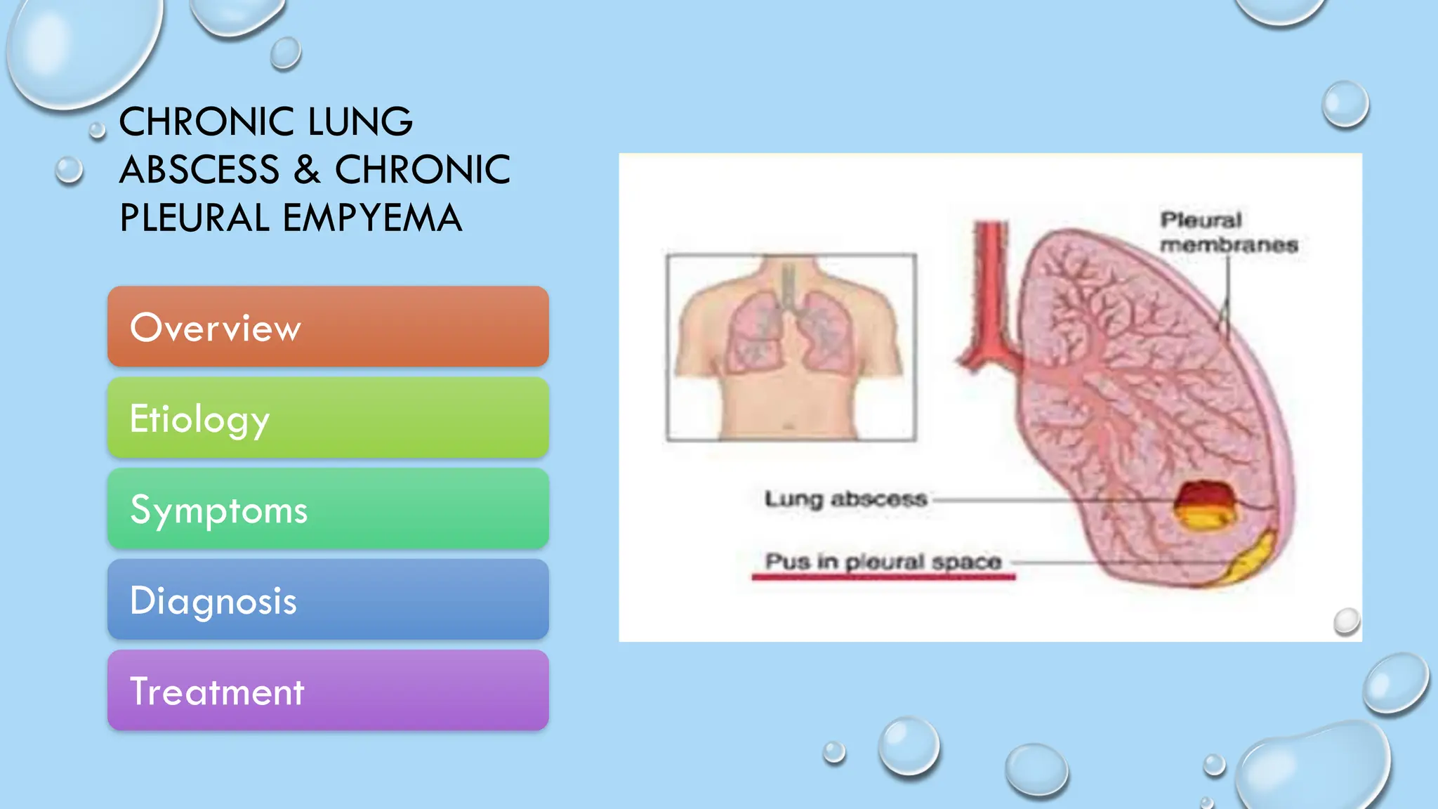
CHRONIC LUNG
ABSCESS & CHRONIC
PLEURAL EMPYEMA
Overview
Etiology
Symptoms
Diagnosis
Treatment
PLEURAL EMPYEMA
• EMPYEMA IS A CONDITION THAT
CAUSES PUS TO DEVELOP IN YOUR
PLEURAL SPACE. PNEUMONIA IS THE
MOST COMMON CAUSE. A
HEALTHCARE PROVIDER CAN
DIAGNOSE EMPYEMA BY LISTENING
TO YOUR LUNGS AND ORDERING
IMAGING AND BLOOD TESTS.
ANTIBIOTICS TREAT MOST CASES OF
EMPYEMA, BUT A PROVIDER MAY
NEED TO DRAIN PUS WITH A
NEEDLE OR THROUGH SURGERY.
EMPYEMA-
ETIOLOGY
EMPYEMA IS USUALLY CAUSED BY AN INFECTION THAT SPREADS DIRECTLY
FROM THE LUNG. IT LEADS TO A BUILDUP OF PUS IN THE PLEURAL SPACE.
THERE CAN BE 2 CUPS (1/2 LITER) OR MORE OF INFECTED FLUID. THIS
FLUID PUTS PRESSURE ON THE LUNGS.
RISK FACTORS INCLUDE:
BACTERIAL PNEUMONIA
TUBERCULOSIS
CHEST SURGERY
LUNG ABSCESS
TRAUMA OR INJURY TO THE CHEST
• IN RARE CASES, EMPYEMA CAN OCCUR AFTER THORACENTESIS. THIS IS
A PROCEDURE IN WHICH A NEEDLE IS INSERTED THROUGH THE CHEST
WALL TO REMOVE FLUID IN THE PLEURAL SPACE FOR MEDICAL
DIAGNOSIS OR TREATMENT
SYMPTOMS
EMPYEMA SYMPTOMS ARE NONSPECIFIC AND MAY
INCLUDE:
CHEST PAIN OR DISCOMFORT. WHICH WORSENS WHEN
YOU BREATHE IN DEEPLY
FEVER.
TROUBLE BREATHING.
COUGH.
EXTREME EXHAUSTION (FATIGUE).
UNEXPLAINED WEIGHT LOSS.
• EXCESSIVE SWEATING, ESPECIALLY NIGHT SWEATS
DIAGNOSIS
Imaging tests. X-rays and a CT scan help your provider see if there’s fluid in your
pleural space.
An ultrasound helps show how much fluid you have in your pleural space and helps
guide the fluid sample collection..
Blood tests. During a blood test, a provider will use a thin needle to withdraw a small
amount of blood from a vein in your arm. They’ll use the sample to look for a high white
blood cell count and conduct a C-reactive protein (CRP) and a bacteria culture.
Thoracentesis
Pleural fluid analysis
TREATMENT
THE GOAL OF TREATMENT IS TO CURE THE INFECTION. THIS INVOLVES
THE FOLLOWING:
• PLACING A TUBE IN YOUR CHEST TO DRAIN THE PUS
• GIVING YOU ANTIBIOTICS TO CONTROL THE INFECTION
• AMOXICILLIN-CLAVULANATE.
• PIPERACILLIN-TAZOBACTAM.
• IMIPENEM.
1. MEROPENEM.
• A PROVIDER WILL REMOVE PUS THROUGH THORACENTESIS IN THE
EARLY STAGES OF EMPYEMA. THEY’LL USE A THIN NEEDLE TO INJECT
A LOCAL ANESTHETIC INTO YOUR SKIN AND DEEPER TISSUE. THEY’LL
THEN ATTACH A LARGER NEEDLE/CATHETER DEVICE TO WITHDRAW
THE PUS AND DRAINAGE TUBING TO ALLOW THE PUS TO FLOW
OUTSIDE YOUR BODY. IF DRAINAGE ISN’T ENOUGH, A PROVIDER MAY
TRY TO BREAK UP THE PUS THROUGH FIBRINOLYTIC THERAPY.
LUNG ABSCESS
• A LUNG ABSCESS IS A PUS-FILLED
CAVITY IN YOUR LUNG
SURROUNDED BY INFLAMED
TISSUE. IT USUALLY RESULTS
FROM BREATHING BACTERIA THAT
NORMALLY LIVE IN YOUR MOUTH
OR THROAT INTO THE LUNGS,
LEADING TO AN INFECTION.
ETIOLOGY
A lung abscess is a pus-filled cavity in your lung surrounded by inflamed tissue. It usually results from
breathing bacteria that normally live in your mouth or throat into the lungs, leading to an infection.
Poor oral health: People with gum disease are more likely to get an abscess.
Your immune system isn’t working well: This can let in germs that aren’t usually found in your mouth or
throat, like fungi or the bacteria that cause tuberculosis, strep throat, and MRSA.
Blocked airway:Mucus can form behind a tumor or foreign object in your windpipe and lead to an
abscess. If bacteria get into the mucus, the blockage stops you from coughing it out.
Blood-borne causes: It’s rare, but bacteria or infected blood clots from an infected part of your body
can travel through your bloodstream and into your lung, where they cause an abscess.
SYMPTOMS
SYMPTOMS OF A LUNG ABSCESS COMMONLY COME
SLOWLY OVER WEEKS. THEY MAY INCLUDE:
CHEST PAIN, ESPECIALLY WHEN YOU BREATHE IN
COUGH
FATIGUE
FEVER
LOSS OF APPETITE
NIGHT SWEATS
SPUTUM (A MIXTURE OF SALIVA AND MUCUS) WITH PUS
THAT’S OFTEN SOUR-TASTING, FOUL-SMELLING, OR
STREAKED WITH BLOOD
• WEIGHT LOSS
DIAGNOSIS
A lung abscess is typically diagnosed
in two ways:
Chest X-ray: This shows your doctor
where the abscess is.
CT scan of the chest: Your doctor is
looking for an air- and fluid-filled
cavity in the middle of your lung
The doctor might also use a device
called a bronchoscope, a thin tube
with a light and a camera on the
end, to get samples of sputum or
lung tissue for more tests if:
Antibiotics aren’t helping.
They think your airways are blocked.
Your immune system is damagded
TREATMENT
Antibiotics: Most people get antibiotics into a vein for up to
3-8 weeks. You might switch to oral antibiotics after that.
You’ll take them until a chest X-ray shows the abscess is
gone.
Drainage: You may need this if your abscess is 6
centimeters or more in diameter. Your doctor will use a CT
scan to guide them as they insert the drain through your
chest wall into the abscess.
Surgery: It’s rare, but some people need surgery to remove
the part of the lung with the abscess Sometimes the entire
lung has to come out to get rid of the infection. Surgery can
also help remove a foreign object
THANK YOU

CASE DISCUSSION Pleural Emphyma & Lungs Abcess.pptx

  • 1.
    CHRONIC LUNG ABSCESS &CHRONIC PLEURAL EMPYEMA Overview Etiology Symptoms Diagnosis Treatment
  • 2.
    PLEURAL EMPYEMA • EMPYEMAIS A CONDITION THAT CAUSES PUS TO DEVELOP IN YOUR PLEURAL SPACE. PNEUMONIA IS THE MOST COMMON CAUSE. A HEALTHCARE PROVIDER CAN DIAGNOSE EMPYEMA BY LISTENING TO YOUR LUNGS AND ORDERING IMAGING AND BLOOD TESTS. ANTIBIOTICS TREAT MOST CASES OF EMPYEMA, BUT A PROVIDER MAY NEED TO DRAIN PUS WITH A NEEDLE OR THROUGH SURGERY.
  • 3.
    EMPYEMA- ETIOLOGY EMPYEMA IS USUALLYCAUSED BY AN INFECTION THAT SPREADS DIRECTLY FROM THE LUNG. IT LEADS TO A BUILDUP OF PUS IN THE PLEURAL SPACE. THERE CAN BE 2 CUPS (1/2 LITER) OR MORE OF INFECTED FLUID. THIS FLUID PUTS PRESSURE ON THE LUNGS. RISK FACTORS INCLUDE: BACTERIAL PNEUMONIA TUBERCULOSIS CHEST SURGERY LUNG ABSCESS TRAUMA OR INJURY TO THE CHEST • IN RARE CASES, EMPYEMA CAN OCCUR AFTER THORACENTESIS. THIS IS A PROCEDURE IN WHICH A NEEDLE IS INSERTED THROUGH THE CHEST WALL TO REMOVE FLUID IN THE PLEURAL SPACE FOR MEDICAL DIAGNOSIS OR TREATMENT
  • 4.
    SYMPTOMS EMPYEMA SYMPTOMS ARENONSPECIFIC AND MAY INCLUDE: CHEST PAIN OR DISCOMFORT. WHICH WORSENS WHEN YOU BREATHE IN DEEPLY FEVER. TROUBLE BREATHING. COUGH. EXTREME EXHAUSTION (FATIGUE). UNEXPLAINED WEIGHT LOSS. • EXCESSIVE SWEATING, ESPECIALLY NIGHT SWEATS
  • 5.
    DIAGNOSIS Imaging tests. X-raysand a CT scan help your provider see if there’s fluid in your pleural space. An ultrasound helps show how much fluid you have in your pleural space and helps guide the fluid sample collection.. Blood tests. During a blood test, a provider will use a thin needle to withdraw a small amount of blood from a vein in your arm. They’ll use the sample to look for a high white blood cell count and conduct a C-reactive protein (CRP) and a bacteria culture. Thoracentesis Pleural fluid analysis
  • 7.
    TREATMENT THE GOAL OFTREATMENT IS TO CURE THE INFECTION. THIS INVOLVES THE FOLLOWING: • PLACING A TUBE IN YOUR CHEST TO DRAIN THE PUS • GIVING YOU ANTIBIOTICS TO CONTROL THE INFECTION • AMOXICILLIN-CLAVULANATE. • PIPERACILLIN-TAZOBACTAM. • IMIPENEM. 1. MEROPENEM. • A PROVIDER WILL REMOVE PUS THROUGH THORACENTESIS IN THE EARLY STAGES OF EMPYEMA. THEY’LL USE A THIN NEEDLE TO INJECT A LOCAL ANESTHETIC INTO YOUR SKIN AND DEEPER TISSUE. THEY’LL THEN ATTACH A LARGER NEEDLE/CATHETER DEVICE TO WITHDRAW THE PUS AND DRAINAGE TUBING TO ALLOW THE PUS TO FLOW OUTSIDE YOUR BODY. IF DRAINAGE ISN’T ENOUGH, A PROVIDER MAY TRY TO BREAK UP THE PUS THROUGH FIBRINOLYTIC THERAPY.
  • 8.
    LUNG ABSCESS • ALUNG ABSCESS IS A PUS-FILLED CAVITY IN YOUR LUNG SURROUNDED BY INFLAMED TISSUE. IT USUALLY RESULTS FROM BREATHING BACTERIA THAT NORMALLY LIVE IN YOUR MOUTH OR THROAT INTO THE LUNGS, LEADING TO AN INFECTION.
  • 9.
    ETIOLOGY A lung abscessis a pus-filled cavity in your lung surrounded by inflamed tissue. It usually results from breathing bacteria that normally live in your mouth or throat into the lungs, leading to an infection. Poor oral health: People with gum disease are more likely to get an abscess. Your immune system isn’t working well: This can let in germs that aren’t usually found in your mouth or throat, like fungi or the bacteria that cause tuberculosis, strep throat, and MRSA. Blocked airway:Mucus can form behind a tumor or foreign object in your windpipe and lead to an abscess. If bacteria get into the mucus, the blockage stops you from coughing it out. Blood-borne causes: It’s rare, but bacteria or infected blood clots from an infected part of your body can travel through your bloodstream and into your lung, where they cause an abscess.
  • 10.
    SYMPTOMS SYMPTOMS OF ALUNG ABSCESS COMMONLY COME SLOWLY OVER WEEKS. THEY MAY INCLUDE: CHEST PAIN, ESPECIALLY WHEN YOU BREATHE IN COUGH FATIGUE FEVER LOSS OF APPETITE NIGHT SWEATS SPUTUM (A MIXTURE OF SALIVA AND MUCUS) WITH PUS THAT’S OFTEN SOUR-TASTING, FOUL-SMELLING, OR STREAKED WITH BLOOD • WEIGHT LOSS
  • 11.
    DIAGNOSIS A lung abscessis typically diagnosed in two ways: Chest X-ray: This shows your doctor where the abscess is. CT scan of the chest: Your doctor is looking for an air- and fluid-filled cavity in the middle of your lung The doctor might also use a device called a bronchoscope, a thin tube with a light and a camera on the end, to get samples of sputum or lung tissue for more tests if: Antibiotics aren’t helping. They think your airways are blocked. Your immune system is damagded
  • 13.
    TREATMENT Antibiotics: Most peopleget antibiotics into a vein for up to 3-8 weeks. You might switch to oral antibiotics after that. You’ll take them until a chest X-ray shows the abscess is gone. Drainage: You may need this if your abscess is 6 centimeters or more in diameter. Your doctor will use a CT scan to guide them as they insert the drain through your chest wall into the abscess. Surgery: It’s rare, but some people need surgery to remove the part of the lung with the abscess Sometimes the entire lung has to come out to get rid of the infection. Surgery can also help remove a foreign object
  • 14.