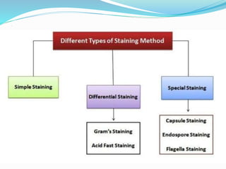
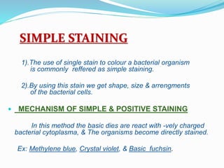
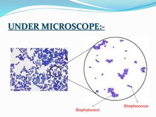
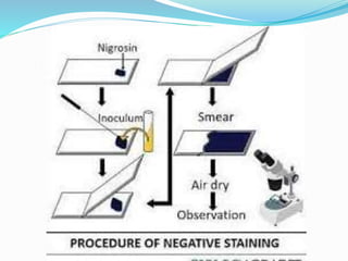
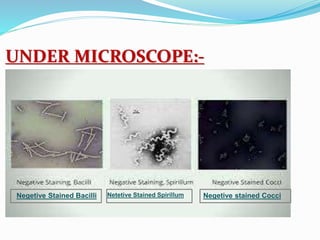
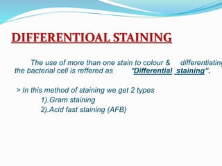
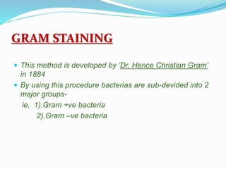
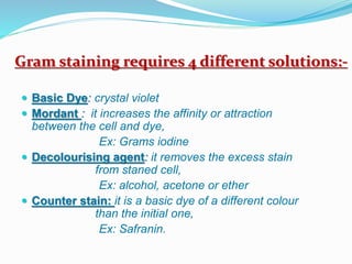
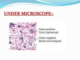
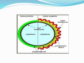
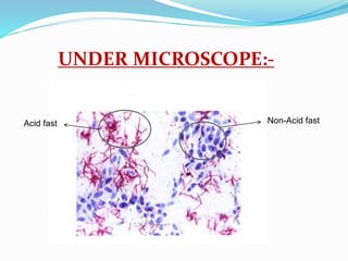
.
.
CLASS PRESENTATION
TOPIC
STINING AND STANING TECHNIQUES
PRESENTED BY:
Eshwrappa BSc 1st sem, ( MbZC)
Govt. degree college
kalaburgi.
CONTENTS
 INTODUCTION
 DEFINATION
 PURPOSE OF STAING
 TYPES OF STINING
 PROCEDURE
INTRODUCTION
1). Stains are nothing but colours/dyes
2). Dyes are classified into two types
> Natural dye : Ex- Hematoxylin ---Hertwood,
> Synthetic dye: Ex- Crystal violet ---Aniline.
3). A dye which contains benzene ring + chromophore &
auxochrome
4). Dyes are may be acidic, basic or neutral.
5). Acidic dyes such as, nigrosine, indian ink, acid fuchsin,
& eosin
6). Basic dyes such as, methylene blue, crystal violet & basic
fuchsin
7). Neutral dyes are formed by mixing together.
DEFINATION
“It is the process of imparting colour to
the microbial/bacterial cells”
or
Stains are the coloured organic
compounds, By which we have to visualize the
microbes.
PURPOSE OF STAINING
 Bacterias are -
 Microscopic organisms
 Lack of contrast/ poor resolution when they have
suspended in th eaqueous medium
 In order to visualize them to study their structur,
shape, & other structural charecteristics, it
becomes necessory to make them more easily
visible
.
…
REQUIRMENTS FOR STAINING:-
Glass slide Inoculating loop Dropper
CULTURES:-
Liquid medium
Solid medium
containing Mycobacteria
agar
STAINS/DYES:-
.
Staining tray Wash bottle Spirit lamp
Microscope
SIMPLE STAINING
1).The use of single stain to colour a bacterial organism
is commonly reffered as simple staining.
2).By using this stain we get shape, size & arrengments
of the bacterial cells.
 MECHANISM OF SIMPLE & POSITIVE STAINING
In this method the basic dies are react with -vely charged
bacterial cytoplasma, & The organisms become directly stained.
Ex: Methylene blue, Crystal violet, & Basic fuchsin.
METHODS OF SIMPLE & POSITIVE
STAINING
UNDER MICROSCOPE:-
Staphylococci
Streptococcus
.
MECHANISM OF SIMPLE &NEGETIVE
STAINING
1). In this method of staining process instead of cells
“Background” is get stained.
2). Acidic stain carries –ve charge & repelled by the
bacterial cells
3). Which also carries –ve charge on there surface
hence, acidic stains don’t stain bateria
4). It deposite around the organism, living organism
remains colourless
Ex: Nigrosine, Indian ink & acid fuchsin.
.
UNDER MICROSCOPE:-
Negetive Stained Bacilli Netetive Stained Spirillum Negetive stained Cocci
DIFFERENTIOAL STAINING
The use of more than one stain to colour & differentiating
the bacterial cell is reffered as “Differential staining”.
> In this method of staining we get 2 types
1).Gram staining
2).Acid fast staining (AFB)
GRAM STAINING
 This method is developed by ‘Dr. Hence Christian Gram’
in 1884
 By using this procedure bacterias are sub-devided into 2
major groups-
ie, 1).Gram +ve bacteria
2).Gram –ve bacteria
Gram staining requires 4 different solutions:-
 Basic Dye: crystal violet
 Mordant : it increases the affinity or attraction
between the cell and dye,
Ex: Grams iodine
 Decolourising agent: it removes the excess stain
from staned cell,
Ex: alcohol, acetone or ether
 Counter stain: it is a basic dye of a different colour
than the initial one,
Ex: Safranin.
PROCEDURE:
 Prepare smear on a clean glass slide.
 Heat fix & air dry the smear.
 Stain with crystal violet for 1 min.
 Wash with tap water
 Add grams iodine for 1 min
 Wash with tap water
 Decolourise with 95% of alcohol for 10-20 sec
 Rinse with water
 Counter stain with safernin for 20-30 sec
 Wash with water
 Air dry and examine under oil immersion objective
UNDER MICROSCOPE:-
ACID-FAST STAINING
 Developed by ‘Poul Ehrlich’ in 1882 & later on modified
by ‘Ziehl-Neelsen’
 These method employs carbol fuchsin, acid-alcohol &
methylene blue as a counter stain.
 By using this method bacterias are classified into 2 types-
(1). Acid-fast :
(2). Non acid-fast :
 The most common type of acid fast bacteria is
‘mycobacteria’. A strain of ‘mycobacteria’ is responsible
for the disease “tuberculosis”.
PROCEDURE:-
UNDER MICROSCOPE:-
Non-Acid fast
Acid fast
Staining and staining techniques 1.pptx

Staining and staining techniques 1.pptx

  • 1.
  • 2.
    CLASS PRESENTATION TOPIC STINING ANDSTANING TECHNIQUES PRESENTED BY: Eshwrappa BSc 1st sem, ( MbZC) Govt. degree college kalaburgi.
  • 3.
    CONTENTS  INTODUCTION  DEFINATION PURPOSE OF STAING  TYPES OF STINING  PROCEDURE
  • 4.
    INTRODUCTION 1). Stains arenothing but colours/dyes 2). Dyes are classified into two types > Natural dye : Ex- Hematoxylin ---Hertwood, > Synthetic dye: Ex- Crystal violet ---Aniline. 3). A dye which contains benzene ring + chromophore & auxochrome 4). Dyes are may be acidic, basic or neutral. 5). Acidic dyes such as, nigrosine, indian ink, acid fuchsin, & eosin 6). Basic dyes such as, methylene blue, crystal violet & basic fuchsin 7). Neutral dyes are formed by mixing together.
  • 5.
    DEFINATION “It is theprocess of imparting colour to the microbial/bacterial cells” or Stains are the coloured organic compounds, By which we have to visualize the microbes.
  • 6.
    PURPOSE OF STAINING Bacterias are -  Microscopic organisms  Lack of contrast/ poor resolution when they have suspended in th eaqueous medium  In order to visualize them to study their structur, shape, & other structural charecteristics, it becomes necessory to make them more easily visible
  • 7.
  • 8.
    REQUIRMENTS FOR STAINING:- Glassslide Inoculating loop Dropper
  • 9.
  • 10.
  • 11.
    . Staining tray Washbottle Spirit lamp
  • 12.
  • 13.
    SIMPLE STAINING 1).The useof single stain to colour a bacterial organism is commonly reffered as simple staining. 2).By using this stain we get shape, size & arrengments of the bacterial cells.  MECHANISM OF SIMPLE & POSITIVE STAINING In this method the basic dies are react with -vely charged bacterial cytoplasma, & The organisms become directly stained. Ex: Methylene blue, Crystal violet, & Basic fuchsin.
  • 14.
    METHODS OF SIMPLE& POSITIVE STAINING
  • 15.
  • 16.
    . MECHANISM OF SIMPLE&NEGETIVE STAINING 1). In this method of staining process instead of cells “Background” is get stained. 2). Acidic stain carries –ve charge & repelled by the bacterial cells 3). Which also carries –ve charge on there surface hence, acidic stains don’t stain bateria 4). It deposite around the organism, living organism remains colourless Ex: Nigrosine, Indian ink & acid fuchsin.
  • 17.
  • 18.
    UNDER MICROSCOPE:- Negetive StainedBacilli Netetive Stained Spirillum Negetive stained Cocci
  • 19.
    DIFFERENTIOAL STAINING The useof more than one stain to colour & differentiating the bacterial cell is reffered as “Differential staining”. > In this method of staining we get 2 types 1).Gram staining 2).Acid fast staining (AFB)
  • 20.
    GRAM STAINING  Thismethod is developed by ‘Dr. Hence Christian Gram’ in 1884  By using this procedure bacterias are sub-devided into 2 major groups- ie, 1).Gram +ve bacteria 2).Gram –ve bacteria
  • 21.
    Gram staining requires4 different solutions:-  Basic Dye: crystal violet  Mordant : it increases the affinity or attraction between the cell and dye, Ex: Grams iodine  Decolourising agent: it removes the excess stain from staned cell, Ex: alcohol, acetone or ether  Counter stain: it is a basic dye of a different colour than the initial one, Ex: Safranin.
  • 22.
    PROCEDURE:  Prepare smearon a clean glass slide.  Heat fix & air dry the smear.  Stain with crystal violet for 1 min.  Wash with tap water  Add grams iodine for 1 min  Wash with tap water  Decolourise with 95% of alcohol for 10-20 sec  Rinse with water  Counter stain with safernin for 20-30 sec  Wash with water  Air dry and examine under oil immersion objective
  • 23.
  • 25.
    ACID-FAST STAINING  Developedby ‘Poul Ehrlich’ in 1882 & later on modified by ‘Ziehl-Neelsen’  These method employs carbol fuchsin, acid-alcohol & methylene blue as a counter stain.  By using this method bacterias are classified into 2 types- (1). Acid-fast : (2). Non acid-fast :  The most common type of acid fast bacteria is ‘mycobacteria’. A strain of ‘mycobacteria’ is responsible for the disease “tuberculosis”.
  • 26.
  • 27.