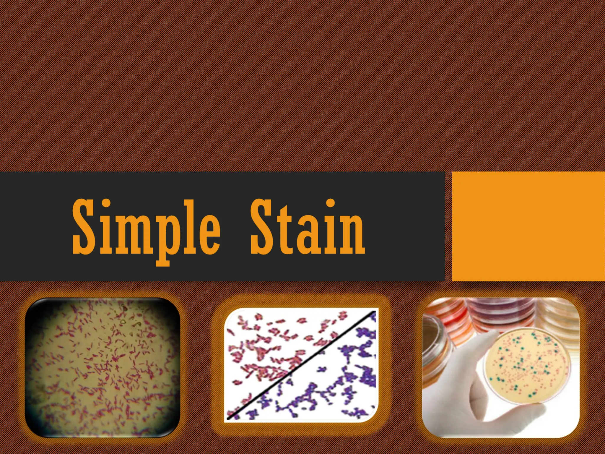
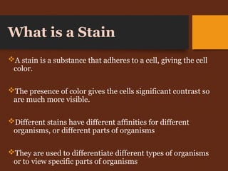
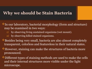
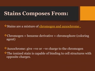
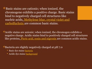
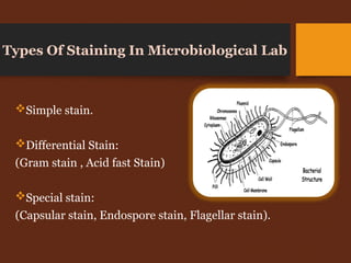
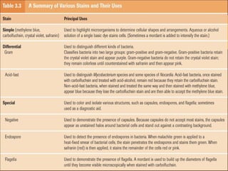
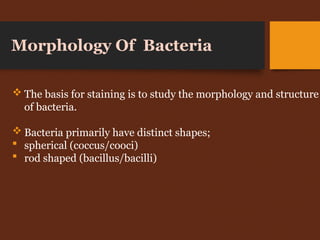
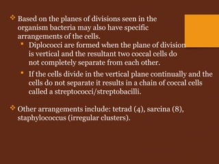
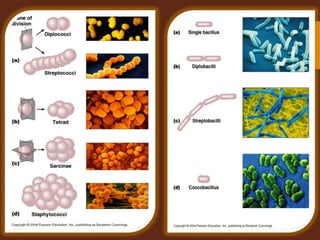
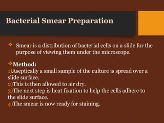
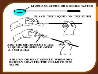
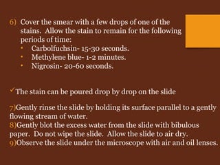
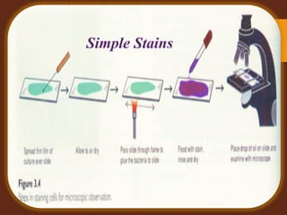
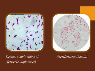
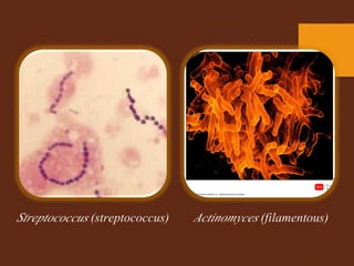
What is a Stain
A stain is a substance that adheres to a cell, giving the cell
color.
The presence of color gives the cells significant contrast so
are much more visible.
Different stains have different affinities for different
organisms, or different parts of organisms
They are used to differentiate different types of organisms
or to view specific parts of organisms
Why we should be Stain Bacteria
In our laboratory, bacterial morphology (form and structure)
may be examined in two ways:
1) by observing living unstained organisms (wet mount).
2) by observing killed stained organisms.
Besides being very small, bacteria are also almost completely
transparent, colorless and featureless in their natural states.
However, staining can make the structures of bacteria more
pronounced.
Different types of staining methods are used to make the cells
and their internal structures more visible under the light
microscope.
Stains Composes From:
Stains are a mixture of chromogen and auxochrome .
Chromogen = benzene derivative + chromophore (coloring
agent)
Auxochrome: give +ve or –ve charge to the chromogen
The ionized stain is capable of binding to cell structures with
opposite charges.
Acidic stains are anionic; when ionized, the chromogen exhibits a
negative charge. Acidic stains bind to positively charged cell structures
like proteins. Picric acid, eosin and nigrosin are common acidic stains.
Bacteria are slightly negatively charged at pH 7.0
 Basic dye stains bacteria
 Acidic dye stains background
Basic stains are cationic; when ionized, the
chromogene exhibits a positive charge. Basic stains
bind to negatively charged cell structures like
nucleic acids. Methylene blue, crystal violet and
carbolfuchsin are common basic stains.
Types Of Staining In Microbiological Lab
Simple stain.
Differential Stain:
(Gram stain , Acid fast Stain)
Special stain:
(Capsular stain, Endospore stain, Flagellar stain).
 The basis for staining is to study the morphology and structure
of bacteria.
 Bacteria primarily have distinct shapes;
 spherical (coccus/cooci)
 rod shaped (bacillus/bacilli)
Morphology Of Bacteria
 If the cells divide in the vertical plane continually and the
cells do not separate it results in a chain of coccal cells
called a streptococci/streptobacilli.
 Other arrangements include: tetrad (4), sarcina (8),
staphylococcus (irregular clusters).
 Based on the planes of divisions seen in the
organism bacteria may also have specific
arrangements of the cells.
 Diplococci are formed when the plane of division
is vertical and the resultant two coccal cells do
not completely separate from each other.
Method:
1)Aseptically a small sample of the culture is spread over a
slide surface.
2)This is then allowed to air dry.
3)The next step is heat fixation to help the cells adhere to
the slide surface.
4)The smear is now ready for staining.
Bacterial Smear Preparation
 Smear is a distribution of bacterial cells on a slide for the
purpose of viewing them under the microscope.
Simple Stain
In this exercise, we will use simple
stains to show the general structures
of some bacteria. Usually, a single
basic stain is used in the procedure.
Simple stains do not usually provide
any data for identification of the
bacterium; they simply make the
bacterium easier to see.
To observe basic external structures
of cell with bright field scope (cellular
morphology)
Procedure of Test
1) Obtain broth cultures of the bacteria.
2) Using an inoculating loop, remove a loopful of suspension
from one of the tubes. Remember to use sterile technique.
3) Smear the bacteria across the center of the slide with the
loop. If the bacterial suspension is very thick, add a drop
of water and mix the bacteria and the water on the slide.
4) Allow the smear to completely air dry.
 Air dry first to prevent lysis (boiling)
5) Heat-fix the smear by quickly passing the slide
through a Bunsen burner flame three times.
This causes partial melting of the cell walls and
membranes of the bacteria, and makes them
stick to the slide. Do not overheat the slide as
this will destroy the bacteria.
The stain can be poured drop by drop on the slide
7)Gently rinse the slide by holding its surface parallel to a gently
flowing stream of water.
8)Gently blot the excess water from the slide with bibulous
paper. Do not wipe the slide. Allow the slide to air dry.
9)Observe the slide under the microscope with air and oil lenses.
6) Cover the smear with a few drops of one of the
stains. Allow the stain to remain for the following
periods of time:
• Carbolfuchsin- 15-30 seconds.
• Methylene blue- 1-2 minutes.
• Nigrosin- 20-60 seconds.
simple stain gram positive gram negative

simple stain gram positive gram negative

  • 2.
    What is aStain A stain is a substance that adheres to a cell, giving the cell color. The presence of color gives the cells significant contrast so are much more visible. Different stains have different affinities for different organisms, or different parts of organisms They are used to differentiate different types of organisms or to view specific parts of organisms
  • 3.
    Why we shouldbe Stain Bacteria In our laboratory, bacterial morphology (form and structure) may be examined in two ways: 1) by observing living unstained organisms (wet mount). 2) by observing killed stained organisms. Besides being very small, bacteria are also almost completely transparent, colorless and featureless in their natural states. However, staining can make the structures of bacteria more pronounced. Different types of staining methods are used to make the cells and their internal structures more visible under the light microscope.
  • 4.
    Stains Composes From: Stainsare a mixture of chromogen and auxochrome . Chromogen = benzene derivative + chromophore (coloring agent) Auxochrome: give +ve or –ve charge to the chromogen The ionized stain is capable of binding to cell structures with opposite charges.
  • 5.
    Acidic stains areanionic; when ionized, the chromogen exhibits a negative charge. Acidic stains bind to positively charged cell structures like proteins. Picric acid, eosin and nigrosin are common acidic stains. Bacteria are slightly negatively charged at pH 7.0  Basic dye stains bacteria  Acidic dye stains background Basic stains are cationic; when ionized, the chromogene exhibits a positive charge. Basic stains bind to negatively charged cell structures like nucleic acids. Methylene blue, crystal violet and carbolfuchsin are common basic stains.
  • 6.
    Types Of StainingIn Microbiological Lab Simple stain. Differential Stain: (Gram stain , Acid fast Stain) Special stain: (Capsular stain, Endospore stain, Flagellar stain).
  • 8.
     The basisfor staining is to study the morphology and structure of bacteria.  Bacteria primarily have distinct shapes;  spherical (coccus/cooci)  rod shaped (bacillus/bacilli) Morphology Of Bacteria
  • 9.
     If thecells divide in the vertical plane continually and the cells do not separate it results in a chain of coccal cells called a streptococci/streptobacilli.  Other arrangements include: tetrad (4), sarcina (8), staphylococcus (irregular clusters).  Based on the planes of divisions seen in the organism bacteria may also have specific arrangements of the cells.  Diplococci are formed when the plane of division is vertical and the resultant two coccal cells do not completely separate from each other.
  • 11.
    Method: 1)Aseptically a smallsample of the culture is spread over a slide surface. 2)This is then allowed to air dry. 3)The next step is heat fixation to help the cells adhere to the slide surface. 4)The smear is now ready for staining. Bacterial Smear Preparation  Smear is a distribution of bacterial cells on a slide for the purpose of viewing them under the microscope.
  • 14.
    Simple Stain In thisexercise, we will use simple stains to show the general structures of some bacteria. Usually, a single basic stain is used in the procedure. Simple stains do not usually provide any data for identification of the bacterium; they simply make the bacterium easier to see. To observe basic external structures of cell with bright field scope (cellular morphology)
  • 15.
    Procedure of Test 1)Obtain broth cultures of the bacteria. 2) Using an inoculating loop, remove a loopful of suspension from one of the tubes. Remember to use sterile technique. 3) Smear the bacteria across the center of the slide with the loop. If the bacterial suspension is very thick, add a drop of water and mix the bacteria and the water on the slide. 4) Allow the smear to completely air dry.  Air dry first to prevent lysis (boiling)
  • 16.
    5) Heat-fix thesmear by quickly passing the slide through a Bunsen burner flame three times. This causes partial melting of the cell walls and membranes of the bacteria, and makes them stick to the slide. Do not overheat the slide as this will destroy the bacteria.
  • 17.
    The stain canbe poured drop by drop on the slide 7)Gently rinse the slide by holding its surface parallel to a gently flowing stream of water. 8)Gently blot the excess water from the slide with bibulous paper. Do not wipe the slide. Allow the slide to air dry. 9)Observe the slide under the microscope with air and oil lenses. 6) Cover the smear with a few drops of one of the stains. Allow the stain to remain for the following periods of time: • Carbolfuchsin- 15-30 seconds. • Methylene blue- 1-2 minutes. • Nigrosin- 20-60 seconds.