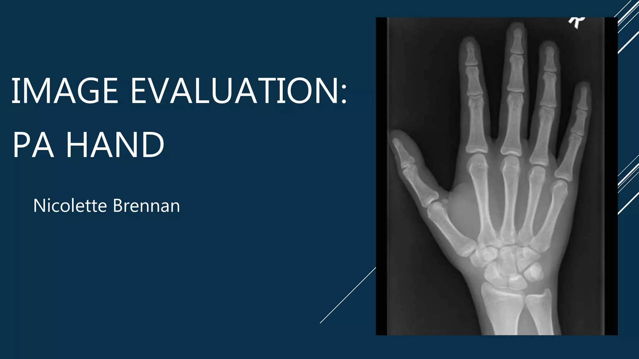
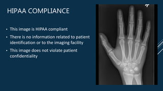
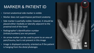
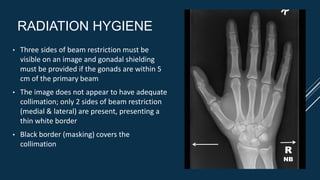
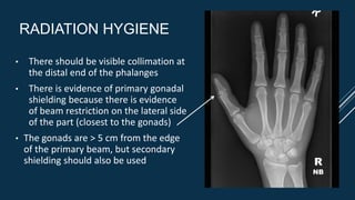
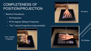
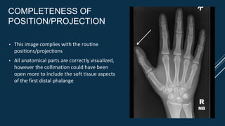
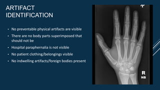
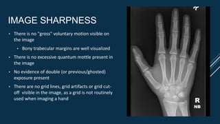
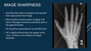
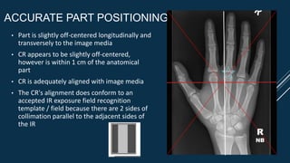
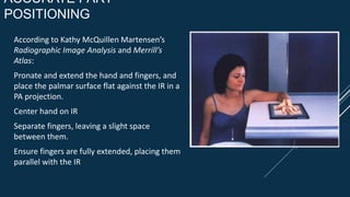
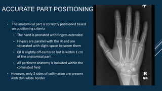
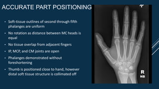
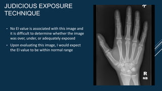
IMAGE EVALUATION:
PA HAND
Nicolette Brennan
HIPAA COMPLIANCE
• This image is HIPAA compliant
• There is no information related to patient
identification or to the imaging facility
• This image does not violate patient
confidentiality
MARKER & PATIENT ID
• Correct anatomical side marker is visible
• Marker does not superimpose pertinent anatomy
• Side marker is partially visible; however, it should be
placed either medially or laterally adjacent to the
proximal end of the hand
• Radiographer's identification number
(initials/numbers) are not present
• An arrow marker can be used to point to an area of
pain/trauma, but is not present in this image
• Image is displayed correctly, oriented as if the patient
is hanging from the distal phalanges
R
NB
RADIATION HYGIENE
• Three sides of beam restriction must be
visible on an image and gonadal shielding
must be provided if the gonads are within 5
cm of the primary beam
• The image does not appear to have adequate
collimation; only 2 sides of beam restriction
(medial & lateral) are present, presenting a
thin white border
• Black border (masking) covers the
collimation
R
NB
RADIATION HYGIENE
• There should be visible collimation at
the distal end of the phalanges
• There is evidence of primary gonadal
shielding because there is evidence
of beam restriction on the lateral side
of the part (closest to the gonads)
• The gonads are > 5 cm from the edge
of the primary beam, but secondary
shielding should also be used R
NB
COMPLETENESS OF
POSITION/PROJECTION
• Routine Procedures:
• PA Projection
• PA 45 degree Oblique Projection
• "Fan" Lateral Projection (Lateromedial)
PA Hand
PA Oblique
"Fan" Lateral
Projection
COMPLETENESS OF
POSITION/PROJECTION
• This image complies with the routine
positions/projections
• All anatomical parts are correctly visualized,
however the collimation could have been
open more to include the soft tissue aspects
of the first distal phalange
R
NB
ARTIFACT
IDENTIFICATION
• No preventable physical artifacts are visible
• There are no body parts superimposed that
should not be
• Hospital paraphernalia is not visible
• No patient clothing/belongings visible
• No indwelling artifacts/foreign bodies present
R
NB
ARTIFACT
IDENTIFICATION
• There is no excess fog visible or
degrading overall image quality
• No CR/DR artifacts visible in the image
R
NB
IMAGE SHARPNESS
• There is no "gross" voluntary motion visible on
the image
• Bony trabecular margins are well visualized
• There is no excessive quantum mottle present in
the image
• No evidence of double (or previous/ghosted)
exposure present
• There are no grid lines, grid artifacts or grid cut-
off visible in the image, as a grid is not routinely
used when imaging a hand R
NB
IMAGE SHARPNESS
• Size distortion does not appear to be greater
than expected for this image
• OID should be minimal when imaging a PA
hand. Phalanges should be extended, palm is
flat against IR plate
• CR should be perpendicular to 3rd MCP joint
• CR is slightly off but does not appear to be
>1cm, so there is no evidence of shape
distortion
R
NB
ACCURATE PART POSITIONING
• Part is slightly off-centered longitudinally and
transversely to the image media
• CR appears to be slightly off-centered,
however is within 1 cm of the anatomical
part
• CR is adequately aligned with image media
• The CR's alignment does conform to an
accepted IR exposure field recognition
template / field because there are 2 sides of
collimation parallel to the adjacent sides of
the IR
R
NB
ACCURATE PART
POSITIONING
According to Kathy McQuillen Martensen’s
Radiographic Image Analysis and Merrill’s
Atlas:
Pronate and extend the hand and fingers, and
place the palmar surface flat against the IR in a
PA projection.
Center hand on IR
Separate fingers, leaving a slight space
between them.
Ensure fingers are fully extended, placing them
parallel with the IR
ACCURATE PART
POSITIONING
According to Kathy McQuillen Martensen’s
Radiographic Image Analysis and Merrill’s
Atlas:
Position thumb a small distance from the hand
Center a perpendicular CR to third MCP joint
Open the longitudinal collimation to include
the distal phalanges and 1 inch of the distal
forearm
Transversely collimate to within 0.5 inch of the
first and fifth digits' skin line
ACCURATE PART
POSITIONING
According to Kathy McQuillen Martensen’s
Radiographic Image Analysis and Merrill’s
Atlas:
Soft tissue outlines of the second through
fifth phalanges are uniform
Distance between the MC heads is equal
Equal midshaft concavity is seen on both
sides of the phalanges and MCs of the second
through fifth fingers
IP, MCP, and CM joints are demonstrated as
open spaces
Phalanges are demonstrated without
foreshortening
Thumb demonstrates a 45-degree-
oblique projection
Thumb is positioned close to the hand
Third MCP joint is at the center of the
exposure field
Phalanges, MCs, carpals, and 1 inch of
the distal radius and ulna are included
within the exposure field
ACCURATE PART POSITIONING
• The anatomical part is correctly positioned based
on positioning criteria
• The hand is pronated with fingers extended
• Fingers are parallel with the IR and are
separated with slight space between them
• CR is slightly off-centered but is within 1 cm
of the anatomical part
• All pertinent anatomy is included within the
collimated field
• However, only 2 sides of collimation are present
with thin white border
R
NB
ACCURATE PART POSITIONING
• Soft-tissue outlines of second through fifth
phalanges are uniform
• No rotation as distance between MC heads is
equal
• No tissue overlap from adjacent fingers
• IP, MCP, and CM joints are open
• Phalanges demonstrated without
foreshortening
• Thumb is positioned close to hand, however
distal soft tissue structure is collimated off R
NB
JUDICIOUS EXPOSURE
TECHNIQUE
• The most radiolucent structures are the soft
tissue surrounding the bony structure and
the joint spaces they are visible on the image
• The most radiopaque structure is the bony
cortex and it is visible
• The image's scale of contrast is short scale
and appears adequate
• The image's brightness is adequate to
visualize all anatomical structures necessary R
NB
JUDICIOUS EXPOSURE
TECHNIQUE
• No EI value is associated with this image and
it is difficult to determine whether the image
was over, under, or adequately exposed
• Upon evaluating this image, I would expect
the EI value to be within normal range
R
NB
ACCEPT/REJECT IMAGE
This image meets the minimum established
standards for acceptance criteria – ACCEPT
Required Corrections:
• Include soft tissue structure of the first distal
phalanx
• "Right" marker including technologist's
initials or identification number
• Proper collimation ≥ 3 sides
• Center a perpendicular CR to third MCP joint R
NB
REFERENCES
https://radiopaedia.org/cases/hand-annotated-x-rays
Kathy McQuillen Martensen Radiographic Image Analysis and Merrill’s
Atlas

Final Image Evaluation

  • 1.
  • 2.
    HIPAA COMPLIANCE • Thisimage is HIPAA compliant • There is no information related to patient identification or to the imaging facility • This image does not violate patient confidentiality
  • 3.
    MARKER & PATIENTID • Correct anatomical side marker is visible • Marker does not superimpose pertinent anatomy • Side marker is partially visible; however, it should be placed either medially or laterally adjacent to the proximal end of the hand • Radiographer's identification number (initials/numbers) are not present • An arrow marker can be used to point to an area of pain/trauma, but is not present in this image • Image is displayed correctly, oriented as if the patient is hanging from the distal phalanges R NB
  • 4.
    RADIATION HYGIENE • Threesides of beam restriction must be visible on an image and gonadal shielding must be provided if the gonads are within 5 cm of the primary beam • The image does not appear to have adequate collimation; only 2 sides of beam restriction (medial & lateral) are present, presenting a thin white border • Black border (masking) covers the collimation R NB
  • 5.
    RADIATION HYGIENE • Thereshould be visible collimation at the distal end of the phalanges • There is evidence of primary gonadal shielding because there is evidence of beam restriction on the lateral side of the part (closest to the gonads) • The gonads are > 5 cm from the edge of the primary beam, but secondary shielding should also be used R NB
  • 6.
    COMPLETENESS OF POSITION/PROJECTION • RoutineProcedures: • PA Projection • PA 45 degree Oblique Projection • "Fan" Lateral Projection (Lateromedial) PA Hand PA Oblique "Fan" Lateral Projection
  • 7.
    COMPLETENESS OF POSITION/PROJECTION • Thisimage complies with the routine positions/projections • All anatomical parts are correctly visualized, however the collimation could have been open more to include the soft tissue aspects of the first distal phalange R NB
  • 8.
    ARTIFACT IDENTIFICATION • No preventablephysical artifacts are visible • There are no body parts superimposed that should not be • Hospital paraphernalia is not visible • No patient clothing/belongings visible • No indwelling artifacts/foreign bodies present R NB
  • 9.
    ARTIFACT IDENTIFICATION • There isno excess fog visible or degrading overall image quality • No CR/DR artifacts visible in the image R NB
  • 10.
    IMAGE SHARPNESS • Thereis no "gross" voluntary motion visible on the image • Bony trabecular margins are well visualized • There is no excessive quantum mottle present in the image • No evidence of double (or previous/ghosted) exposure present • There are no grid lines, grid artifacts or grid cut- off visible in the image, as a grid is not routinely used when imaging a hand R NB
  • 11.
    IMAGE SHARPNESS • Sizedistortion does not appear to be greater than expected for this image • OID should be minimal when imaging a PA hand. Phalanges should be extended, palm is flat against IR plate • CR should be perpendicular to 3rd MCP joint • CR is slightly off but does not appear to be >1cm, so there is no evidence of shape distortion R NB
  • 12.
    ACCURATE PART POSITIONING •Part is slightly off-centered longitudinally and transversely to the image media • CR appears to be slightly off-centered, however is within 1 cm of the anatomical part • CR is adequately aligned with image media • The CR's alignment does conform to an accepted IR exposure field recognition template / field because there are 2 sides of collimation parallel to the adjacent sides of the IR R NB
  • 13.
    ACCURATE PART POSITIONING According toKathy McQuillen Martensen’s Radiographic Image Analysis and Merrill’s Atlas: Pronate and extend the hand and fingers, and place the palmar surface flat against the IR in a PA projection. Center hand on IR Separate fingers, leaving a slight space between them. Ensure fingers are fully extended, placing them parallel with the IR
  • 14.
    ACCURATE PART POSITIONING According toKathy McQuillen Martensen’s Radiographic Image Analysis and Merrill’s Atlas: Position thumb a small distance from the hand Center a perpendicular CR to third MCP joint Open the longitudinal collimation to include the distal phalanges and 1 inch of the distal forearm Transversely collimate to within 0.5 inch of the first and fifth digits' skin line
  • 15.
    ACCURATE PART POSITIONING According toKathy McQuillen Martensen’s Radiographic Image Analysis and Merrill’s Atlas: Soft tissue outlines of the second through fifth phalanges are uniform Distance between the MC heads is equal Equal midshaft concavity is seen on both sides of the phalanges and MCs of the second through fifth fingers IP, MCP, and CM joints are demonstrated as open spaces Phalanges are demonstrated without foreshortening Thumb demonstrates a 45-degree- oblique projection Thumb is positioned close to the hand Third MCP joint is at the center of the exposure field Phalanges, MCs, carpals, and 1 inch of the distal radius and ulna are included within the exposure field
  • 16.
    ACCURATE PART POSITIONING •The anatomical part is correctly positioned based on positioning criteria • The hand is pronated with fingers extended • Fingers are parallel with the IR and are separated with slight space between them • CR is slightly off-centered but is within 1 cm of the anatomical part • All pertinent anatomy is included within the collimated field • However, only 2 sides of collimation are present with thin white border R NB
  • 17.
    ACCURATE PART POSITIONING •Soft-tissue outlines of second through fifth phalanges are uniform • No rotation as distance between MC heads is equal • No tissue overlap from adjacent fingers • IP, MCP, and CM joints are open • Phalanges demonstrated without foreshortening • Thumb is positioned close to hand, however distal soft tissue structure is collimated off R NB
  • 18.
    JUDICIOUS EXPOSURE TECHNIQUE • Themost radiolucent structures are the soft tissue surrounding the bony structure and the joint spaces they are visible on the image • The most radiopaque structure is the bony cortex and it is visible • The image's scale of contrast is short scale and appears adequate • The image's brightness is adequate to visualize all anatomical structures necessary R NB
  • 19.
    JUDICIOUS EXPOSURE TECHNIQUE • NoEI value is associated with this image and it is difficult to determine whether the image was over, under, or adequately exposed • Upon evaluating this image, I would expect the EI value to be within normal range R NB
  • 20.
    ACCEPT/REJECT IMAGE This imagemeets the minimum established standards for acceptance criteria – ACCEPT Required Corrections: • Include soft tissue structure of the first distal phalanx • "Right" marker including technologist's initials or identification number • Proper collimation ≥ 3 sides • Center a perpendicular CR to third MCP joint R NB
  • 21.