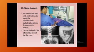
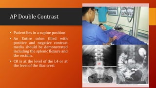
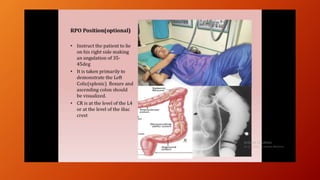
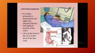
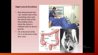
A barium enema is a radiographic procedure used to examine the large intestine. During the procedure, a liquid suspension of barium sulfate is introduced into the rectum to coat the intestinal walls. X-rays are then taken to visualize the colon. It allows visualization of the entire colon and can detect conditions like colon cancer, inflammatory bowel disease, and diverticulitis. Proper preparation and positioning of the patient is important. Double contrast technique involves injecting air in addition to barium to better visualize the mucosal lining. Findings are evaluated for abnormalities that may indicate diseases of the colon.
2. It is the radiographic study of the large bowel by administration of barium
through the rectum.
The major advantage of barium enema is its ability to examine the entire
colon.
It is reasonably accurate, minimally invasive and requires no sedation on
routine basis.
3. INDICATION
Screening for colon cancer
Inflammatory bowel disease
Diverticular disease
Inconclusive colonoscopy
Tocheck patency of distal loop
5. METHODS
• Double contrast
• the method of choice to demonstrate mucosal pattern.
The primary aim in a double contrast study is to achieve good mucosal
coating.
Preferred in high risk patients- rectal bleeding, anemia, weight loss,
family history of carcinoma / polyp, suspected IBD
6. Single contrast
• simpler, shorter and does not require rigorous maneuvers.
Preferred in very young, very old, sick and disabled patients.
In suspected obstruction and in evaluation of distal colon after
colostomy.
7. contrast medium
• For SCBE- low density barium suspension - 12-25% w/v, and a kilo voltage
of 100 -110 is used.
For DCBE- high density barium suspension – 60-120% w/v and a kilo
voltage of about 90 is used.
8. patient preparation
• For 3 days prior to examination
Low residue diet.
• On the day prior to examination
Fluids only
Drink plenty of water to prevent dehydration.
Magnesium citrate solution or Bisacodyl tablets for 2 days.
A tap water cleansing enema of 1500 ml on the morning of the barium enema
examination.
9. Procedure of double-contrast enema
The quality of the images depends on
- mucosal coating which in turn depends on the barium
suspension.
- distension ( should just efface the normal mucosal folds )
• - projection ( ideally without any overlapping loops and
with lesions in profile )
10. procedure
A scout film is taken of the AP abdomen- if retained stool is present
consider rescheduling.
The patient lies on their left side with right leg flexed in Sims position,
and the catheter tip is lubricated and is inserted gently into the rectum.
The insertion should not exceed 3 to 4 cm. It is taped firmly in position.
12. SIM'S POSITION
• The patient is asked to roll onto the left side and lean forward. The
right leg is flexed at the knee and hip and is placed in front of the
left leg. The left knee is comfortably flexed. This is called the Sims
position. The goal is to relax the abdominal muscles and decrease
pressure within the abdomen.
13. Connections are made to the barium reservoir and the hand pump for injecting air.
An intravenous injection of Buscopan (20 mg) or glucagon (1mg) may be given.
The infusion of barium is commenced. Intermittent screening is required to
check the progress of the barium.
The infusion is terminated when the barium reaches the hepatic flexure.
The column of barium within the sigmoid colon is run back out by either lowering
the infusion bag to the floor or tilting the table to the erect position.
Air is gently pumped into the bowel, forcing the column of barium round towards
the caecum, and producing the double contrast effect.
CO2 can be used as an alternative to air.
14. •Spot films of all areas of the large bowel are taken including oblique views.
Rectum: PAand left lateral view
Sigmoid: LPO and right lateral
Splenic flexure: RPO view
Hepatic flexure: LPO view
Caecum:AP and LPOview
15.
16.
17.
18.
19. AP Double Contrast
• Patient lies in a supine position
• An Entire colon filled with
positive and negative contrast
media should be demonstrated
including the splenic flexure and
the rectum.
• CR is at the level of the L4 or at
the level of the iliac crest
20.
21.
22.
23.
24.
25.
26. Supine position
• A postevacuation radiograph view
of the colon is taken after the
procedure is done
• If inadequate satisfactory
delineation of the mucus the
patient may be given hot beverage
(tea/coffee) to stimulate
evacuation
27. structure Shown for Right Anterior ObliqueStructures Shown for PA or AP Barium Enema
28. Aftercare
Patients should be warned that their bowel motions will be white for a few days
after the examination, and to eat and drink normally to avoid barium
impaction.
The patient must not leave the department until any blurring of vision
produced by the Buscopan has resolved.
29. • What are the Advantages & disadvantages?
• ADVANTAGES:
Non absorbable, hence barium does not degrade in the bowel.
Barium is not absorbed into the blood, allergic reactions are extremely rare.
• DISADVANTAGES:
Chemical peritonitis due to extravasation of additives of barium sulphate.
Rarely, the barium that remains in the colon can harden into clumps & can
cause an obstruction in the gastrointestinal tract, called barium inspissation.
This risk is reduced by taking plenty of fluids by mouth after the test.
Aggressive bowel cleansing may cause hypokalemia (low potassium) and/or
dehydration in some patients.
Rarely, small clumps of barium retained in the bowel, termed barium
granulomas, may cause inflammation in the colon.
Barium encephalopathy in case of perforation.
32. COLORECTAL CARCINOMA
Apple core sign - This sign is typical of
colorectal carcinoma. Obstruction is a
common finding.
Double-contrast barium enema demonstrates a
focal narrowing in the sigmoid colon and
resulting in a severe stenosis called apple-core
stenosis.
33. Rectal carcinoma: the narrowed segment showing
mucosal irregularity indicate a circumferential lesion.