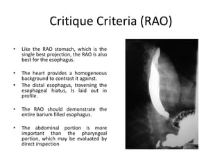
This document provides information about barium swallow studies, including:
- The learning objectives which are to describe the anatomy and physiology of the esophagus and the procedure for barium swallow studies.
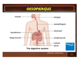
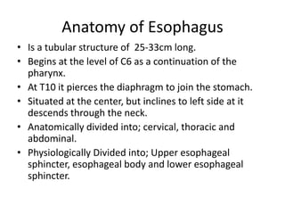
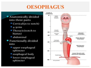
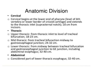
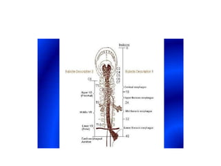
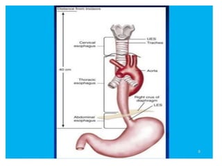
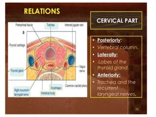
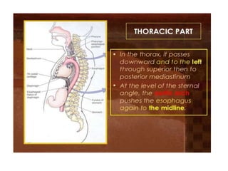
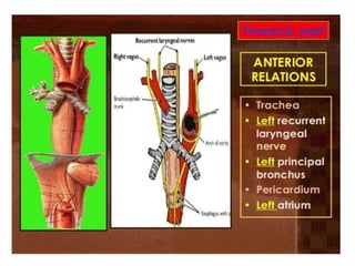
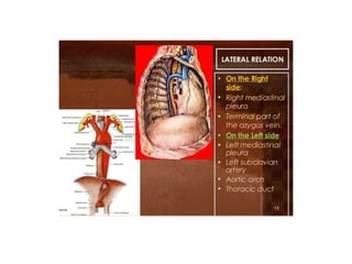
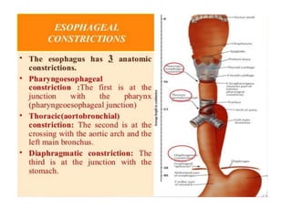
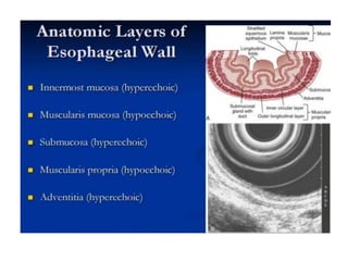
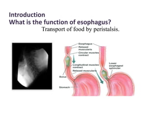
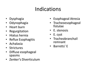
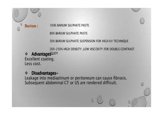
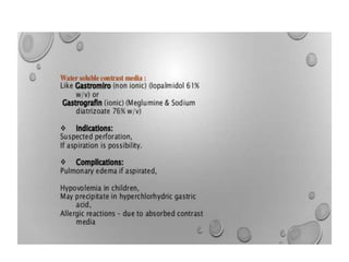
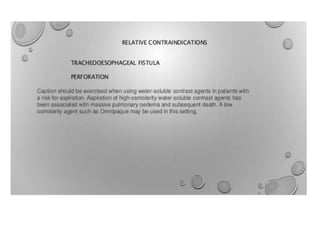
- Details of the anatomy of the esophagus and the indications for barium swallow studies such as dysphagia and heartburn.
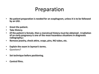
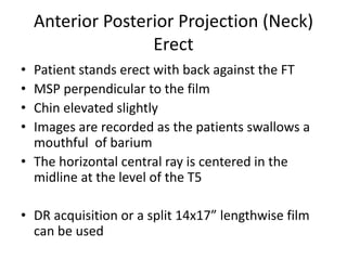
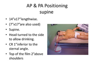
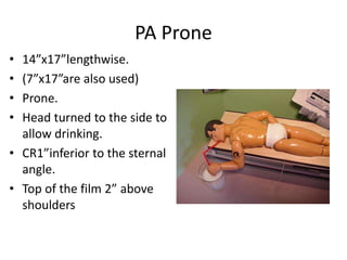
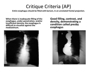
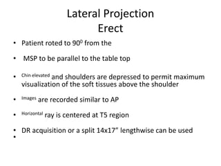
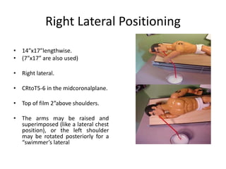
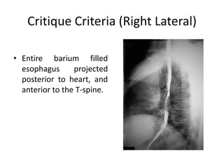
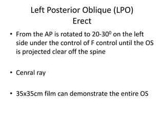
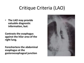
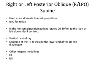
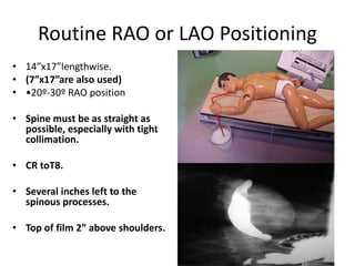
- The preparation, examination process, positioning, and critique criteria for barium swallow imaging including anterior-posterior, lateral, and oblique views.
- Additional imaging modalities that can be used such as CT and MRI are also mentioned.
the simple technique you can do barium swallow by yourself if you have any complications.......you can also help many people with this simple technique. just give it a look and try it.....<a>Click Here!</a>
Basic Chest X ray Views - AP, PA & Lateral etc . pptxDr Abna J
PA PROJECTION
Sit or stand upright.
Positioned to minimize magnification of the anteriorly positioned heart and consequent obscuration of the lungs.
Make sure the patient is standing straight and is equally distributing the weight of the body on both feet.
The upright position is preferred for the following reasons: It prevents engorgement (an excess of blood) of pulmonary vessels.
It allows full expansion of the lungs
To visualize possible air and fluid levels in the chest.
An upright chest film is preferred over an upright abdominal film for the diagnosis of pneumoperitoneum (free air in the abdominal cavity).
Ask the patient to move the shoulders forward and downward, so that the chest wall and both shoulders are in contact with the cassette. This helps to carry the clavicles below the lung apices.
It is very important to minimize breast shadows.
Ask the patient to pull the breasts upward and laterally (outwards), then remove her hands as she leans against the cassette holder to keep them in position.
Rotation
Even a small degree of rotation distorts the mediastinal borders, and the lung nearest the film will appear less translucent.
The following points should be stressed to obtain a true PA view (without rotation):
Ensure that the patient is standing evenly on both feet.
Both shoulders should be rolled forward and downward.
The chest radiograph should be well centred so that the medial ends of the clavicle are equidistant from the vertebral spinous processes at T4/5.
CENTRAL RAY
Over T7 vertebra
SID: 72 inches
Central ray
Film holder (image receptor) placement
The horizontal dimension of an average chest is greater than the vertical dimension.
This requires that a 14 x 17-inch film holder or image receptor (IR) be placed crosswise.
Or lengthwise depending on body type.
Collimation
The upper border of the illuminated field should be at the level of vertebra prominence (4 cm above the apex of lungs).
This will result in a lower collimation border of 1-2 inches below the costophrenic angle, if the central ray was correctly centred.
A general rule for average adult patients is to show a minimum of 10 ribs on a good PA chest radiograph.
Evaluation criteria for a good PA projection
Entire lung fields from apices to costophrenic angles should be clearly demonstrated.
No rotation. (both the right and left sternal ends of the clavicle will be the same distance from the center line of the spine.)
The direction of rotation can be determined by which sternal end of the clavicle is closest to the spine.
Trachea is visible in midline.
Scapula projected outside the lung fields.
Ten posterior ribs are visible above the diaphragm.
There is a sharp outline of the heart and diaphragm.
A faint shadow of the ribs and superior thoracic vertebrae is visible through the heart shadow.
Lung markings are visible from the hilum to the periphery of the lung.
Variations
An expiratory film may be helpful under some circumstances.
Learn Chest X-Ray With Its Normal Positioning & Radio-AnatomyDr.Santosh Atreya
Learn Chest X-Ray With Its Normal Positioning & Radio-Anatomy..For some image description please go through the text book "David Sutton" because i have described these image during my presentation Verbally..There are many animations used inside this presentation so to see all the pictures which are placed layer by layer with the help of animations you simple need to download this presentation first.... Thanx.
HERE IT REVIWES ABOUT THE X RAY OF CHEST IN DIFFERENT VIEWS OTHER THAN THE SPECIAL VIEWS OF CHEST.IT SHOWS THE ANATOMY OF CHEST IMPORTANCE OF PA CHEST X RAY OVER AP.DIFFERNEC BETTWEEN X RAY PA AND AP VIEW
Introducing Diagnostic Ultrasound in General Practiceupstatevet
Chris Ryan, DVM, DACVR
This lecture will begin by reviewing the basic operation of ultrasound equipment with a focus on hardware and software features common to almost all machines. The various settings and controls will be reviewed, along with the effects that these have on overall image quality, and how to utilize these settings to optimize image quality. A roadmap will then be developed for applying ultrasound in everyday general practice, beginning with the basics of evaluation for abdominal or pleural cavity fluid, and proceeding to perform a complete basic abdominal ultrasound exam. Normal sonographic anatomy and measurements will be reviewed, along with a recommended acquisition protocol for submission to teleradiology services.
Title: Sense of Taste
Presenter: Dr. Faiza, Assistant Professor of Physiology
Qualifications:
MBBS (Best Graduate, AIMC Lahore)
FCPS Physiology
ICMT, CHPE, DHPE (STMU)
MPH (GC University, Faisalabad)
MBA (Virtual University of Pakistan)
Learning Objectives:
Describe the structure and function of taste buds.
Describe the relationship between the taste threshold and taste index of common substances.
Explain the chemical basis and signal transduction of taste perception for each type of primary taste sensation.
Recognize different abnormalities of taste perception and their causes.
Key Topics:
Significance of Taste Sensation:
Differentiation between pleasant and harmful food
Influence on behavior
Selection of food based on metabolic needs
Receptors of Taste:
Taste buds on the tongue
Influence of sense of smell, texture of food, and pain stimulation (e.g., by pepper)
Primary and Secondary Taste Sensations:
Primary taste sensations: Sweet, Sour, Salty, Bitter, Umami
Chemical basis and signal transduction mechanisms for each taste
Taste Threshold and Index:
Taste threshold values for Sweet (sucrose), Salty (NaCl), Sour (HCl), and Bitter (Quinine)
Taste index relationship: Inversely proportional to taste threshold
Taste Blindness:
Inability to taste certain substances, particularly thiourea compounds
Example: Phenylthiocarbamide
Structure and Function of Taste Buds:
Composition: Epithelial cells, Sustentacular/Supporting cells, Taste cells, Basal cells
Features: Taste pores, Taste hairs/microvilli, and Taste nerve fibers
Location of Taste Buds:
Found in papillae of the tongue (Fungiform, Circumvallate, Foliate)
Also present on the palate, tonsillar pillars, epiglottis, and proximal esophagus
Mechanism of Taste Stimulation:
Interaction of taste substances with receptors on microvilli
Signal transduction pathways for Umami, Sweet, Bitter, Sour, and Salty tastes
Taste Sensitivity and Adaptation:
Decrease in sensitivity with age
Rapid adaptation of taste sensation
Role of Saliva in Taste:
Dissolution of tastants to reach receptors
Washing away the stimulus
Taste Preferences and Aversions:
Mechanisms behind taste preference and aversion
Influence of receptors and neural pathways
Impact of Sensory Nerve Damage:
Degeneration of taste buds if the sensory nerve fiber is cut
Abnormalities of Taste Detection:
Conditions: Ageusia, Hypogeusia, Dysgeusia (parageusia)
Causes: Nerve damage, neurological disorders, infections, poor oral hygiene, adverse drug effects, deficiencies, aging, tobacco use, altered neurotransmitter levels
Neurotransmitters and Taste Threshold:
Effects of serotonin (5-HT) and norepinephrine (NE) on taste sensitivity
Supertasters:
25% of the population with heightened sensitivity to taste, especially bitterness
Increased number of fungiform papillae
HOT NEW PRODUCT! BIG SALES FAST SHIPPING NOW FROM CHINA!! EU KU DB BK substit...GL Anaacs
Contact us if you are interested:
Email / Skype : kefaya1771@gmail.com
Threema: PXHY5PDH
New BATCH Ku !!! MUCH IN DEMAND FAST SALE EVERY BATCH HAPPY GOOD EFFECT BIG BATCH !
Contact me on Threema or skype to start big business!!
Hot-sale products:
NEW HOT EUTYLONE WHITE CRYSTAL!!
5cl-adba precursor (semi finished )
5cl-adba raw materials
ADBB precursor (semi finished )
ADBB raw materials
APVP powder
5fadb/4f-adb
Jwh018 / Jwh210
Eutylone crystal
Protonitazene (hydrochloride) CAS: 119276-01-6
Flubrotizolam CAS: 57801-95-3
Metonitazene CAS: 14680-51-4
Payment terms: Western Union,MoneyGram,Bitcoin or USDT.
Deliver Time: Usually 7-15days
Shipping method: FedEx, TNT, DHL,UPS etc.Our deliveries are 100% safe, fast, reliable and discreet.
Samples will be sent for your evaluation!If you are interested in, please contact me, let's talk details.
We specializes in exporting high quality Research chemical, medical intermediate, Pharmaceutical chemicals and so on. Products are exported to USA, Canada, France, Korea, Japan,Russia, Southeast Asia and other countries.
Tom Selleck Health: A Comprehensive Look at the Iconic Actor’s Wellness Journeygreendigital
Tom Selleck, an enduring figure in Hollywood. has captivated audiences for decades with his rugged charm, iconic moustache. and memorable roles in television and film. From his breakout role as Thomas Magnum in Magnum P.I. to his current portrayal of Frank Reagan in Blue Bloods. Selleck's career has spanned over 50 years. But beyond his professional achievements. fans have often been curious about Tom Selleck Health. especially as he has aged in the public eye.
Follow us on: Pinterest
Introduction
Many have been interested in Tom Selleck health. not only because of his enduring presence on screen but also because of the challenges. and lifestyle choices he has faced and made over the years. This article delves into the various aspects of Tom Selleck health. exploring his fitness regimen, diet, mental health. and the challenges he has encountered as he ages. We'll look at how he maintains his well-being. the health issues he has faced, and his approach to ageing .
Early Life and Career
Childhood and Athletic Beginnings
Tom Selleck was born on January 29, 1945, in Detroit, Michigan, and grew up in Sherman Oaks, California. From an early age, he was involved in sports, particularly basketball. which played a significant role in his physical development. His athletic pursuits continued into college. where he attended the University of Southern California (USC) on a basketball scholarship. This early involvement in sports laid a strong foundation for his physical health and disciplined lifestyle.
Transition to Acting
Selleck's transition from an athlete to an actor came with its physical demands. His first significant role in "Magnum P.I." required him to perform various stunts and maintain a fit appearance. This role, which he played from 1980 to 1988. necessitated a rigorous fitness routine to meet the show's demands. setting the stage for his long-term commitment to health and wellness.
Fitness Regimen
Workout Routine
Tom Selleck health and fitness regimen has evolved. adapting to his changing roles and age. During his "Magnum, P.I." days. Selleck's workouts were intense and focused on building and maintaining muscle mass. His routine included weightlifting, cardiovascular exercises. and specific training for the stunts he performed on the show.
Selleck adjusted his fitness routine as he aged to suit his body's needs. Today, his workouts focus on maintaining flexibility, strength, and cardiovascular health. He incorporates low-impact exercises such as swimming, walking, and light weightlifting. This balanced approach helps him stay fit without putting undue strain on his joints and muscles.
Importance of Flexibility and Mobility
In recent years, Selleck has emphasized the importance of flexibility and mobility in his fitness regimen. Understanding the natural decline in muscle mass and joint flexibility with age. he includes stretching and yoga in his routine. These practices help prevent injuries, improve posture, and maintain mobilit
Lung Cancer: Artificial Intelligence, Synergetics, Complex System Analysis, S...Oleg Kshivets
RESULTS: Overall life span (LS) was 2252.1±1742.5 days and cumulative 5-year survival (5YS) reached 73.2%, 10 years – 64.8%, 20 years – 42.5%. 513 LCP lived more than 5 years (LS=3124.6±1525.6 days), 148 LCP – more than 10 years (LS=5054.4±1504.1 days).199 LCP died because of LC (LS=562.7±374.5 days). 5YS of LCP after bi/lobectomies was significantly superior in comparison with LCP after pneumonectomies (78.1% vs.63.7%, P=0.00001 by log-rank test). AT significantly improved 5YS (66.3% vs. 34.8%) (P=0.00000 by log-rank test) only for LCP with N1-2. Cox modeling displayed that 5YS of LCP significantly depended on: phase transition (PT) early-invasive LC in terms of synergetics, PT N0—N12, cell ratio factors (ratio between cancer cells- CC and blood cells subpopulations), G1-3, histology, glucose, AT, blood cell circuit, prothrombin index, heparin tolerance, recalcification time (P=0.000-0.038). Neural networks, genetic algorithm selection and bootstrap simulation revealed relationships between 5YS and PT early-invasive LC (rank=1), PT N0—N12 (rank=2), thrombocytes/CC (3), erythrocytes/CC (4), eosinophils/CC (5), healthy cells/CC (6), lymphocytes/CC (7), segmented neutrophils/CC (8), stick neutrophils/CC (9), monocytes/CC (10); leucocytes/CC (11). Correct prediction of 5YS was 100% by neural networks computing (area under ROC curve=1.0; error=0.0).
CONCLUSIONS: 5YS of LCP after radical procedures significantly depended on: 1) PT early-invasive cancer; 2) PT N0--N12; 3) cell ratio factors; 4) blood cell circuit; 5) biochemical factors; 6) hemostasis system; 7) AT; 8) LC characteristics; 9) LC cell dynamics; 10) surgery type: lobectomy/pneumonectomy; 11) anthropometric data. Optimal diagnosis and treatment strategies for LC are: 1) screening and early detection of LC; 2) availability of experienced thoracic surgeons because of complexity of radical procedures; 3) aggressive en block surgery and adequate lymph node dissection for completeness; 4) precise prediction; 5) adjuvant chemoimmunoradiotherapy for LCP with unfavorable prognosis.
Pulmonary Thromboembolism - etilogy, types, medical- Surgical and nursing man...VarunMahajani
Disruption of blood supply to lung alveoli due to blockage of one or more pulmonary blood vessels is called as Pulmonary thromboembolism. In this presentation we will discuss its causes, types and its management in depth.
These lecture slides, by Dr Sidra Arshad, offer a quick overview of physiological basis of a normal electrocardiogram.
Learning objectives:
1. Define an electrocardiogram (ECG) and electrocardiography
2. Describe how dipoles generated by the heart produce the waveforms of the ECG
3. Describe the components of a normal electrocardiogram of a typical bipolar leads (limb II)
4. Differentiate between intervals and segments
5. Enlist some common indications for obtaining an ECG
Study Resources:
1. Chapter 11, Guyton and Hall Textbook of Medical Physiology, 14th edition
2. Chapter 9, Human Physiology - From Cells to Systems, Lauralee Sherwood, 9th edition
3. Chapter 29, Ganong’s Review of Medical Physiology, 26th edition
4. Electrocardiogram, StatPearls - https://www.ncbi.nlm.nih.gov/books/NBK549803/
5. ECG in Medical Practice by ABM Abdullah, 4th edition
6. ECG Basics, http://www.nataliescasebook.com/tag/e-c-g-basics
Anti ulcer drugs and their Advance pharmacology ||
Anti-ulcer drugs are medications used to prevent and treat ulcers in the stomach and upper part of the small intestine (duodenal ulcers). These ulcers are often caused by an imbalance between stomach acid and the mucosal lining, which protects the stomach lining.
||Scope: Overview of various classes of anti-ulcer drugs, their mechanisms of action, indications, side effects, and clinical considerations.
Flu Vaccine Alert in Bangalore Karnatakaaddon Scans
As flu season approaches, health officials in Bangalore, Karnataka, are urging residents to get their flu vaccinations. The seasonal flu, while common, can lead to severe health complications, particularly for vulnerable populations such as young children, the elderly, and those with underlying health conditions.
Dr. Vidisha Kumari, a leading epidemiologist in Bangalore, emphasizes the importance of getting vaccinated. "The flu vaccine is our best defense against the influenza virus. It not only protects individuals but also helps prevent the spread of the virus in our communities," he says.
This year, the flu season is expected to coincide with a potential increase in other respiratory illnesses. The Karnataka Health Department has launched an awareness campaign highlighting the significance of flu vaccinations. They have set up multiple vaccination centers across Bangalore, making it convenient for residents to receive their shots.
To encourage widespread vaccination, the government is also collaborating with local schools, workplaces, and community centers to facilitate vaccination drives. Special attention is being given to ensuring that the vaccine is accessible to all, including marginalized communities who may have limited access to healthcare.
Residents are reminded that the flu vaccine is safe and effective. Common side effects are mild and may include soreness at the injection site, mild fever, or muscle aches. These side effects are generally short-lived and far less severe than the flu itself.
Healthcare providers are also stressing the importance of continuing COVID-19 precautions. Wearing masks, practicing good hand hygiene, and maintaining social distancing are still crucial, especially in crowded places.
Protect yourself and your loved ones by getting vaccinated. Together, we can help keep Bangalore healthy and safe this flu season. For more information on vaccination centers and schedules, residents can visit the Karnataka Health Department’s official website or follow their social media pages.
Stay informed, stay safe, and get your flu shot today!
Maxilla, Mandible & Hyoid Bone & Clinical Correlations by Dr. RIG.pptx
Barium studies aminu abubakar a
1. Barium Studies
Barium Swallow (BS)
Rad. Aminu Abubakar Abubakar
Radiography Department
Bayero University Kano
October, 2020
2. • By the end of this lecture student will be able to;
• Describe the anatomy and physiology of Esophagus
• Describe the methodology of BS.
• List the indications and contraindications of BS.
• Describe patient care prior, during and after the
procedure
• Describe the imaging procedure (Single or double
contrast).
• Describe the routine and alternative positioning.
• Film critique; positioning, image quality, radiographic
anatomy and pathology
• Describe other alternative imaging modalities.
Learning Objectives
3.
4. Anatomy of Esophagus
• Is a tubular structure of 25-33cm long.
• Begins at the level of C6 as a continuation of the
pharynx.
• At T10 it pierces the diaphragm to join the stomach.
• Situated at the center, but inclines to left side at it
descends through the neck.
• Anatomically divided into; cervical, thoracic and
abdominal.
• Physiologically Divided into; Upper esophageal
sphincter, esophageal body and lower esophageal
sphincter.
20. Introduction
• BS; Special radiologic contrast examination that involves the oral
cavity to the fundus of the stomach.
• Barium Sulpahate used as contrast.
• Digital fluoroscopy
• Spot films
• Double or single contrast studies
• Barium flows from the mouth to the stomach
• Erect or recumbent
28. Preparation
• No patient preparation is needed for an esophagram, unless it is to be followed
by an UGI.
• Greet the patient.
• Take History.
• (If the patient is female, then a menstrual history must be obtained . Irradiation
of an early pregnancy is one of the most hazardous situations in diagnostic
radiography.)
• Remove jewelry, check attire, snaps, pins, NG tubes, etc.
• Explain the exam in layman’s terms.
• Questions?
• Set technique before positioning.
• Control films.
29. Examination
Single Contrast Study
• Esophagrams begin under fluoroscopy, in the upright position.
• The patient holds a cup of barium, with a straw, In the right hand.
• Instructs the patient to drink, and films in the AP, RPO, and LPO positions.
• The patient is often put into an RAO before the table is lowered to horizontal. The
examination continues in the recumbent position.
• The patient is instructed to take three large bolus swallows.
• On the fourth, breathing is suspended and the exposure is made at the moment
the patient swallows.
• A shallow trendelenburg position will help keep the esophagus full.
30. Anterior Posterior Projection (Neck)
Erect
• Patient stands erect with back against the FT
• MSP perpendicular to the film
• Chin elevated slightly
• Images are recorded as the patients swallows a
mouthful of barium
• The horizontal central ray is centered in the
midline at the level of the T5
• DR acquisition or a split 14x17” lengthwise film
can be used
31. AP & PA Positioning
supine
• 14”x17”lengthwise.
• (7”x17”are also used)
• Supine.
• Head turned to the side
to allow drinking.
• CR 1”inferior to the
sternal angle.
• Top of the film 2”above
shoulders
32. PA Prone
• 14”x17”lengthwise.
• (7”x17”are also used)
• Prone.
• Head turned to the side to
allow drinking.
• CR1”inferior to the sternal
angle.
• Top of the film 2” above
shoulders
33. Critique Criteria (AP)
Entire esophagus should be filled with barium, in an unrotated frontal projection.
When there is inadequate filling of the
esophagus, under-penetration, and/or
insufficient density, the esophagus is
difficult to visualize against the
mediastinum.
Good filling, contrast, and
density, demonstrating a
condition called presby
esophagus
34. Lateral Projection
Erect
• Patient roted to 900 from the
• MSP to be parallel to the table top
• Chin elevated and shoulders are depressed to permit maximum
visualization of the soft tissues above the shoulder
• Images are recorded similar to AP
• Horizontal ray is centered at T5 region
• DR acquisition or a split 14x17” lengthwise can be used
•
35. Right Lateral Positioning
• 14”x17”lengthwise.
• (7”x17” are also used)
• Right lateral.
• CRtoT5-6 in the midcoronalplane.
• Top of film 2”above shoulders.
• The arms may be raised and
superimposed (like a lateral chest
position), or the left shoulder
may be rotated posteriorly for a
“swimmer’s lateral
36. Critique Criteria (Right Lateral)
• Entire barium filled
esophagus projected
posterior to heart, and
anterior to the T-spine.
37. Left Posterior Oblique (LPO)
Erect
• From the AP is rotated to 20-300 on the left
side under the control of F control until the OS
is projected clear off the spine
• Cenral ray
• 35x35cm film can demonstrate the entire OS
38. Critique Criteria (LAO)
• The LAO may provide
valuable diagnostic
information, but:
Contrasts the esophagus
against the hilar area of the
right lung.
Foreshortens the abdominal
esophagus at the
gasteroesophageal junction
39. Right or Left Posterior Oblique (R/LPO)
Supine
• Used as an alternate to erect projections
• RPO for reflux
• In the horizontal position patient rotated 20-300 on to the right or
left side under F control…
• Vertical central ray
• Centered at the T8 to include the lower end of the Os and
diaphragm
• Other imaging modalities
• CT
• RNI
40. Routine RAO or LAO Positioning
• 14”x17”lengthwise.
• (7”x17”are also used)
• •20º-30º RAO position
• Spine must be as straight as
possible, especially with tight
collimation.
• CR toT8.
• Several inches left to the
spinous processes.
• Top of film 2” above shoulders.
41. Critique Criteria (RAO)
• Like the RAO stomach, which is the
single best projection, the RAO is also
best for the esophagus.
• The heart provides a homogeneous
background to contrast it against.
• The distal esophagus, traversing the
esophageal hiatus, Is laid out in
profile.
• The RAO should demonstrate the
entire barium filled esophagus.
• The abdominal portion is more
important than the pharyngeal
portion, which may be evaluated by
direct inspection
42. Examination
Double Contrast (DC)
• Similar to SC except patient is given an
effervescent agent prior to the administration
of the barium
• IV hypotonic agent may also be given
• Patient is then instructed to drink several
mouthfuls of barium rapidly and is examined
using a similar protocol to that of SC
Editor's Notes
Upper=Anatomical :Cricopharyngeal spinter formed by inferior pharyngeal constrictor. Lower=Physiological: Gastroesophageal , cardiac s, cardioesophageal s…gastroesophageal reflux…damage to the esophageal muscle