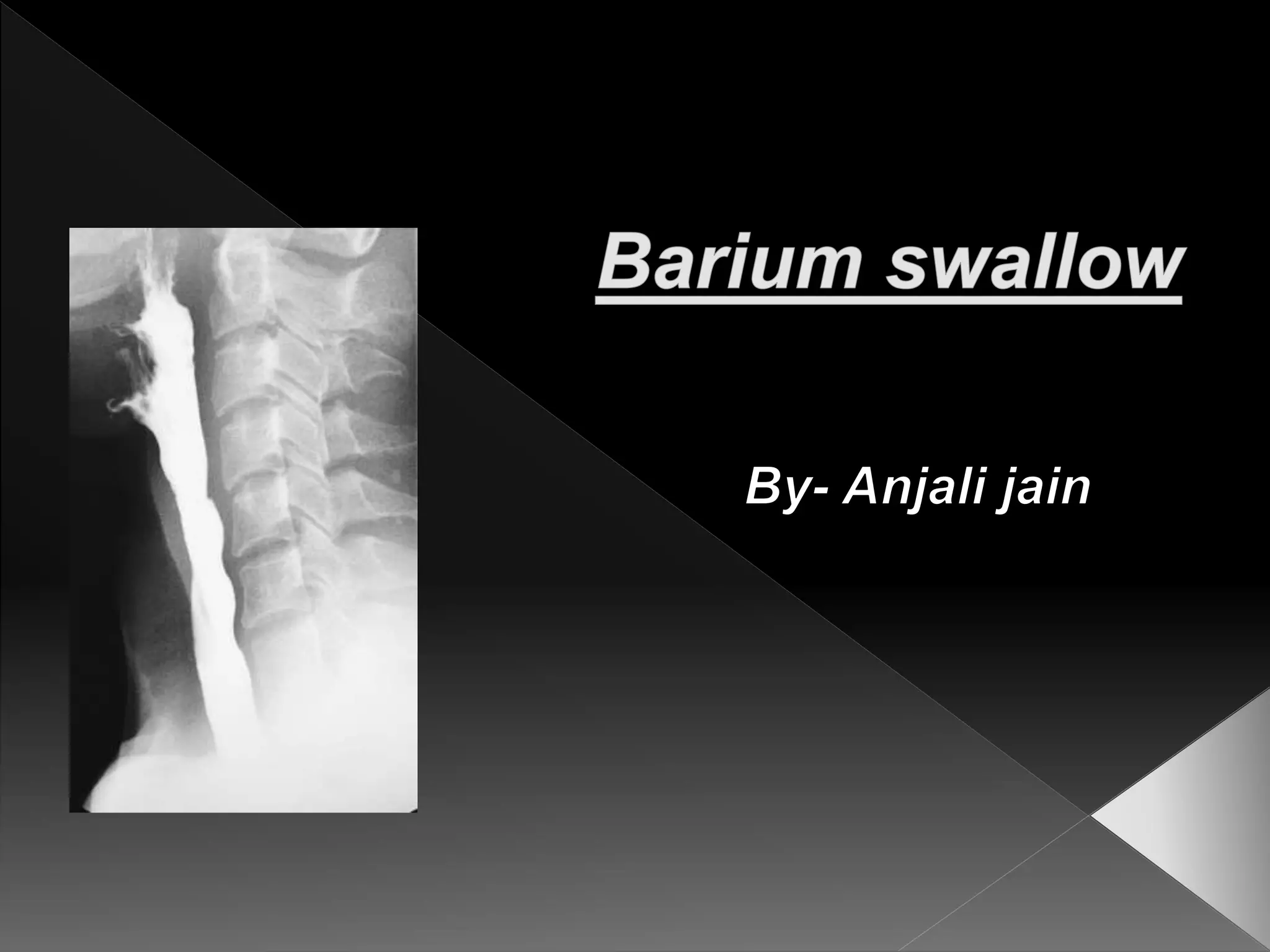
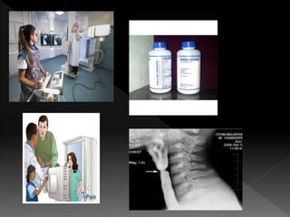
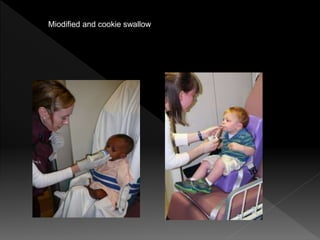
Miodified and cookie swallow
 Indication
 Dysphagia and obstruction
 Pain during swallowing
 Assessment of mediastinal masses
 Pre assessment of carcinoma bronchus
 Motility disorders of oesophagus e.g. aclasia
and diffuse oesophageal spasm, scleroderma
 Assessment of site of perforation
 Zenker’s diverticulum and cricoid webs. In these
cases water soluble contrast media are used.
E.g. Gastrograffin or dinosial aqueous
 Relative contraindications-
 Tracheo oesophageal fistula
 perforation
 Contrast-
 100% barium sulphate paste
 80% barium sulphate suspension
 30% barium sulphate suspension for high kv
technique
 200-250% high density, low viscosity for
double contrast study
 Pharynx-
 One mouthful( 10-15 ml) of contrast media
(barium sulphate paste) is given and
fluoroscopic observation of act of deglutition
is observed in frontal and lateral view with
patient erect.
 To get optimum distension of the pharynx,
exposure is triggered at the time when hyoid
bone is at highest point during swallowing.
 For this , a string is tied just above the level
of larynx. The rotor is kept running and
patient is asked to swallow.
 Exposure is released when larynx comes
above the string.
 Lateral film is taken erect and front film in
supine position.
 One mouthful of contrast media ( barium
sulphate paste) is given to patient and
patient is instructed to swallow once and
stop swallowing thereafter.
 Spot films are taken in frontal and lateral
projection.
 Patient performs valsalva maneuver in erect
position with nose closed.
 Frontal and lateral spots are taken to show
distended pyriform sinuses and vallecullae.
 Oesophagus-
 Single contrast- multiple mouthful of 80%
W/V barium suspension are given.
 Follow the barium bolus down the
oesophagus and observe the peristalsis
always in supine position.
 Films are exposed in erect position – RAO,
LAO, frontal and lateral views when the
oesophagus is well distended.
 The escape of contrast at the level of the
diaphragmatic hiatus should not be confused
for reflux.
 Mucosal film is taken in RAO after the
oesophagus is empty.
 Then fundus of stomach and GO junction are
assessed with spot film in different obliquities
in erect and recumbent position.
 Double contrast-
 Barium contrast should be high density, low
viscosity(200-250%).
 15-20 ml barium is given in the mouth and
patient is asked to swallow.
 Then , effervescent powder is given with
another mouthful of barium.
 In erect position , gas tends to stay up,
resulting in adequate distension which stays
foe longer time as compared to supine
position.
 Prone position also retains more gas within
the esophagus and gives adequate
distension.
 Hypotonia using Buscopan or glucagon
keeps the oesophagus keeps the
oesophagus distended for longer time .
 Filming is done in frontal , lateral, RAO, and
LAO.
 Introduction of gas for double contrast
studies can also be done through a tube
passed into upper oesophagus.
 Specific Condition –
 Dysphagia for both solids and liquid.
 Pharyngeal web
 Foreign body impaction ( marsh mallow
coated with barium is swallowed as whole).
The passage of marsh mallow will be
hindered at the level of obstruction
 In carcinoma ( high viscosity, normal density
liquid barium is given).
 Achlasia – the oesophagus should be
cleaned thoroughly( aspirate and wash) so
that secondary achlasia due to ca
oesophagus not be missed.
 Barium 80% W/V is used and patient is
studied in erect position .
 To differentiate achlasia, from other condition
showing abnormal peristalsis , mecholyl test
is done.
 Hiatus hernia- high abdominal pressure
required .
 Patient is asked to lie down , straighten legs
and then raise them up.
 Stomach should be well distended,
otherwise hiatus hernia may not be
demonstrated.
 Gastro – oesophageal reflux
Please like, share
and subscribe
radiology solution
for lectures
Meet u
soon

Barium swallow

  • 3.
  • 4.
     Indication  Dysphagiaand obstruction  Pain during swallowing  Assessment of mediastinal masses  Pre assessment of carcinoma bronchus  Motility disorders of oesophagus e.g. aclasia and diffuse oesophageal spasm, scleroderma  Assessment of site of perforation
  • 5.
     Zenker’s diverticulumand cricoid webs. In these cases water soluble contrast media are used. E.g. Gastrograffin or dinosial aqueous  Relative contraindications-  Tracheo oesophageal fistula  perforation  Contrast-  100% barium sulphate paste  80% barium sulphate suspension  30% barium sulphate suspension for high kv technique  200-250% high density, low viscosity for double contrast study
  • 6.
     Pharynx-  Onemouthful( 10-15 ml) of contrast media (barium sulphate paste) is given and fluoroscopic observation of act of deglutition is observed in frontal and lateral view with patient erect.  To get optimum distension of the pharynx, exposure is triggered at the time when hyoid bone is at highest point during swallowing.
  • 7.
     For this, a string is tied just above the level of larynx. The rotor is kept running and patient is asked to swallow.  Exposure is released when larynx comes above the string.  Lateral film is taken erect and front film in supine position.
  • 8.
     One mouthfulof contrast media ( barium sulphate paste) is given to patient and patient is instructed to swallow once and stop swallowing thereafter.  Spot films are taken in frontal and lateral projection.  Patient performs valsalva maneuver in erect position with nose closed.  Frontal and lateral spots are taken to show distended pyriform sinuses and vallecullae.
  • 9.
     Oesophagus-  Singlecontrast- multiple mouthful of 80% W/V barium suspension are given.  Follow the barium bolus down the oesophagus and observe the peristalsis always in supine position.  Films are exposed in erect position – RAO, LAO, frontal and lateral views when the oesophagus is well distended.  The escape of contrast at the level of the diaphragmatic hiatus should not be confused for reflux.
  • 10.
     Mucosal filmis taken in RAO after the oesophagus is empty.  Then fundus of stomach and GO junction are assessed with spot film in different obliquities in erect and recumbent position.  Double contrast-  Barium contrast should be high density, low viscosity(200-250%).  15-20 ml barium is given in the mouth and patient is asked to swallow.  Then , effervescent powder is given with another mouthful of barium.
  • 11.
     In erectposition , gas tends to stay up, resulting in adequate distension which stays foe longer time as compared to supine position.  Prone position also retains more gas within the esophagus and gives adequate distension.  Hypotonia using Buscopan or glucagon keeps the oesophagus keeps the oesophagus distended for longer time .  Filming is done in frontal , lateral, RAO, and LAO.
  • 12.
     Introduction ofgas for double contrast studies can also be done through a tube passed into upper oesophagus.  Specific Condition –  Dysphagia for both solids and liquid.  Pharyngeal web  Foreign body impaction ( marsh mallow coated with barium is swallowed as whole). The passage of marsh mallow will be hindered at the level of obstruction
  • 13.
     In carcinoma( high viscosity, normal density liquid barium is given).  Achlasia – the oesophagus should be cleaned thoroughly( aspirate and wash) so that secondary achlasia due to ca oesophagus not be missed.  Barium 80% W/V is used and patient is studied in erect position .  To differentiate achlasia, from other condition showing abnormal peristalsis , mecholyl test is done.
  • 14.
     Hiatus hernia-high abdominal pressure required .  Patient is asked to lie down , straighten legs and then raise them up.  Stomach should be well distended, otherwise hiatus hernia may not be demonstrated.  Gastro – oesophageal reflux
  • 15.
    Please like, share andsubscribe radiology solution for lectures Meet u soon