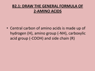
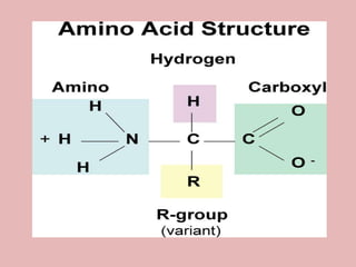
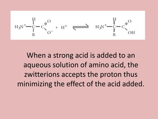
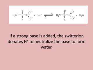
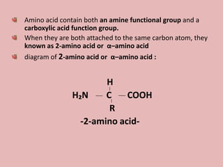
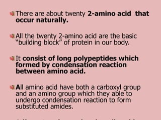
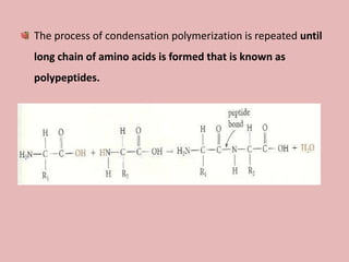
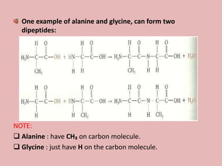
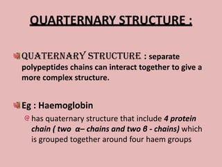
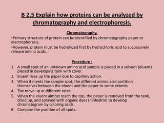
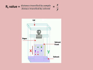
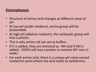
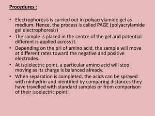
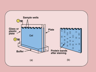
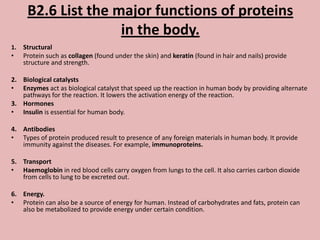
HUMAN
BIOCHEMISTRY
    -fatin
    -hanis
    -bella
B1.1: CALCULATE THE ENERGY VALUE OF A
  FOOD FROM ENTHALPHY OF COMBUSTION
                   DATA
             ∆H = mc∆T
Keys:
m = mass
c = specific heat of water (4.18 Jg-1C°-1)
∆T = change in temperature
Example:
1.00 g cereal raises the temperature of 400 cm3
water in an insulated food calorimeter from
23.7 C° to 33.4 C°. Calculate the energy value
per gram of the cereal, assuming the heat
capacity of the calorimeter is negligible and
given the specific heat of water = 4.18 Jg-1C°-1

(answer: 16.2 kJ per gram of cereal)
B2.1: DRAW THE GENERAL FORMULA OF
             2-AMINO ACIDS


• Central carbon of amino acids is made up of
  hydrogen (H), amino group (-NH), carboxylic
  acid group (-COOH) and side chain (R)
B2.2: DESCRIBE THE CHARACTERISTIC
       PROPERTIES OF 2-AMINO ACIDS
• Amino acids are:
  – Colorless
  – Crystalline solids
  – Exist as zwitterions (dipolar ions)
  – Relatively high melting points
  – More soluble in water than organic solvents
  – Are amphoterics (capable of behaving as acids or
    bases)
H2NCHRCOOH + H2O               H3N+CHRCOOH + OH-


At lower pH, H+ added reacts with OH- and the
forward reaction is favoured to replace some of
the OH- used up. Thus, in an acidic solution, the
-NH2 group is protonated.
H2NCHRCOOH + H2O              H2NCHRCOO- + H3O+


  At higher pH, the base added react with H3O+
and the forward reaction is favoured to replace
some of the H3O+ used up. Thus, in an alkaline
solution, the carboxylic acid group donates a
proton and converted to the carboxylate ion.
When a strong acid is added to an
 aqueous solution of amino acid, the
 zwitterions accepts the proton thus
minimizing the effect of the acid added.
If a strong base is added, the zwitterion
donates H+ to neutralize the base to form
                   water.
B 2.3 : DESCRIBE THE CONDENSATION

           REACTION OF

2 - AMINO ACID TO FORM POLYPEPTIDES
Amino acid contain both an amine functional group and a
carboxylic acid function group.
When they are both attached to the same carbon atom, they
known as 2-amino acid or α–amino acid
diagram of 2-amino acid or α–amino acid :


                      H
              H₂N     C    COOH
                       R
                -2-amino acid-
There are about twenty 2-amino acid that
occur naturally.

All the twenty 2-amino acid are the basic
“building block” of protein in our body.

It consist of long polypeptides which
formed by condensation reaction
between amino acid.

All amino acid have both a carboxyl group
and an amino group which they able to
undergo condensation reaction to form
substituted amides.
This process call as condensation reaction as it involve the formation of
water molecule.


The reaction between two amino acid result in the formation of a
dipeptide and water.


Definition of dipeptide : a product which is a substituted amide made up
of two amino acid joined by a peptide bond or peptide linkage.


The two amino acid residues are joined by a strong carbon - nitrogen
bond.
The process of condensation polymerization is repeated until
long chain of amino acids is formed that is known as
polypeptides.
One example of alanine and glycine, can form two
  dipeptides:


NOTE:
 Alanine : have CH₃ on carbon molecule.
 Glycine : just have H on the carbon molecule.
B 2.4 : DESCRIBE AND
EXPLAIN THE STRUCTURE
      OF PROTEIN
PRIMARY STRUCTURE :
 Definition : Linear sequences of amino acids in
  polypeptides chain.
 Primary structure of protein is determined by the
  sequence of bases in gene that codes for protein.
 If there is changing if a single amino acid due to a
  mutation in DNA of its gene will alter its properties
  drastically. Example: sickle anaemia cell.
 Each protein has its own unique primary sequence if
  amino acid.
 Primary structure of protein gives the protein ability to
  carry out its characteristic function.
 Eg : a tripeptide containing the amino acid :
 lysine, glycine and leucine           lys-gly-leu
SECONDARY STRUCTURE :
  Secondary structure is the chain of amino acid that
  folds itself due to the intramolecular hydrogen bond.

  Two type of secondary structure :
 α-helix : the protein is twist in a spiralling manner like
  coiled spring.
 β pleated sheet : give a sheet- like structure.

  Example of :
 α-helix : keratin that found in nail and hair
 β pleated sheet : silk fibre
TERTIARY STRUCTURE :
 Tertiary structure is the overall folding of chain by interaction between
  distant amino acid and gives protein its three-dimensional shape of
  single protein.
  (when the secondary structure is folded further to form 3D shape of
  globule)

 Tertiary sturucture of protein is folded each other because of they have
  attraction between amino acid in polypetides

   The interaction that involve :
   Hydrogen bond
   Van Der Waal’s between non polar side group
   Ionic attraction between polar groups
   Disulphide bridge

   Example of tertiary structure of protein :
   Immunoglobin
   Antibody
   Enzyme
   Insulin (Protein based hormone)
QUARTERNARY STRUCTURE :

Quaternary structure : separate
polypeptides chains can interact together to give a
more complex structure.


Eg : Haemoglobin
  has quaternary structure that include 4 protein
  chain ( two α– chains and two β - chains) which
  is grouped together around four haem groups
B 2.5 Explain how proteins can be analyzed by
           chromatography and electrophoresis.
                                Chromatography.
•Primary structure of protein can be identified by chromatography paper or
electrophoresis.
•However, protein must be hydrolyzed first by hydrochloric acid to successively
release amino acids.

                                     Procedure :
1.   A small spot of an unknown amino acid sample is placed in a solvent (eluent)
     placed in developing tank with cover.
2.   Eluent rises up the paper due to capillary action.
3.   When it meets the sample spot, the different amino acid partition
     themselves between the eluent and the paper to some extents
4.   The move up at different rates.
5.   When the eluent almost reach the top, the paper is removed from the tank,
     dried up, and sprayed with organic dyes (ninhydrin) to develop
     chromatogram by coloring acids.
6.   Compare the position of all spots.
Rf value =           =


             x   y
Alternative ways :
If sample of known amino acid is not available, Rf
value (retention factor) can be measured and
compared with known values as each amino acid
had a different Rf value.
Sometimes, we may found that two acids have the
same Rf value using the same solvent, but using a
different solvent may result different Rf value.
Chromatogram can be turned through 90o and run
again using the second solvent.
Electrophoresis

• Structure of amino acid changes at different value of
  pH.
• At low pH (acidic medium), amine group will be
  protonated.
• At high pH (alkaline medium), the carboxylic group will
  lose a proton.
• This is why amino cid can act as buffers.
• If H is added, they are removed as –NH and if OH is
  added, -COOH will lose a proton to remove OH- ions in
  water.
• For each amino acid, there is a unique pH value named
  isoelectric point where the acid exists as zwitterions.
Procedures :

• Electrophoresis is carried out in polyacrylamide gel as
  medium. Hence, the process is called PAGE (polyacrylamide
  gel electrophoresis)
• The sample is placed in the centre of the gel and potential
  different is applied across it.
• Depending on the pH of amino acid, the sample will move
  at different rates toward the negative and positive
  electrodes.
• At isolelectric point, a particular amino acid will stop
  moving as its charge is balanced already.
• When separation is completed, the acids can be sprayed
  with ninhydrin and identified by comparing distances they
  have travelled with standard samples or from comparison
  of their isoelectric point.
B2.6 List the major functions of proteins
                     in the body.
1.   Structural
•    Protein such as collagen (found under the skin) and keratin (found in hair and nails) provide
     structure and strength.

2. Biological catalysts
• Enzymes act as biological catalyst that speed up the reaction in human body by providing alternate
   pathways for the reaction. It lowers the activation energy of the reaction.
3. Hormones
• Insulin is essential for human body.

4. Antibodies
• Types of protein produced result to presence of any foreign materials in human body. It provide
   immunity against the diseases. For example, immunoproteins.

5. Transport
• Haemoglobin in red blood cells carry oxygen from lungs to the cell. It also carries carbon dioxide
   from cells to lung to be excreted out.

6. Energy.
• Protein can also be a source of energy for human. Instead of carbohydrates and fats, protein can
   also be metabolized to provide energy under certain condition.

B1 & b2 energy + protein

  • 1.
    HUMAN BIOCHEMISTRY -fatin -hanis -bella
  • 2.
    B1.1: CALCULATE THEENERGY VALUE OF A FOOD FROM ENTHALPHY OF COMBUSTION DATA ∆H = mc∆T Keys: m = mass c = specific heat of water (4.18 Jg-1C°-1) ∆T = change in temperature
  • 3.
    Example: 1.00 g cerealraises the temperature of 400 cm3 water in an insulated food calorimeter from 23.7 C° to 33.4 C°. Calculate the energy value per gram of the cereal, assuming the heat capacity of the calorimeter is negligible and given the specific heat of water = 4.18 Jg-1C°-1 (answer: 16.2 kJ per gram of cereal)
  • 4.
    B2.1: DRAW THEGENERAL FORMULA OF 2-AMINO ACIDS • Central carbon of amino acids is made up of hydrogen (H), amino group (-NH), carboxylic acid group (-COOH) and side chain (R)
  • 6.
    B2.2: DESCRIBE THECHARACTERISTIC PROPERTIES OF 2-AMINO ACIDS • Amino acids are: – Colorless – Crystalline solids – Exist as zwitterions (dipolar ions) – Relatively high melting points – More soluble in water than organic solvents – Are amphoterics (capable of behaving as acids or bases)
  • 7.
    H2NCHRCOOH + H2O H3N+CHRCOOH + OH- At lower pH, H+ added reacts with OH- and the forward reaction is favoured to replace some of the OH- used up. Thus, in an acidic solution, the -NH2 group is protonated.
  • 8.
    H2NCHRCOOH + H2O H2NCHRCOO- + H3O+ At higher pH, the base added react with H3O+ and the forward reaction is favoured to replace some of the H3O+ used up. Thus, in an alkaline solution, the carboxylic acid group donates a proton and converted to the carboxylate ion.
  • 9.
    When a strongacid is added to an aqueous solution of amino acid, the zwitterions accepts the proton thus minimizing the effect of the acid added.
  • 10.
    If a strongbase is added, the zwitterion donates H+ to neutralize the base to form water.
  • 11.
    B 2.3 :DESCRIBE THE CONDENSATION REACTION OF 2 - AMINO ACID TO FORM POLYPEPTIDES
  • 12.
    Amino acid containboth an amine functional group and a carboxylic acid function group. When they are both attached to the same carbon atom, they known as 2-amino acid or α–amino acid diagram of 2-amino acid or α–amino acid : H H₂N C COOH R -2-amino acid-
  • 13.
    There are abouttwenty 2-amino acid that occur naturally. All the twenty 2-amino acid are the basic “building block” of protein in our body. It consist of long polypeptides which formed by condensation reaction between amino acid. All amino acid have both a carboxyl group and an amino group which they able to undergo condensation reaction to form substituted amides.
  • 14.
    This process callas condensation reaction as it involve the formation of water molecule. The reaction between two amino acid result in the formation of a dipeptide and water. Definition of dipeptide : a product which is a substituted amide made up of two amino acid joined by a peptide bond or peptide linkage. The two amino acid residues are joined by a strong carbon - nitrogen bond.
  • 15.
    The process ofcondensation polymerization is repeated until long chain of amino acids is formed that is known as polypeptides.
  • 16.
    One example ofalanine and glycine, can form two dipeptides: NOTE:  Alanine : have CH₃ on carbon molecule.  Glycine : just have H on the carbon molecule.
  • 17.
    B 2.4 :DESCRIBE AND EXPLAIN THE STRUCTURE OF PROTEIN
  • 18.
    PRIMARY STRUCTURE : Definition : Linear sequences of amino acids in polypeptides chain.  Primary structure of protein is determined by the sequence of bases in gene that codes for protein.  If there is changing if a single amino acid due to a mutation in DNA of its gene will alter its properties drastically. Example: sickle anaemia cell.  Each protein has its own unique primary sequence if amino acid.  Primary structure of protein gives the protein ability to carry out its characteristic function.  Eg : a tripeptide containing the amino acid :  lysine, glycine and leucine lys-gly-leu
  • 19.
    SECONDARY STRUCTURE : Secondary structure is the chain of amino acid that folds itself due to the intramolecular hydrogen bond. Two type of secondary structure :  α-helix : the protein is twist in a spiralling manner like coiled spring.  β pleated sheet : give a sheet- like structure. Example of :  α-helix : keratin that found in nail and hair  β pleated sheet : silk fibre
  • 20.
    TERTIARY STRUCTURE : Tertiary structure is the overall folding of chain by interaction between distant amino acid and gives protein its three-dimensional shape of single protein. (when the secondary structure is folded further to form 3D shape of globule)  Tertiary sturucture of protein is folded each other because of they have attraction between amino acid in polypetides  The interaction that involve :  Hydrogen bond  Van Der Waal’s between non polar side group  Ionic attraction between polar groups  Disulphide bridge  Example of tertiary structure of protein :  Immunoglobin  Antibody  Enzyme  Insulin (Protein based hormone)
  • 21.
    QUARTERNARY STRUCTURE : Quaternarystructure : separate polypeptides chains can interact together to give a more complex structure. Eg : Haemoglobin has quaternary structure that include 4 protein chain ( two α– chains and two β - chains) which is grouped together around four haem groups
  • 22.
    B 2.5 Explainhow proteins can be analyzed by chromatography and electrophoresis. Chromatography. •Primary structure of protein can be identified by chromatography paper or electrophoresis. •However, protein must be hydrolyzed first by hydrochloric acid to successively release amino acids. Procedure : 1. A small spot of an unknown amino acid sample is placed in a solvent (eluent) placed in developing tank with cover. 2. Eluent rises up the paper due to capillary action. 3. When it meets the sample spot, the different amino acid partition themselves between the eluent and the paper to some extents 4. The move up at different rates. 5. When the eluent almost reach the top, the paper is removed from the tank, dried up, and sprayed with organic dyes (ninhydrin) to develop chromatogram by coloring acids. 6. Compare the position of all spots.
  • 23.
    Rf value = = x y
  • 24.
    Alternative ways : Ifsample of known amino acid is not available, Rf value (retention factor) can be measured and compared with known values as each amino acid had a different Rf value. Sometimes, we may found that two acids have the same Rf value using the same solvent, but using a different solvent may result different Rf value. Chromatogram can be turned through 90o and run again using the second solvent.
  • 25.
    Electrophoresis • Structure ofamino acid changes at different value of pH. • At low pH (acidic medium), amine group will be protonated. • At high pH (alkaline medium), the carboxylic group will lose a proton. • This is why amino cid can act as buffers. • If H is added, they are removed as –NH and if OH is added, -COOH will lose a proton to remove OH- ions in water. • For each amino acid, there is a unique pH value named isoelectric point where the acid exists as zwitterions.
  • 26.
    Procedures : • Electrophoresisis carried out in polyacrylamide gel as medium. Hence, the process is called PAGE (polyacrylamide gel electrophoresis) • The sample is placed in the centre of the gel and potential different is applied across it. • Depending on the pH of amino acid, the sample will move at different rates toward the negative and positive electrodes. • At isolelectric point, a particular amino acid will stop moving as its charge is balanced already. • When separation is completed, the acids can be sprayed with ninhydrin and identified by comparing distances they have travelled with standard samples or from comparison of their isoelectric point.
  • 28.
    B2.6 List themajor functions of proteins in the body. 1. Structural • Protein such as collagen (found under the skin) and keratin (found in hair and nails) provide structure and strength. 2. Biological catalysts • Enzymes act as biological catalyst that speed up the reaction in human body by providing alternate pathways for the reaction. It lowers the activation energy of the reaction. 3. Hormones • Insulin is essential for human body. 4. Antibodies • Types of protein produced result to presence of any foreign materials in human body. It provide immunity against the diseases. For example, immunoproteins. 5. Transport • Haemoglobin in red blood cells carry oxygen from lungs to the cell. It also carries carbon dioxide from cells to lung to be excreted out. 6. Energy. • Protein can also be a source of energy for human. Instead of carbohydrates and fats, protein can also be metabolized to provide energy under certain condition.