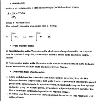
1. Amino acids are organic compounds that contain amino and carboxyl groups and form proteins through peptide bonds. There are essential and non-essential amino acids.
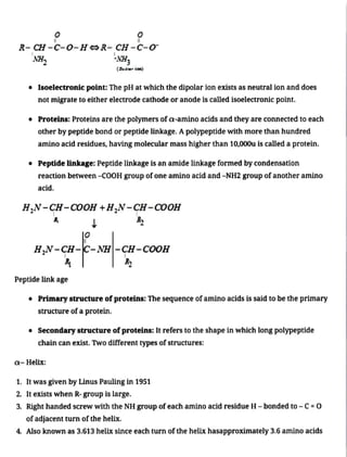
2. In aqueous solutions, amino acids exist as zwitterions with both positive and negative charges which allows them to react as both acids and bases.

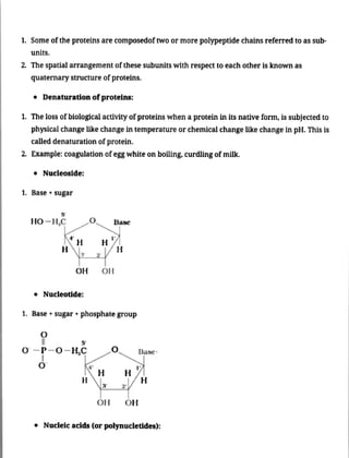
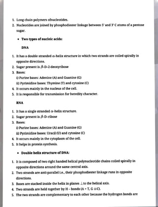


![Bio Molecules [Autosaved]- essential.pptx](https://cdn.slidesharecdn.com/ss_thumbnails/biomoleculesautosaved-240516215401-592caf25-thumbnail.jpg?width=640&height=640&fit=bounds)


