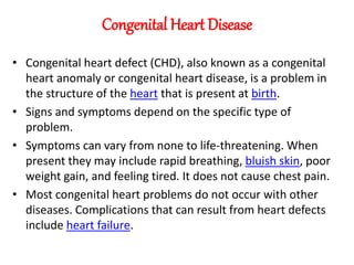
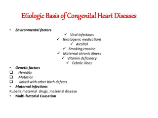
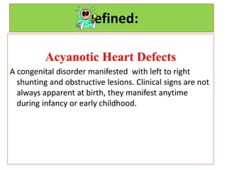
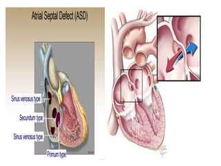
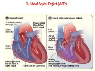
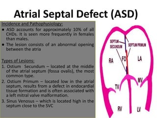
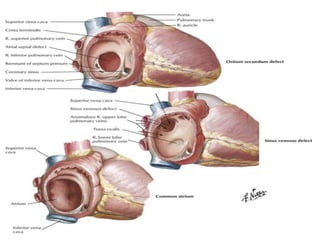
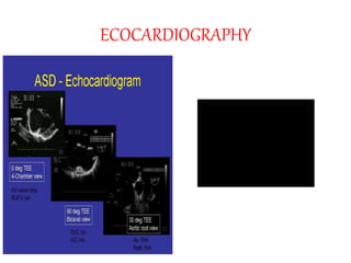
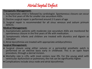
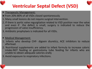
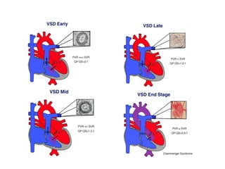
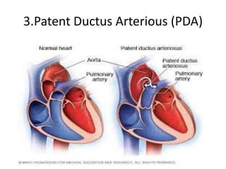
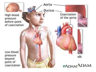
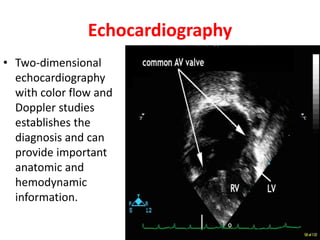
The document discusses acyanotic heart disease, specifically defining acyanotic heart defects as congenital disorders with left to right shunting and obstructive lesions that are not always apparent at birth. It provides details on atrial septal defects (ASD) and ventricular septal defects (VSD), including their incidence, pathophysiology, clinical manifestations, diagnostic testing, and treatment options. The objectives are to define acyanotic heart defects, describe different types of defects, and discuss their etiology, clinical presentation, diagnosis, management, and relevant nursing care.
This document presents the case of a 4-year-old boy with tetralogy of Fallot. The key details are:
- He has had cyanosis, breathlessness and fainting episodes since infancy.
- Examination found cyanosis, clubbing and a grade IV/VI systolic murmur. Echocardiogram showed severe pulmonary stenosis, VSD, and RV hypertrophy consistent with tetralogy of Fallot.
- If left untreated, tetralogy of Fallot can cause serious complications like strokes from anoxic spells or cerebral thrombosis. Surgical repair is usually recommended in childhood.
Tetralogy of Fallot (TOF) is the most common cyanotic congenital heart disease, occurring in approximately 1 in 3,000 live births. It involves four abnormalities - a ventricular septal defect, right ventricular outflow tract obstruction, right ventricular hypertrophy, and overriding of the aorta. Without surgical repair, only 10% of patients survive beyond 20 years of age. Clinical manifestations include cyanosis, clubbing of the fingers and toes, and hypoxic spells in infants. Diagnosis is made through echocardiography, chest x-ray, and cardiac catheterization. Treatment involves palliative shunt procedures for infants or complete repair surgery to close the ventricular septal defect and widen the right ventricular
A ventricular septal defect (VSD) is a hole in the septum separating the left and right ventricles of the heart. VSDs are the most common type of congenital heart defect, occurring in about 2 out of every 1000 live births. They can range from small to large in size. Echocardiography is the primary way to diagnose a VSD and determine its location and size. Small VSDs may close on their own, but larger defects often require surgery to repair.
Ebstein's anomaly is a rare heart defect where the tricuspid valve, which separates the right atrium and ventricle, is abnormally located low in the right ventricle. This causes the right atrium to enlarge and the right ventricle to have two sections - an upper atrialized section and a lower functional section. Symptoms range from none in mild cases to cyanosis, heart failure and arrhythmias in severe cases. Diagnosis involves echocardiogram, ECG and chest x-ray. Treatment options include medications, surgery to repair or replace the valve, and management of complications like heart failure. Prognosis depends on severity of symptoms, with earlier presentation indicating poorer prognosis.
This document discusses cyanotic congenital heart disease (CCHD), which is defined as a cardiovascular birth defect that results in systemic arterial desaturation due to a right-to-left shunt. CCHDs can be classified based on pulmonary blood flow as having reduced, increased, or near normal flow. Common types of CCHDs with reduced pulmonary blood flow include tetralogy of Fallot and pulmonary atresia. CCHDs with increased pulmonary blood flow can present with features of congestive heart failure. The clinical approach to CCHDs involves delineating the anatomical and physiological abnormalities through assessment of anatomy, pulmonary circulation, systemic circulation, and ventricular function.
Patent Ductus Arteroisus, PDA, Cardiology, Paediatrics, Pedicatrics, Critical Care, Emergency medicine, Medicine, Internal Medicine, MBBD, MD, India, CMC Vellore, Christian Medical College
- Tricuspid atresia is a congenital heart defect where the tricuspid valve is absent, preventing blood flow from the right atrium to the right ventricle. It occurs in approximately 1-2.4% of congenital heart disease cases.
- There are several types depending on anatomy and relationship of arteries. The most common type has normally related arteries and a small ventricular septal defect.
- Without intervention, few infants survive beyond 6 months due to hypoxia. Surgical options include shunts to increase/decrease pulmonary blood flow or corrective surgeries like the Fontan procedure.
- Long term complications can include heart failure, arrhythmias, and liver
This document provides information on the approach and assessment of acyanotic congenital heart diseases in children. It discusses:
1. The typical presenting complaints which include feeding difficulties, respiratory distress, easy fatigability, recurrent infections, and failure to thrive.
2. The physical exam findings to assess including inspection, palpation of pulses, blood pressure, jugular venous pressure, auscultation of heart sounds and murmurs.
3. The classification of acyanotic heart diseases which include left-to-right shunts and outflow obstructions. The most common types are also listed.
The document discusses patent ductus arteriosus (PDA), which is the failure of the ductus arteriosus to close after birth. It defines PDA, discusses its incidence, risk factors, embryology, fetal circulation, closure at birth, classification, natural history, clinical manifestations including signs and symptoms, investigations including echocardiogram and cardiac catheterization, and management including medical treatment with drugs and nonsurgical closure using devices. PDA is usually diagnosed using echocardiogram and can often be closed nonsurgically using devices like the Amplatzer duct occluder.
This document provides an overview of coarctation of the aorta, including its definition, pathophysiology, classification, diagnosis, treatment, and prognosis. It defines coarctation of the aorta as a narrowing of the aorta that most commonly occurs just below the origin of the left subclavian artery. The pathophysiology involves increased blood pressure in vessels proximal to the narrowing and decreased blood pressure distal to it. Diagnosis involves physical exam findings like blood pressure discrepancies and imaging like echocardiography and MRI. Treatment is typically surgical repair of the narrowed segment. Complications can include rebound hypertension after surgery. Prognosis is generally good if repaired, but risks include recoarctation and aneurysm
This document discusses Eisenmenger syndrome, a condition where pulmonary hypertension develops due to increased blood flow through defects between the systemic and pulmonary circulations. It provides details on causes, clinical features, pathology findings, and treatments. Key points include:
- Eisenmenger syndrome is caused by defects like VSDs, ASDs, and PDA that allow high blood flow to the lungs and cause pulmonary hypertension over time.
- Common causes of death include hemoptysis from pulmonary artery ruptures, heart failure, and complications from attempted defect repair surgery.
- Pathological findings show thickened pulmonary arteries that resemble the fetal pattern and contribute to high pulmonary vascular resistance.
- Medical treatments are generally ineffective once int
Approach to child with congenital heart diseaseAnkur Puri
This document provides guidance on evaluating a child with congenital heart disease. It outlines key questions to answer, including whether the condition is cyanotic or acyanotic. A thorough history is important, including prenatal, natal, and postnatal details. A physical exam involves assessing vital signs, growth, precordial examination, palpation of pulses and thrills, and auscultation of heart sounds and murmurs. The goal is to characterize the nature and severity of the congenital heart condition.
1. A ventricular septal defect (VSD) is an opening in the wall separating the ventricles that allows blood to shunt between them.
2. VSDs are the most common congenital heart defect in children and can be classified based on their location as membranous, perimembranous, muscular, inlet, or outlet.
3. A complete echocardiogram is needed to evaluate the location, size, direction of shunting, and effects of the defect. Three-dimensional echocardiography can help further define the anatomy and guide potential transcatheter closure of the VSD.
This document discusses congenital heart defects, specifically atrial septal defects (ASD). It describes the types of ASDs, including ostium secundum, patent foramen ovale, and ostium primum. Clinical manifestations are typically asymptomatic for infants and children, with possible findings of a heart murmur, EKG changes, and enlarged heart on xray. Echocardiogram can diagnose and show the defect. Small defects may close on their own, while larger defects are treated with devices or surgery if causing heart failure or pulmonary hypertension. Surgical repair involves closing the defect with sutures or a patch.
The document discusses acyanotic heart disease, which refers to congenital heart defects that do not cause cyanosis. It defines acyanotic heart disease as a circulatory problem present at birth where blood contains enough oxygen but is pumped abnormally. The main types of acyanotic heart disease discussed are atrial septal defect (ASD), ventricular septal defect (VSD), patent ductus arteriosus (PDA), pulmonary stenosis, and aortic stenosis. Causes are often unknown but may include infections, genetic factors, or maternal health conditions during pregnancy.
Restrictive cardiomyopathy is characterized by stiff ventricles that do not fill properly, though systolic function is usually preserved initially. It can be caused by infiltrative diseases, fibrosis, or other processes that restrict ventricular filling. On echocardiogram, restrictive cardiomyopathy shows impaired ventricular filling and enlarged atria, while cardiac catheterization reveals elevated diastolic pressures and a distinctive "square root sign" pressure tracing. Treatment focuses on managing symptoms and underlying causes if identifiable, though prognosis is often poor without transplantation.
Approach to Cyanotic Congenital Heart DiseasesCSN Vittal
Cyanotic congenital heart diseases are those with decreased pulmonary blood flow leading to central cyanosis. Tetralogy of Fallot is the commonest cyanotic heart disease, accounting for 10% of congenital heart defects. It is characterized by four anatomical features - pulmonary stenosis, ventricular septal defect, right ventricular hypertrophy, and overriding of the aorta. Patients typically present with cyanosis, clubbing of fingers, and cyanotic spells which can be relieved by squatting. Investigations reveal findings like boot-shaped heart on chest X-ray and right axis deviation on ECG. Without intervention, patients experience complications like infections, stroke, and delayed growth and development.
commonly used for medical students, and helpful to use this ppt to study for them, and also a common man can understand very easily what is coarctation of aorta.
Congenital heart diseases are structural abnormalities present at birth that affect the normal formation and function of the heart. The most common types are ventricular septal defect (VSD), atrial septal defect (ASD), patent ductus arteriosus (PDA), tetralogy of Fallot, pulmonary stenosis, coarctation of the aorta, and transposition of the great arteries (TGA). Symptoms range from none to signs of heart failure. Treatment options include medical management, catheter-based procedures such as angioplasty or device closure, and open heart surgery to repair or palliate the defects. Prognosis depends on the specific type of congenital heart disease and whether complete repair is possible.
Atrial septal defect (ASD) is an abnormal opening in the wall separating the left and right atria of the heart. There are several types of ASDs including secundum, ostium primum, sinus venosus, and coronary sinus defects. ASDs are usually diagnosed through echocardiography which can determine the size and location of the defect. Small, asymptomatic ASDs may not require treatment, but larger defects with evidence of right heart strain often warrant closure either through open heart surgery or a nonsurgical approach using an implantable device delivered through catheters. Both methods effectively close the defect to prevent long-term complications like heart failure and pulmonary hypertension.
Diagnosis and management of acute heart failureAlaa Ateya
Acute heart failure (AHF) can be defined as new or worsening symptoms of heart failure requiring urgent medical care or hospitalization. Common triggers include non-adherence to medications or diet, infections, or worsening of underlying comorbidities like hypertension. This leads to worsening congestion through mechanisms like neurohormonal activation and myocardial injury. Around half of AHF patients have preserved ejection fraction. Ongoing myocardial damage, worsening kidney function, and elevated filling pressures all contribute to poor outcomes of AHF patients.
Tetralogy of Fallot is a congenital heart defect characterized by four abnormalities: pulmonary stenosis, ventricular septal defect, right ventricular hypertrophy, and overriding of the aorta. It occurs in approximately 1 in 2,500 live births. Without treatment, it can cause cyanosis and heart failure in infants. The definitive treatment is open-heart surgery to repair the abnormalities. After successful surgery, patients typically enjoy an active life without symptoms.
Patent ductus arteriosus A long case presentationNizam Uddin
The document discusses the history, diagnosis, and management of patent ductus arteriosus (PDA), including the timeline of surgical and transcatheter closure techniques. PDA is a persistent opening between the aorta and pulmonary artery that normally closes shortly after birth; if it remains open, closure may be recommended depending on the size of the shunt. Transcatheter closure has largely replaced surgery as the treatment of choice, with high success rates using devices designed to occlude the opening in the PDA.
The document discusses atrioventricular septal defects (AVSDs), which are characterized by the complete absence of the atrioventricular septum. It describes the anatomy, classification, epidemiology, presentation, investigations, and management of AVSDs. Key points include that AVSDs can be partial or complete, account for 4-5% of congenital heart disease, and require surgical repair in early infancy to prevent congestive heart failure and pulmonary hypertension. Left ventricular outflow tract obstruction is a potential postoperative complication.
Ventricular septal defect (VSD) is a congenital heart defect where there is an abnormal opening in the wall separating the left and right ventricles of the heart. There are four main types of VSD. VSDs can be small or large depending on size, and large VSDs typically require surgical closure. Symptoms include heart murmur, fatigue, sweating, and cyanosis. Echocardiogram and ECG are used for diagnosis. Treatment involves medication for heart failure symptoms or closure of the defect via catheterization or open-heart surgery. Post-operative nursing care focuses on monitoring for dysrhythmias and improving cardiac output and gas exchange.
Tetralogy of Fallot is the most common cyanotic heart disease characterized by four features: ventricular septal defect, overriding aorta, infundibular pulmonary stenosis, and right ventricular hypertrophy. It presents with cyanosis that is more frequent in the second half of the first year and hypercyanotic spells after exercise or positions like crying and squatting. Treatment involves palliative surgery like Blalock-Taussig shunt initially, followed by complete repair around 6 months of age. Management of hypercyanotic spells includes medications like morphine, oxygen, and positioning changes.
Truncus arteriosus is a rare congenital heart defect where a single arterial trunk arises from the heart to supply the pulmonary and systemic circulations. It occurs when the embryonic truncus arteriosus fails to divide into the aorta and pulmonary artery. Left untreated, it causes cyanosis and heart failure in newborns. Surgical repair is now possible to connect the pulmonary artery to the right ventricle, improving survival rates to over 80% at one year of age compared to just 15% for uncorrected patients.
ACYANOTIC DISEASE- Non cyanotic heart diseasesNelsonNgulube
ETIOLOGY AND EPIDEMIOLOGY
Congenital heart disease occurs in 8 per 1,000 births. The spectrum of lesions ranges from asymptomatic to fatal. Although most cases of congenital heart disease are multifactorial, some lesions are associated with chromosomal disorders, single gene defects, teratogens, or maternal metabolic disease (see Table139-2).
Congenital heart defects can be divided into three pathophysiological groups (Table 143.1).
1. Left-to-right shunts
2. Right-to-left shunts
3. Obstructive, stenotic lesions
Acyanotic congenital heart disease includes left-to-right shunts resulting in an increase in pulmonary blood flow (patent ductus arteriosus [PDA], ventricular septal defect [VSD], atrial septal defect [ASD]) and obstructive lesions (aortic stenosis, pulmonary stenosis, coarctation of the aorta), which usually have normal pulmonary blood flow.
VENTRICULAR SEPTAL DEFECTEtiology and Epidemiology
The ventricular septum is a complex structure that can be divided
into four components. The largest component is the muscular
septum. The inlet or posterior septum comprises endocardial
cushion tissue. The subarterial or supracristal septum com
prises conotruncal tissue. The membranous septum is below
the aortic valve and is relatively small. VSDs occur when any of these components fail to develop normally (Fig. 143.1). VSD,
the most common congenital heart defect, accounts for 25% of all congenital heart disease. Perimembranous VSD
1. Acyanotic heart defects are congenital heart disorders involving left to right shunting of blood without cyanosis. Common defects include atrial septal defects (ASD) and ventricular septal defects (VSD).
2. ASDs involve an abnormal opening between the left and right atria, increasing pulmonary blood flow. VSDs involve an opening between the left and right ventricles, also increasing pulmonary flow.
3. Clinical manifestations vary depending on defect size but may include fatigue, palpitations, infections. Diagnosis involves echocardiogram, EKG, chest x-ray. Surgical repair is often recommended.
The lecture is for medical student. It is from Dr RUSINGIZA Emmanuel, MD, senior lecture at UR( UNIVERSITY OF RWANDA) .
It will help to understand heart diseases in newborn, infants and children.
This document discusses acyanotic heart defects, which are congenital heart defects that cause left-to-right shunting of blood or obstructive lesions without cyanosis. It focuses on two specific defects: atrial septal defect (ASD) and ventricular septal defect (VSD). For ASD, it describes the types of lesions, altered hemodynamics, manifestations, diagnostics, and medical and surgical management. For VSD, it similarly describes the pathophysiology, manifestations, diagnostics, and therapeutic management including medical, surgical, and nursing interventions.
cyanotic and acyanotic Congenital heart disease for undergraduated student uo...Azad Haleem
This document provides information on various types of congenital heart defects (CHDs), including descriptions, classifications, pathophysiology, clinical manifestations, investigations, and management. It discusses ventricular septal defects (VSDs), atrial septal defects (ASDs), patent ductus arteriosus (PDA), coarctation of the aorta, tetralogy of Fallot, and cyanosis. VSDs, ASDs, and PDA are examples of acyanotic left-to-right shunt lesions, while tetralogy of Fallot is a common cyanotic heart defect. Clinical features, imaging findings, and treatment approaches are described for each condition.
The document discusses congenital heart diseases, which occur in approximately 1% of live births. It describes several types of congenital heart defects including atrial septal defect (ASD), ventricular septal defect (VSD), atrioventricular canal defect, and patent ductus arteriosus - all of which involve increased pulmonary blood flow. It also discusses obstructive defects like aortic stenosis and pulmonary stenosis. The document provides details on the pathophysiology, clinical manifestations, diagnosis, and treatment of these various congenital heart conditions.
This document discusses congenital heart disease, specifically atrial septal defect (ASD), ventricular septal defect (VSD), and patent ductus arteriosus (PDA). It defines each condition, describes their signs and symptoms, risk factors, pathophysiology, diagnosis, and management including both medical and surgical treatment options. The prognosis for each condition with and without treatment is addressed. The document provides a detailed yet concise overview of these three common types of acyanotic heart disease.
This document discusses congenital heart disease, specifically atrial septal defects and ventricular septal defects. It defines what each condition is, describes the causes and types, and outlines the pathophysiology, clinical features, diagnosis, and management. Atrial septal defects are abnormalities where blood passes from the left atrium to the right atrium, while ventricular septal defects allow blood to pass from the left ventricle to the right ventricle through a hole in the ventricular septum. Treatment may involve medication, surgery to repair the defects, or in some small cases, simply monitoring for spontaneous closure of the hole.
The document discusses several congenital heart diseases including ventricular septal defects (VSD), atrial septal defects (ASD), patent ductus arteriosus (PDA), pulmonary stenosis, aortic stenosis, and coarctation of the aorta. It describes the pathophysiology, clinical presentation, investigations, and management of each condition. Cyanotic heart diseases are defined as those involving a right-to-left or left-to-right shunt leading to low oxygen saturation. The document provides classification, epidemiology, etiology and detailed information about specific lesions causing cyanosis.
1. An atrial septal defect is an opening in the septum between the left and right atria, allowing blood to shunt from the left to the right side of the heart.
2. It is one of the most common congenital heart defects found in adults.
3. Symptoms range from none in small defects to fatigue and shortness of breath from right heart strain in large defects that cause significant shunting of blood from the left to the right atrium.
This document presents information from a presentation on acyanotic congenital heart disease. It begins with objectives that cover fetal circulation, defining CHD and risk factors, classifying CHD, explaining acyanotic heart disease and specific defects. It then provides detailed information on ventricular septal defect, atrial septal defect, patent ductus arteriosus, aortic stenosis, pulmonary stenosis, and coarctation of aorta. For each defect, it discusses clinical manifestation, diagnostic criteria, management, and complications. It also includes summaries of two research papers on neurodevelopmental outcomes after surgery for acyanotic CHD and a comparison of renal function between cyanotic and acyanotic CHD in children.
This document provides information on congenital heart disease (CHD), including epidemiology, classification, diagnosis, and treatment. It discusses several specific types of CHD such as atrial septal defect (ASD), ventricular septal defect (VSD), patent ductus arteriosus (PDA), coarctation of the aorta, tetralogy of Fallot, transposition of the great arteries, tricuspid atresia, truncus arteriosus, and hypoplastic left heart syndrome. It describes the clinical presentation, investigations, and management approaches for each condition.
This document provides information on congenital heart disease. It discusses various types of acyanotic heart defects including atrial septal defects (ASD), ventricular septal defects (VSD), patent ductus arteriosus (PDA), and coarctation of the aorta (CoA). For each condition, it describes the pathophysiology, clinical presentation, diagnostic evaluation, and treatment options including surgical repair. Common anatomical variations are defined for different subtypes of each condition.
Congenital Heart disease or CHD refers to structural abnormalities in the heart present at birth. It is the most common type of birth defect, occurring in 5-8 per 1,000 live births. CHD encompasses several types of defects including atrial septal defect (ASD), ventricular septal defect (VSD), and patent ductus arteriosus (PDA). ASD involves an abnormal opening between the left and right atria, VSD is an opening in the ventricular septum between the ventricles, and PDA is the failure of the ductus arteriosus to close after birth, allowing blood to flow between the aorta and pulmonary artery. Symptoms vary depending on the size and
This document provides an overview of congenital heart disease, including prevalence, circulatory adjustments at birth, hemodynamic classifications, and descriptions of specific conditions like atrial septal defect (ASD) and ventricular septal defect (VSD). It notes that congenital heart defects affect 6-8 per 1000 live births and can range widely in severity. Diagnosis typically occurs by 1 week or 1 month of age. After birth, clamping of the umbilical cord and expansion of the lungs cause pressure changes and closure of passages between circulations. Conditions are classified as acyanotic or cyanotic depending on oxygen saturation levels. ASD and VSD are both described in detail including typical clinical features, imaging findings, and management
This document provides information on various types of acyanotic congenital heart defects, including their anatomy, physiology, clinical features, diagnosis, treatment and prognosis. It discusses atrial septal defects (ASD), ventricular septal defects (VSD), and patent ductus arteriosus (PDA). ASDs are classified based on their location. VSDs account for one-quarter of all congenital heart defects and result in left-to-right shunting. PDA causes left-to-right shunting between the aorta and pulmonary artery. Surgical or catheterization closure is often recommended for larger defects.
This document provides information on various congenital heart diseases including their incidence, etiology, classification, clinical presentation and treatment. It discusses conditions such as atrial septal defect (ASD), ventricular septal defect (VSD), patent ductus arteriosus (PDA), tetralogy of Fallot (TOF) and transposition of the great arteries (TGA). Key information includes their definitions, types, signs and symptoms, diagnostic evaluation through imaging studies, and management approaches including medical therapy or surgical intervention.
ABDOMINAL TRAUMA in pediatrics part one.drhasanrajab
Abdominal trauma in pediatrics refers to injuries or damage to the abdominal organs in children. It can occur due to various causes such as falls, motor vehicle accidents, sports-related injuries, and physical abuse. Children are more vulnerable to abdominal trauma due to their unique anatomical and physiological characteristics. Signs and symptoms include abdominal pain, tenderness, distension, vomiting, and signs of shock. Diagnosis involves physical examination, imaging studies, and laboratory tests. Management depends on the severity and may involve conservative treatment or surgical intervention. Prevention is crucial in reducing the incidence of abdominal trauma in children.
Ozempic: Preoperative Management of Patients on GLP-1 Receptor Agonists Saeid Safari
Preoperative Management of Patients on GLP-1 Receptor Agonists like Ozempic and Semiglutide
ASA GUIDELINE
NYSORA Guideline
2 Case Reports of Gastric Ultrasound
Recomendações da OMS sobre cuidados maternos e neonatais para uma experiência pós-natal positiva.
Em consonância com os ODS – Objetivos do Desenvolvimento Sustentável e a Estratégia Global para a Saúde das Mulheres, Crianças e Adolescentes, e aplicando uma abordagem baseada nos direitos humanos, os esforços de cuidados pós-natais devem expandir-se para além da cobertura e da simples sobrevivência, de modo a incluir cuidados de qualidade.
Estas diretrizes visam melhorar a qualidade dos cuidados pós-natais essenciais e de rotina prestados às mulheres e aos recém-nascidos, com o objetivo final de melhorar a saúde e o bem-estar materno e neonatal.
Uma “experiência pós-natal positiva” é um resultado importante para todas as mulheres que dão à luz e para os seus recém-nascidos, estabelecendo as bases para a melhoria da saúde e do bem-estar a curto e longo prazo. Uma experiência pós-natal positiva é definida como aquela em que as mulheres, pessoas que gestam, os recém-nascidos, os casais, os pais, os cuidadores e as famílias recebem informação consistente, garantia e apoio de profissionais de saúde motivados; e onde um sistema de saúde flexível e com recursos reconheça as necessidades das mulheres e dos bebês e respeite o seu contexto cultural.
Estas diretrizes consolidadas apresentam algumas recomendações novas e já bem fundamentadas sobre cuidados pós-natais de rotina para mulheres e neonatos que recebem cuidados no pós-parto em unidades de saúde ou na comunidade, independentemente dos recursos disponíveis.
É fornecido um conjunto abrangente de recomendações para cuidados durante o período puerperal, com ênfase nos cuidados essenciais que todas as mulheres e recém-nascidos devem receber, e com a devida atenção à qualidade dos cuidados; isto é, a entrega e a experiência do cuidado recebido. Estas diretrizes atualizam e ampliam as recomendações da OMS de 2014 sobre cuidados pós-natais da mãe e do recém-nascido e complementam as atuais diretrizes da OMS sobre a gestão de complicações pós-natais.
O estabelecimento da amamentação e o manejo das principais intercorrências é contemplada.
Recomendamos muito.
Vamos discutir essas recomendações no nosso curso de pós-graduação em Aleitamento no Instituto Ciclos.
Esta publicação só está disponível em inglês até o momento.
Prof. Marcus Renato de Carvalho
www.agostodourado.com
Muktapishti is a traditional Ayurvedic preparation made from Shoditha Mukta (Purified Pearl), is believed to help regulate thyroid function and reduce symptoms of hyperthyroidism due to its cooling and balancing properties. Clinical evidence on its efficacy remains limited, necessitating further research to validate its therapeutic benefits.
These lecture slides, by Dr Sidra Arshad, offer a quick overview of the physiological basis of a normal electrocardiogram.
Learning objectives:
1. Define an electrocardiogram (ECG) and electrocardiography
2. Describe how dipoles generated by the heart produce the waveforms of the ECG
3. Describe the components of a normal electrocardiogram of a typical bipolar lead (limb II)
4. Differentiate between intervals and segments
5. Enlist some common indications for obtaining an ECG
6. Describe the flow of current around the heart during the cardiac cycle
7. Discuss the placement and polarity of the leads of electrocardiograph
8. Describe the normal electrocardiograms recorded from the limb leads and explain the physiological basis of the different records that are obtained
9. Define mean electrical vector (axis) of the heart and give the normal range
10. Define the mean QRS vector
11. Describe the axes of leads (hexagonal reference system)
12. Comprehend the vectorial analysis of the normal ECG
13. Determine the mean electrical axis of the ventricular QRS and appreciate the mean axis deviation
14. Explain the concepts of current of injury, J point, and their significance
Study Resources:
1. Chapter 11, Guyton and Hall Textbook of Medical Physiology, 14th edition
2. Chapter 9, Human Physiology - From Cells to Systems, Lauralee Sherwood, 9th edition
3. Chapter 29, Ganong’s Review of Medical Physiology, 26th edition
4. Electrocardiogram, StatPearls - https://www.ncbi.nlm.nih.gov/books/NBK549803/
5. ECG in Medical Practice by ABM Abdullah, 4th edition
6. Chapter 3, Cardiology Explained, https://www.ncbi.nlm.nih.gov/books/NBK2214/
7. ECG Basics, http://www.nataliescasebook.com/tag/e-c-g-basics
Here is the updated list of Top Best Ayurvedic medicine for Gas and Indigestion and those are Gas-O-Go Syp for Dyspepsia | Lavizyme Syrup for Acidity | Yumzyme Hepatoprotective Capsules etc
Local Advanced Lung Cancer: Artificial Intelligence, Synergetics, Complex Sys...Oleg Kshivets
Overall life span (LS) was 1671.7±1721.6 days and cumulative 5YS reached 62.4%, 10 years – 50.4%, 20 years – 44.6%. 94 LCP lived more than 5 years without cancer (LS=2958.6±1723.6 days), 22 – more than 10 years (LS=5571±1841.8 days). 67 LCP died because of LC (LS=471.9±344 days). AT significantly improved 5YS (68% vs. 53.7%) (P=0.028 by log-rank test). Cox modeling displayed that 5YS of LCP significantly depended on: N0-N12, T3-4, blood cell circuit, cell ratio factors (ratio between cancer cells-CC and blood cells subpopulations), LC cell dynamics, recalcification time, heparin tolerance, prothrombin index, protein, AT, procedure type (P=0.000-0.031). Neural networks, genetic algorithm selection and bootstrap simulation revealed relationships between 5YS and N0-12 (rank=1), thrombocytes/CC (rank=2), segmented neutrophils/CC (3), eosinophils/CC (4), erythrocytes/CC (5), healthy cells/CC (6), lymphocytes/CC (7), stick neutrophils/CC (8), leucocytes/CC (9), monocytes/CC (10). Correct prediction of 5YS was 100% by neural networks computing (error=0.000; area under ROC curve=1.0).
Rasamanikya is a excellent preparation in the field of Rasashastra, it is used in various Kushtha Roga, Shwasa, Vicharchika, Bhagandara, Vatarakta, and Phiranga Roga. In this article Preparation& Comparative analytical profile for both Formulationon i.e Rasamanikya prepared by Kushmanda swarasa & Churnodhaka Shodita Haratala. The study aims to provide insights into the comparative efficacy and analytical aspects of these formulations for enhanced therapeutic outcomes.
3. Objectives:
At the end of active learning discussion, the
group will be able to:
1. Define acyanotic heart defects
2. Know the incidence of acynotic disease
3. Explain different types of acynotic congenital
defects
4. Describe the etiology, clinical manifestation ,
diagnosis and management of acynotic
congenital defects.
5. Formulate nursing diagnoses and
appropriate nursing care
4. Congenital Heart Disease
• Congenital heart defect (CHD), also known as a congenital
heart anomaly or congenital heart disease, is a problem in
the structure of the heart that is present at birth.
• Signs and symptoms depend on the specific type of
problem.
• Symptoms can vary from none to life-threatening. When
present they may include rapid breathing, bluish skin, poor
weight gain, and feeling tired. It does not cause chest pain.
• Most congenital heart problems do not occur with other
diseases. Complications that can result from heart defects
include heart failure.
6. Defined:
Acyanotic Heart Defects
A congenital disorder manifested with left to right
shunting and obstructive lesions. Clinical signs are not
always apparent at birth, they manifest anytime
during infancy or early childhood.
7. Defects:
a. Left to right shunting lesions, increased pulmonary blood
flow
• The blood is shunted through an abnormal opening from the
left side of the heart to the right side of the heart
• Pulmonary blood flow increases because of the extra volume
in the right side. Physiologic effects include increased
pulmonary blood flow, increased cardiac workload (including
ventricular strain, dilation, and hypertrophy).
Examples: Atrial Septal Defect (ASD), Ventricular
Septal Defect (VSD), Patent Ductus Arteriosus
(PDA), and Atrioventricular Septal Defect (AVSD).
8. b. Obstructive or stenotic lesions – stenosis of an opening can
occur in a valve or vessel constricting or obstructing blood
flow through the area. Pressure rises in the area behind the
obstruction; blood flow distal to the obstruction may be
decreased or absent. Physiologic effects of obstructive or
stenotic lesions include increased cardiac workload and
ventricular strain, clinical consequence of CHF, decreased CO
and pump failure.
Example: Pulmonary stenosis, aortic stenosis,
Coarctation of Aorta, and interrupted aortic
archs.
11. Atrial Septal Defect (ASD)
Incidence and Pathophysiology:
● ASD accounts for approximately 10% of all
CHDs. It is seen more frequently in females
than males.
● The lesion consists of an abnormal opening
between the atria
Types of Lesions:
1. Ostium Secundum – located at the middle
of the atrial septum (fossa ovalis), the most
common type.
2. Ostium Primum – located low in the atrial
septum, results from a defect in endocardial
tissue formation and is often associated with
a left mitral valve malformation.
3. Sinus Venosus – which is located high in the
septum close to the SVC
12.
13. Atrial Septal Defect (ASD)
Altered Hemodynamic:
● Lower right ventricular compliance which is the
ease of ventricular diastolic filling, compared with
left ventricular compliance leads to left to right
shunting at the atrial level through the ASD. This
increased blood flow through the ASD leads to an
enlarge RA and RV and increased pulmonary
blood flow.
14. Manifestations:
● Most infants and children are asymptomatic but over years to decades may
experience:
1. Fatigue and SOB
2. Palpitations or atrial dysrythmias – result of atrial enlargement
3. Recurrent respiratory infections can occur when there is a large amount of
pulmonary blood flow
4. Systolic murmur is produced by increased blood flow across the pulmonary
valve.
5. Diastolic murmur is present with large shunts
6. Stroke or major organ damage can occur because of embolization of
thrombus, air or other materials – PARADOXIMAL EMBOLISM
15. Auscultation in ASD
•Increased flow across the
pulmonary valve produces a systolic
ejection murmur and fixed splitting
of the second heart sound
•Increased flow across the TV
produces a diastolic rumble at the
mid to lower right sternal border.
17. Physical examination
• Shown in this video mid/late
DIASTOLIC murmur (unlike
VSD which is heard in the
same place but is a
holoSYSTOLIC murmur) heard
best over the lower left
sternal border
(LLSB). afterwards, there
will be a pulmonary flow
murmur (systolic murmur)
due to increased flow
through the pulmonic valve
after the blood goes through
the tricuspid valve. This is
heard over the 2nd left
intercostal space,
18. Investigations:
• CXR:
– enlarged heart
– Enlarged PA
– increased pulmonary vascular markings,Central plethora
• ECG:
– Right axis in secundum defect
– hallmark of primum defect is extreme Left axis deviation
– RVH,RBBB
• ECHO:RVH,valve anatomy,flow direction.
• Treatment:(indicated if symptoms+,RV overload)
– Device closure during cardiac cathetrization
– surgical closure.
20. Chest X-ray
• Plain radiograph
• can be normal in early stages +/-
when the ASD is small
• signs of increased pulmonary
flow (pulmonary plethora or
shunt vascularity)
– enlarged pulmonary vessels
– upper zone vascular prominence
– vessels visible to the periphery of
the film
– eventual signs of pulmonary
arterial hypertension
• chamber enlargement
– right atrium
– right ventricle
– note: left atrium is normal in size
unlike VSD or PDA
– note: aortic arch is small to
normal
There is bilateral increased pulmonary
vascularity and prominence of the atrial
appendage. The heart size is within normal
limits. No rib or soft tissue abnormality. The
radiological features are compatible with the
known diagnosis of ASD.
21. • chest X-ray of
a patient with
atrial septal
defect
showing an
enlarged right
pulmonary
artery (RPA),
main
pulmonary
artery (MPA)
and right
atrium (RA).
End-on view
of an enlarged
pulmonary
vessel is also
seen.
22. ECG
1. The most frequent ECG
abnormalities in ASD
are right bundle branch
block (RBBB)
pattern or
2. rsR' pattern in lead
C1 . Complete RBBB is
observed less
frequently.
3. In patients with large
ASD, presence of tall R
or R' in lead C1 suggests
development
of pulmonary
hypertension .
4. Size of defect
determines the shunt
flow which in turn
results in most of the
ECG abnormalities.
5. If the ASD is very small,
the ECG may be
normal.
23.
24.
25. Atrial Septal Defect
Therapeutic Management:
1. Asymptomatic child is followed by cardiologist. Spontaneous closure can occur
in the first years of life for smaller size secundum ASDs.
2. Elective surgical repair is performed around 2-5 years of age
3. Surgical repair is recommended for all sinus venosus and ostium primum
defects.
Medical Management:
1. Asymptomatic patients with moderate size secundum ASDs are monitored for
spontaneous closure in the first years of life with medication.
2. Symptomatic infants and children are treated with diuretics and digoxin as
indicated
3. Atrial dysrythmias are treated with appropriate antidysrythmics
Surgical Management:
● Surgical closure using either sutures or a pericardial prosthetic patch is
performed on an elective basis early in childhood. This is an open heart
procedure, through a sternal incision.
● Mortality rate is <2%, with most centers near 0%. For the young adult with
ventricular dysfunction or pulmonary, the risk can be significantly higher.
● Complications include sinus node and atrial dysrythmias
29. ASD: Therapy
• Percutaneous Closure
– only for secundum (contra
in others)
– adequate superior/inferior
rim around ASD
– no R-L shunting
• Surgical Closure
– Good prognosis:
• closure age < 25, PA
pressure <40
• If >25 or PA>40, decreased
survival due to CHF, stroke,
and afib
31. Ventricular Septal Defect (VSD)
Incidence and Pathophysiology:
● VSDs account for approximately 25% of all CHDs.
● VSD is the most common congenital cardiac
lesion and is often accompanied by other cardiac
defects.
● The lesion consists of an abnormal opening
between the right and left ventricles which may
vary in size from a miniscule hole to complete
absence of the septum, resulting in a common
ventricle.
32. Altered Hemodynamics:
● The degree of left to right shunting through the VSD
depends on the size of the defect and the pulmonary
vascular resistance compared with the systemic
vascular resistance. The pulmonary vascular system
is high in the newborn. Over the first few weeks of
life, the resistance decreases. As this occurs, an
increased amount of blood shunts left to right of the
VSD level. The pulmonary vascular circulation
receives increased pulmonary blood flow. With large
defects the pulmonary arteries are exposed to
systemic pressures, causing pulmonary hypertension,
and over time, progressive pulmonary vascular
disease.
Ventricular Septal Defect (VSD)
33.
34. Ventricular Septal Defect (VSD)
Manifestations:
Signs and symptoms vary with the size
of the defect and the presence of
associated cardiac lesions. Clinical
symptoms are usually not seen at birth
because of continued high pulmonary
vascular resistance in the newborn.
Infants with moderate to large defects
will become symptomatic within the
first few weeks of life.
● Children with small defects will remain
asymptomatic.
35. Clinical Manisfestations
• Tachypnea, dyspnea
• Poor growth
• Palpable thrills
• Systolic murmur at left lower sternal border
• Shortness of breath
• Failure to gain weight
• Fast heart rate
• Pounding heart
• Frequent respiratory infections
36. Investigations
• History
• Physical Examination:
– parasternal thrill
– pansystolic murmur at lower left sternal edge(Loud if small defect,if large VSD
increase flow across pulmonary valve ejection systolic murmur
– loud P.(Pulmonary HT)
• CXR:
– cardiomegaly,enlarged LA&LV.
– Enlarged PA,increased pulmonary vascular markings
– Pulmonary oedema
• ECG:
– extreme leftt axis is charecteristic,biventricular hypertrophy.
• ECHO:chamber size & pressures.
• Cardiac catheter:O2 content,PA pressure,size & no of defects.
41. ECG
• signs of right ventricular
hypertrophy are observed
more frequently.
Signs of left ventricular
hypertrophy may also
accompany.
The rhythm is generally
sinus.
Right atrial abnormality may
be seen.
42.
43. Ventricular Septal Defect (VSD)
Therapeutic Management:
● From 20%-80% of all VSDs closed spontaneously.
1. Many small lesions do not require surgical intervention.
2. If there is aortic valve regurgitation related to VSD position near the valve
and even if the defect is small, surgery is indicated to reduce the
progression of valve insufficiency.
3. Antibiotic prophylaxis is indicated for all VSDs.
Medical Management:
1. Infants who develop CHF- digoxin diuretics, ACE inhibitors to reduce
afterload.
2. Nutritional supplements are added to infant formula to increase caloric
intake.NGT feeding or gastrostomy tube feeding for infants who are
unable to obtain adequate calories orally
3. Avoid exposure to respiratory infections.
44. Ventricular Septal Defect (VSD)
Surgical Management:
1. Pulmonary artery banding for children with multiple muscular VSDs. In this palliative
procedure, a band is placed around the main pulmonary artery, decreasing blood flow,
reducing the severity of CHF and decreasing the risk of pulmonary vascular disease.
● The current trend is to perform corrective surgery earlier in life, and consequently,
pulmonary artery banding is performed less frequently than in the past.
3. Total correction is accomplished by placing sutures to close small defects or by placing a
pericardial or prosthetic patch over moderate to large defects.
● The surgical approach is usually through the RA to avoid a right ventricular incision which
could impair the contractility of the ventricle.
● VSDs just below the pulmonary valve are closed through an incision in the main
pulmonary artery. Mortality is 5%-8%, depending on the age and type of VSD.
● Complications include residual VSDs, pulmonary hypertension in the postoperative period,
heart block that may require a pacemaker and an abnormal rhythm called junctional
ectopic tachycardia.
●CO can be significantly decreased if dysrythmias are persistent. Post pericardiotomy
syndrome can also occur.
45.
46. Complications:
• Congestive heart failure.
• Growth failure, especially in infancy.
• Bacterial endocarditis
• Irregular heartbeat or rhythm
• Pulmonary artery hypertension
47. Eisenmenger’s Syndrome
• Final common pathway for all significant LR
shunting in which unrestricted pulmonary
blood flow leads to pulmonary vaso-occlusive
disease (PVOD); RL shunting/cyanosis
devleops
• Generally need Qp:Qs >2:1
50. DEFINITION
Patent ductus arteriosus (PDA) the vessel does
not close and remains "patent" resulting in
irregular transmission of blood between two
of the most important arteries close to the
heart, the aorta and the pulmonary artery.”
51. INCIDENCE
• Patent ductus arteriosus is a common
finding in premature infants, occurring in as
many as 80% of infants born before 28 wk
gestation.
• There is a progressive decrease in frequency
of persistence of the ductus with increasing
gestational age. Persistent patency of the
ductus in term infants occurs in about 1 in
2000 live births.
52. CLINICAL MANIFESTATION
PDA is usually diagnosed after age 6 to 8 wk on the basis of a
continuous murmur at the upper left sternal border. The
peripheral pulses are full, with a widened pulse pressure.
While some cases of PDA are asymptomatic, common
symptoms include:
• tachycardia
• shortness of breath
• Cardiomegaly
• Left subclavicular thrill
• Bounding pulse
• Widened pulse pressure
53. • If the ductus is moderate or large, widened pulse
pressure and bounding peripheral pulses are
frequently present.
• Prominent suprasternal and carotid pulsations may
be noted secondary to increased left ventricular
stroke volume.
• Poor growth
• Frequent respiratory infections
54.
55. Investigations
• History
• Physical examination
• CXR:cardiomegaly,increased pul vascularity.
• ECG:Lt or biventricular hypertrophy.
• ECHO:2D visualises PDA,doppler shows turbulance.
• Cardiac catheter:PA pressures & O2 sats.
56. Physical Examination
• If the pulmonary circulation is markedly overloaded there will be
tachycardia, tachypnoea and a wide pulse pressure.
• The precordium is hyperactive and a systolic thrill may be present
at the upper left sternal border.
• The first heart sound is normal but the second is often obscured by
the murmur.
• A grade 1 to 4/6 continuous ('machinery') murmur is best audible
at the left infraclavicular area or upper left sternal border.
• In the case of a large PDA shunt, a diastolic mitral rumble may be
heard because of the high flow rate across the mitral valve.
• Patients with a small PDA do not have the above-mentioned
findings.
• Peripheral pulses are bounding as the run-off into the pulmonary
circulation drops the diastolic pressure and causes a wide pulse
pressure
58. Echocardiography
Two-dimensional echocardiogram (suprasternal view). This
image shows a large patent ductus arteriosus (arrow) that
runs above the left atrium (LA) between the aorta (Ao) and
the pulmonary artery (PA).
Frontal chest radiograph in a patient with patent ductus
arteriosus. This image shows filling in of the
aortopulmonary window (arrow).
59. ECG changes in PDA
•
Left atrial abnormality.
Left ventricular hypertrophy.
If PDA is large or if there is
aorticopulmonary window, then
pulmonary hypertension may
develop
and result in biventricular
hypertrophy pattern.
Prolongation of PR interval (rarely
seen in adults).
Deep S wave in C1 and/or tall R
waves in C5 and C6.
60. Chest X ray
• Pulmanory plethora
Enlarged Left Atrium
and Left Ventricle
Dilated aorta and
pulmonary artery
61. Mangement
• Medical Mangement
– Endocardial prophylaxis as long as patent
– Indomethacin:a prostaglandin E1 inhibitor may close a PDA.
– Administration of indomethacin(prostaglandin inhibitor) has
proved successful in closing a PDA in preterm infants and some
newborns before the age of 10 days.
Orally dose 0.2mg /kg tds at an interwal of 12 to 24 hours.
• Surgical Management :
ligation /coil/clipping/division
62.
63.
64. 4.Coarctation of Aorta
• Narrowing in proximal descending aorta usually just beyond the
origin of Left subclavian artery.
• Blood flow to the lower body maintained through collateral
vessels
• 98% of all coarctations at segment of aorta adjacent to ductus arteriosus.
• Natural hx:
– poor prognosis if unrepaired
– High BP in UA & Low BP in LA
– Systemic HypertensionLVF,Aortic Aneurysm/dissection,ICH
– murmur (continuous or systolic murmur heard in back or SEM/ejection
click of bicuspid AV)
– weak/delayed LL pulses
– Rib notching on CXR is pathognomonic
• Associated with
– Turner’s syndrome
– Subarachinoid haemorrhage
65.
66. Auscultation of Heart Sounds
• Auscultation on the
posterior thorax just
medial to the right
scapula reveals a
systolic ejection
murmur. Ec
67. Plain Radiograph
• figure of 3 sign: contour
abnormality of the aorta
• inferior rib
notching: Roesler sign
– secondary to dilated
intercostal collateral
vessels which form as a
way to bypass the
coarctation and supply the
descending aorta
– the dilated and tortuous
vessels erode the inferior
margins of the ribs,
resulting in notching
– the descending thoracic
aorta
69. MANGEMENT:Coarctation Repair
• Surgical correction
1) Patch aortoplasty
with removal of
segment and end
to end anastomosis
or subclavian flap
repair
2) bypass tube
grafting around
segment
71. Pulmonary Stenosis
• No symptoms in mild or moderately severe
lesions.
• Cyanosis and RVH, right-sided heart failure in
patients with severe lesions.
• High pitched systolic ejection murmur maximal
in second left interspace.
• Ejection click often present.
• Oligaemic lung fields(Reduced pulmonary
vascular marking)
72. Pulmonary Stenosis
• Usually associated with ASD, symptoms d/t dominant defect.
Valvular dysplasia with Noonan.
• Hypertrophy of Rt ventricle with increased RV pressure
• Arterial oxygen saturation remain normal even in severe
stenosis unless Rt to Lt shunt through ASD/VSD/PDA exists.
• Mild to moderate PS is usually asymptomatic.
• Critical pulmonary stenosis: Severe PS in neonate leading to Rt
to Lt shunting through PFO- cyanosis and CHF.
• Treatment with balloon valvuloplasty
74. • Right axis devition
• If pulmonary stenosis is
severe, the R wave
amplitude in C1 may be >20
mm (>2mV).
Right atrial
abnormality: Pulmonary
stenosis is one of the few
diseases that increase P
wave
• Right ventricular
hypertrophy pattern in
adults.
75. Bicuspid Aortic Valve/AS
• Mostly an incidental finding
• Calcification of bicuspid valve in later life
leads to stenosis/insufficiency or aortic root dilatation.
• Usually systolic murmur with opening snap
• Treatment:
- Stenosis: valvuloplasty, surgery
- Insufficiency: surgery and vasodilators
- Dilatation: ACE inhibitors
• Associated with Turner’s, Williams, CoA
76. Atrioventricular (AV) septal defect
• Antrioventricular septal
defects is characterised
by a deficiency of the
atrioventricular septum
of the heart.
77. Continued…
• Atrioventricular septal defect
accounts for about 5% of
congenital heart anomalies. An AV
septal defect may be
• Complete, with a large
(nonrestrictive) inlet ventricular
septal defect
• Transitional, with a small or
moderate-sized (restrictive)
ventricular septal defect
• Partial, with no ventricular septal
defect
• The majority of patients with the
complete form have Down
syndrome. AV septal defect is also
common among patients with
asplenia or polysplenia
(heterotaxy) syndromes.
78.
79. Symptoms and Sign
• atrioventricular septal defect
with a large left-to-right shunt
causes signs of heart
failure (HF—eg, tachypnea,
dyspnea during feeding, poor
weight gain, diaphoresis) by
age 4 to 6 wk. Pulmonary
vascular obstructive disease
(Eisenmenger syndrome) is
usually a late complication but
may occur earlier, especially in
children with Down syndrome.
81. • Chest X-ray
cardiomegaly with right atrial enlargement, biventricular
enlargement, a prominent main pulmonary artery segment,
and increased pulmonary vascular markings.
echocardiography
• Two-dimensional with color flow and Doppler studies
establishes the diagnosis and can provide important
anatomic and hemodynamic information. Cardiac
catheterization is not usually necessary unless
hemodynamics must be further characterized before
surgical repair (for example, to assess pulmonary vascular
resistance in a patient presenting at an older age).
83. ECG
• ECG shows a
superiorly directed
QRS axis (eg, left
axis deviation or
northwest axis),
frequent 1st-degree
AV block, left or
right ventricular
hypertrophy or
both, and
occasional right
atrial enlargement
and right bundle
branch block.
86. Mangement
• Surgical repair
• For heart failure,
medical therapy (eg,
diuretics, digoxin,
ACE inhibitors)
before surgery
87. Continued …
• Pulmonary artery banding
may be used, particularly
in premature infants or
those with associated
abnormalities that make
complete repair higher
risk.
• For patients with large
shunts and heart failure,
diuretics, digoxin, and
ACE inhibitors may help
to manage symptoms
before surgery.
88. Nursing management
Nursing Assessment
• Family history
• history of pregnancy
• Assessment of manifestation of CHD
• Observe and record Child’s Level of exercise tolerance
• Observe and record Child’s skin and mucus membrane
for color and temperature change
• Observe for clubbing of fingers, especially the thumb
nails with thickening and shininess of the terminal
phalanges may occur in cyanotic children by2-3 months
of age
89. Continued…
• Observe for Chest deformities /Observe for
Respiratory pattern Dyspnea on activity.
• Fatigue
• Palpate the child’s pulses in all extremities
• Auscultate the child’s heart
• Record the vital signs
• Complications or consequences of hypoxemia.
• Construction of a weak body
90. Nursing Dx
• Risk for decreased cardiac output r / t defect
structure.
• Altered Growth and Development r / t
inadequate oxygen and nutrients to the tissues.
• Risk for infection r / t poor physical status.
• Altered family processes r / t have children with
heart disease.
• Risk for injury (complications) r / t the heart
condition and therapy.
91. Slide 91
Impaired gas exchange
• Monitor intake and output
• Limit fluids as ordered
• Administer diuretics as ordered
• Position changes every 2 hours or as ordered
92. Slide 92
)
Risk for impaired growth and development
• Treat child as normally as possible
• Teach parents that children are more comfortable
when they know what to expect
• Promote age-appropriate activities as condition
allows
93. Slide 93
)Altered nutrition: less than body requirements
• Offer small, frequent feedings
• Use soft nipple for infant to ease the stress
of sucking
• NG Feedings
• Organize care to allow for rest
94. Slide 94
Continued..
Risk for infection
• Limit exposure to individuals with infections
• Promote good pulmonary hygiene
• Prophylactic antibiotics when undergoing
surgical or dental treatment to prevent
subacute bacterial endocarditis