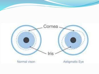
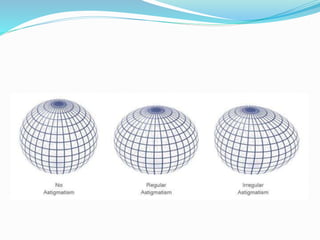
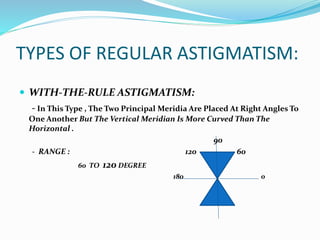
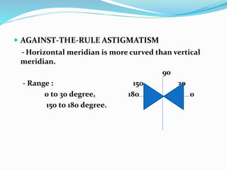
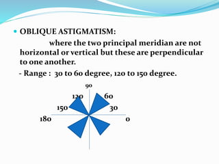
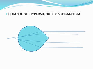
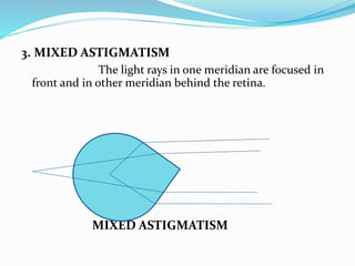
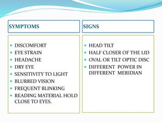
ASTIGMATISM
BAIDHNATH DAS
B. OPTOM
WHAT IS ASTIGMASTISM ?
 When parallel rays of light come from infinity and create
multiple focal point with accomodation at rest is called
astigmatism.
THERE ARE TWO TYPES OF ASTIGMATISM :
1. REGULAR ASTIGMATISM:
The refractive power changes uniformly from one meridian to
another ( there are two principal meridian).
TYPES OF REGULAR ASTIGMATISM:
 WITH-THE-RULE ASTIGMATISM:
- In This Type , The Two Principal Meridia Are Placed At Right Angles To
One Another But The Vertical Meridian Is More Curved Than The
Horizontal .
90
- RANGE : 120 60
60 TO 120 DEGREE
180 0
 AGAINST-THE-RULE ASTIGMATISM
- Horizontal meridian is more curved than vertical
meridian.
90
- Range : 150 30
0 to 30 degree, 180 0
150 to 180 degree.
 OBLIQUE ASTIGMATISM:
where the two principal meridian are not
horizontal or vertical but these are perpendicular
to one another.
- Range : 30 to 60 degree, 120 to 150 degree.
90
120 60
150 30
180 0
OPTICS OF REGULAR ASTIGMATISM
 The parallel rays of light are not focused on a point but
from two focal lines.
 The configuration of rays refracted through the
astigmatic surface (Toric surface ) is called strum’s
conoid and distance between the two focal lines is
known as focal interval of strum.
 The length of this focal interval is measure of the
degree of astigmatism.
REFRACTIVE TYPRS OF REGULAR
ASTIGMATISM
 Depending upon the position of the two focal lines in
relation to retina, Regular astigmatism is classified
into three types:
1. SIMPLE ASTIGMATISM
The rays focused on the retina in one meridian
and either in front (SMA) or behind (SHA) the retina in the
other meridian.
 SIMPLE MYOPIC ASTIGMATISM.
 SIMPLE HYPERMETROPIC ASTIGMATISM.
2. COMPOUND ASTIGMATISM.
The rays of light in the both the meridia
are focused either infront or behind the retina.
compound myopic astigmatism.
 COMPOUND HYPERMETROPIC ASTIGMATISM
3. MIXED ASTIGMATISM
The light rays in one meridian are focused in
front and in other meridian behind the retina.
MIXED ASTIGMATISM
SYMPTOMS SIGNS
 DISCOMFORT
 EYE STRAIN
 HEADACHE
 DRY EYE
 SENSITIVITY TO LIGHT
 BLURRED VISION
 FREQUENT BLINKING
 READING MATERIAL HOLD
CLOSE TO EYES.
 HEAD TILT
 HALF CLOSER OF THE LID
 OVAL OR TILT OPTIC DISC
 DIFFERENT POWER IN
DIFFERENT MERIDIAN
INVESTIGATION
 RETINOSCOPY
 KERATOMETRY
 ASTIGMATIC FAN TEST
 JACKSON CROSS CYLINDER
 STENOPAEIC SLIT
2. IRREGULAR ASTIGMATISM
It is characterized by an irregular changes of refractive
power in different meridia. There are multiple meridia
which admit no geometrical analysis.
ATIOLOGICAL TYPES
1.CORNEAL IRREGULAR ASTIGMATISM
Found in patient with extensive corneal scars
or keratoconus.
2.LENTICULAR IRREGULAR ASTIGMATISM
Occur during maturation of cataract and
seen due to variable refractive index in different parts
of crystalline lens.
3. RETINAL IRREGULAR ASTIGMATISM
Seen due to distortion of macular area.
SYMPTOMS SIGNS
 Defective vision
 Distortion of objects
 polyopia
 Irregular pupillary reflex.
 Corneal irregularity of
keratoconus.
 Plano disc test reveals
distorted circles.
TREATMENT
1. Optical treatment
2. Surgical treatment

Astigmatism

  • 1.
  • 2.
    WHAT IS ASTIGMASTISM?  When parallel rays of light come from infinity and create multiple focal point with accomodation at rest is called astigmatism.
  • 4.
    THERE ARE TWOTYPES OF ASTIGMATISM : 1. REGULAR ASTIGMATISM: The refractive power changes uniformly from one meridian to another ( there are two principal meridian).
  • 6.
    TYPES OF REGULARASTIGMATISM:  WITH-THE-RULE ASTIGMATISM: - In This Type , The Two Principal Meridia Are Placed At Right Angles To One Another But The Vertical Meridian Is More Curved Than The Horizontal . 90 - RANGE : 120 60 60 TO 120 DEGREE 180 0
  • 7.
     AGAINST-THE-RULE ASTIGMATISM -Horizontal meridian is more curved than vertical meridian. 90 - Range : 150 30 0 to 30 degree, 180 0 150 to 180 degree.
  • 8.
     OBLIQUE ASTIGMATISM: wherethe two principal meridian are not horizontal or vertical but these are perpendicular to one another. - Range : 30 to 60 degree, 120 to 150 degree. 90 120 60 150 30 180 0
  • 9.
    OPTICS OF REGULARASTIGMATISM  The parallel rays of light are not focused on a point but from two focal lines.  The configuration of rays refracted through the astigmatic surface (Toric surface ) is called strum’s conoid and distance between the two focal lines is known as focal interval of strum.  The length of this focal interval is measure of the degree of astigmatism.
  • 11.
    REFRACTIVE TYPRS OFREGULAR ASTIGMATISM  Depending upon the position of the two focal lines in relation to retina, Regular astigmatism is classified into three types: 1. SIMPLE ASTIGMATISM The rays focused on the retina in one meridian and either in front (SMA) or behind (SHA) the retina in the other meridian.
  • 12.
     SIMPLE MYOPICASTIGMATISM.  SIMPLE HYPERMETROPIC ASTIGMATISM.
  • 13.
    2. COMPOUND ASTIGMATISM. Therays of light in the both the meridia are focused either infront or behind the retina. compound myopic astigmatism.
  • 14.
  • 15.
    3. MIXED ASTIGMATISM Thelight rays in one meridian are focused in front and in other meridian behind the retina. MIXED ASTIGMATISM
  • 16.
    SYMPTOMS SIGNS  DISCOMFORT EYE STRAIN  HEADACHE  DRY EYE  SENSITIVITY TO LIGHT  BLURRED VISION  FREQUENT BLINKING  READING MATERIAL HOLD CLOSE TO EYES.  HEAD TILT  HALF CLOSER OF THE LID  OVAL OR TILT OPTIC DISC  DIFFERENT POWER IN DIFFERENT MERIDIAN
  • 17.
    INVESTIGATION  RETINOSCOPY  KERATOMETRY ASTIGMATIC FAN TEST  JACKSON CROSS CYLINDER  STENOPAEIC SLIT
  • 18.
    2. IRREGULAR ASTIGMATISM Itis characterized by an irregular changes of refractive power in different meridia. There are multiple meridia which admit no geometrical analysis.
  • 19.
    ATIOLOGICAL TYPES 1.CORNEAL IRREGULARASTIGMATISM Found in patient with extensive corneal scars or keratoconus. 2.LENTICULAR IRREGULAR ASTIGMATISM Occur during maturation of cataract and seen due to variable refractive index in different parts of crystalline lens. 3. RETINAL IRREGULAR ASTIGMATISM Seen due to distortion of macular area.
  • 20.
    SYMPTOMS SIGNS  Defectivevision  Distortion of objects  polyopia  Irregular pupillary reflex.  Corneal irregularity of keratoconus.  Plano disc test reveals distorted circles.
  • 21.