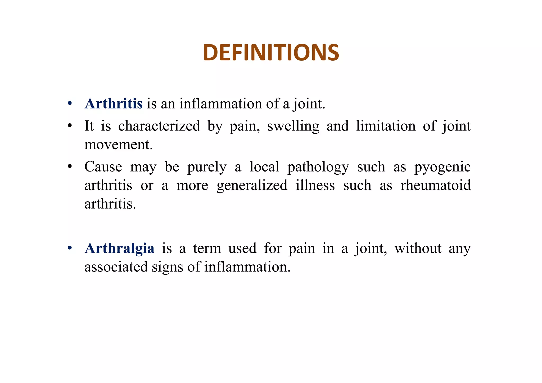
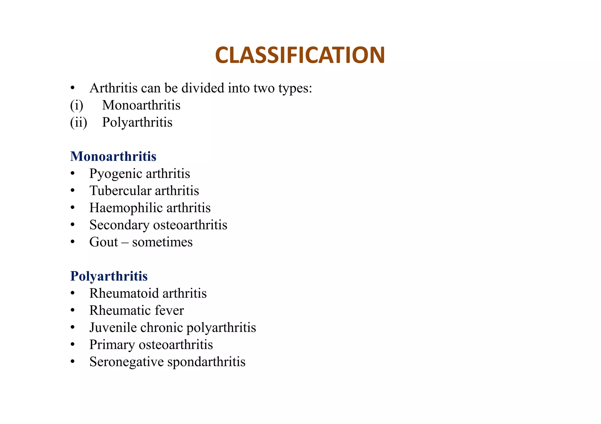
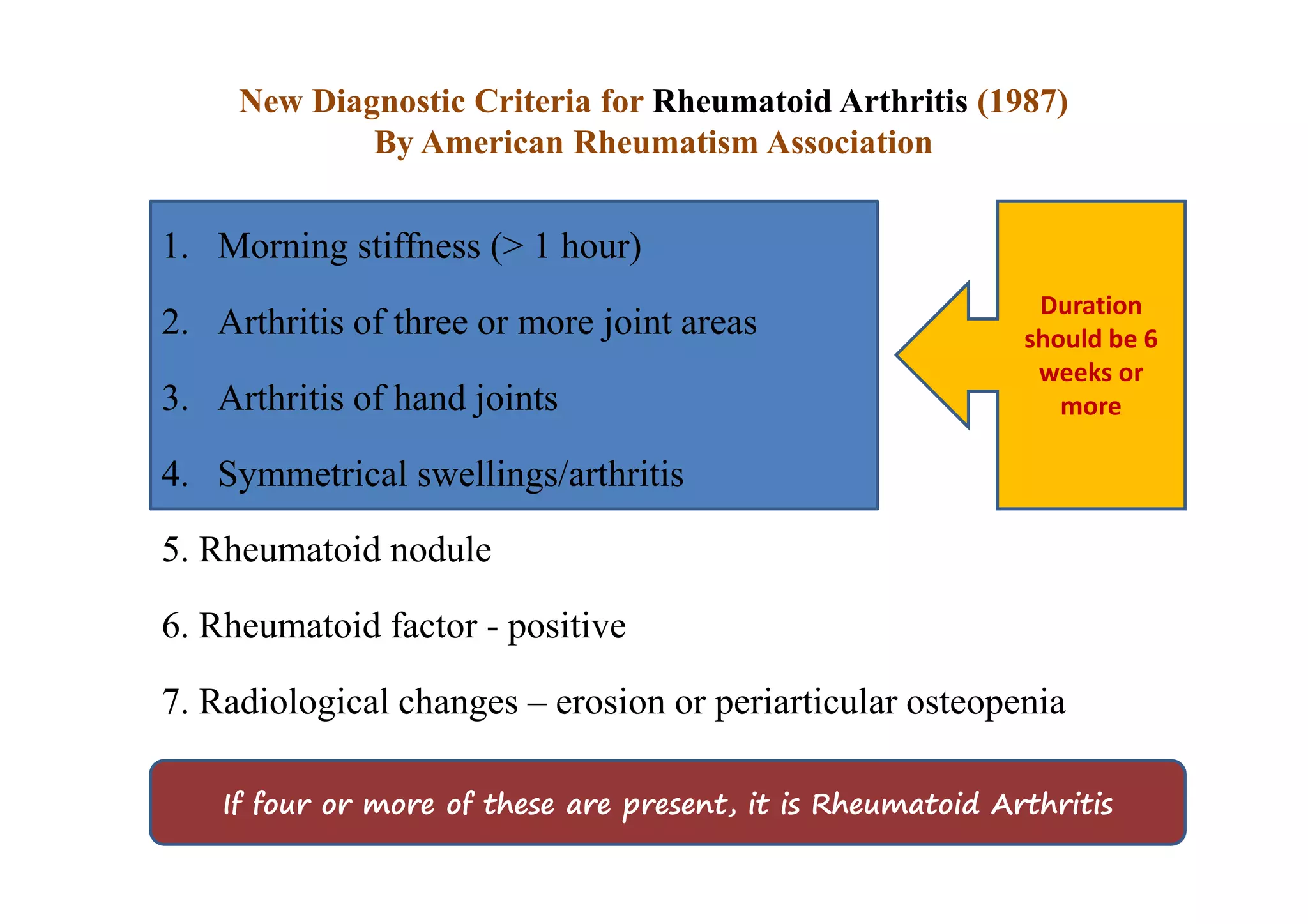
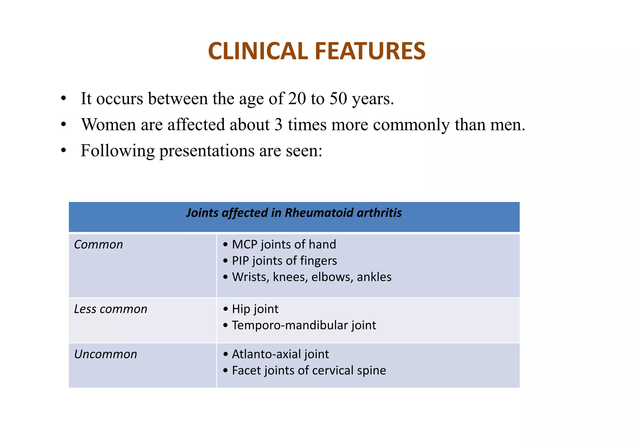
SlideShare a Scribd company logo
Arthritis and Related
Diseases
Dr. Sanjib Kumar Das, PhDDr. Sanjib Kumar Das, PhD
DEFINITIONS
• Arthritis is an inflammation of a joint.
• It is characterized by pain, swelling and limitation of joint
movement.
• Cause may be purely a local pathology such as pyogenic
arthritis or a more generalized illness such as rheumatoid
arthritis.
• Arthralgia is a term used for pain in a joint, without any
associated signs of inflammation.
CLASSIFICATION
• Arthritis can be divided into two types:
(i) Monoarthritis
(ii) Polyarthritis
Monoarthritis
• Pyogenic arthritis
• Tubercular arthritis
• Haemophilic arthritis
• Secondary osteoarthritis
• Gout – sometimes
Polyarthritis
• Rheumatoid arthritis
• Rheumatic fever
• Juvenile chronic polyarthritis
• Primary osteoarthritis
• Seronegative spondarthritis
Rheumatoid Arthritis is a chronic inflammatory joint disease . It is
symmetrical, destructive, deforming polyarthritis affecting small and large
synovial joints with association of systemic disturbances, variety of extra
articular features and presence of rheumatoid factors.
New Diagnostic Criteria for Rheumatoid Arthritis (1987)
By American Rheumatism Association
5. Rheumatoid nodule
6. Rheumatoid factor - positive
7. Radiological changes – erosion or periarticular osteopenia
1. Morning stiffness (> 1 hour)
2. Arthritis of three or more joint areas
3. Arthritis of hand joints
4. Symmetrical swellings/arthritis
Duration
should be 6
weeks or
more
If four or more of these are present, it is Rheumatoid Arthritis
PATHOLOGY
• Initially the synovium becomes oedematous, filled with fibrin
exudates and cellular infiltrates. There is an increase in synovial
fluid.
• As the inflammation persists, the synovium gets hypertrophied
and surrounds the periphery of the articular cartilage to form a
pannus.
• The articular cartilage loses its smooth shiny appearance.
• The pannus extends over the cartilage from the periphery and
burrows into the subchondral bone.
• With further progress of the disease, the cartilage becomes worn
off and the bone surfaces become raw.
• The joints gets deformed, initially because of severe muscle
spasm associated with pain, but later due to fibrosis of the
capsule and other soft tissue structures.
• In some cases, adhesions develop between apposing layers of
pannus, leading to fibrous ankylosis, and later bony ankylosis.
PATHOLOGY
CLINICAL FEATURES
• It occurs between the age of 20 to 50 years.
• Women are affected about 3 times more commonly than men.
• Following presentations are seen:
Joints affected in Rheumatoid arthritis
Common • MCP joints of hand
• PIP joints of fingers
• Wrists, knees, elbows, ankles
Less common • Hip joint
• Temporo-mandibular joint
Uncommon • Atlanto-axial joint
• Facet joints of cervical spine
ON EXAMINATION
• One finds swollen boggy joints as a result of intra-articular effusion,
synovial hypertrophy and oedema of the periarticular structures. The
joints may be deformed.
Deformities in Rheumatoid arthritis
Hand • Ulnar drift of the hand
• Boutonniere deformity
• Swan neck deformity
Elbow • Flexion deformity
Knee • Flexion deformity
Ankle • Equinus deformity
Foot • Hallux valgus, Hammer toe, etc.
Extra-articular manifestations of Rheumatoid arthritis
Vasculitis • Digital arteritis
• Raynaud's phenomenon
• Fever, skin lesions, chronic leg ulcers
• Peripheral neuritis (mononeuritis multiplex)
• Necrotising arteritis involving coronary, mesentric or renal vessels
Rheumatoid
nodules
• Commonest site - olecranon
• Other sites - dorsal surface of forearm, tendoachilles
Serositis • Lung and pleura - pleurisy, parenchymatous nodules, Caplan's syndrome, Honey
comb lung
• Heart - cardiomyopathy, pericarditis
• Eye - iridocyclitis
• Nervous system - peripheral neuritis, carpal tunnel syndrome
Others • Anaemia
• Felty's syndrome
• Sjogren's syndrome
• Amyloidosis
INVESTIGATIONS
1) Radiological examination: This consists of X-
rays of both hands and of the affected joints.
• Reduced joint space
• Irregular subchondral erosions of affected
joints.
• Subchondral cysts formation
• Juxta-articular rarefaction
• Soft tissue shadow at the level of the joint
because of joint effusion or synovial
hypertrophy
• Deformities of the hand and fingers
Fig: X-rays of both hands, AP view, showing
juxta-articular rarefaction
2) Blood: It shows the following changes:
• Elevated ESR
• Low haemoglobin value
• Rheumatoid factor (RF): Positive
3) Synovial fluid examination and Synovial biopsy: Arthroscopically or
by open methods.
INVESTIGATIONS
• All patients with positive rheumatoid factor do not have RA.
• Conversely, all patients with RA do not have a positive rheumatoid factor.
DIFFERENTIAL DIAGNOSIS
a) Systemic lupus erythematosus (SLE):
• In SLE, the joint involvement is not symmetrical; nor are ankylosis
and erosions common.
• Absence of anti-nuclear antibody factor (ANF) is in favour of
rheumatoid arthritis, although its presence does not confirm SLE. It
is present in 25 per cent cases of RA.
b) Osteoarthritis:
• This occurs in older patients.
• There is complete lack of the systemic features of RA such as fever,
weight loss, fatigue, etc.
• Distal inter-phalangeal (DIP) joints are often involved.
• Duration of morning stiffness, joint swelling, ESR etc., are less
compared to RA.
c) Psoriatic arthropathy:
• Characteristic skin and nail lesions may be present.
• Distal inter-phalangeal joints are usually involved.
• Rheumatoid factor is negative.
DIFFERENTIAL DIAGNOSIS
Principles of treatment
a) Induction of remission and its maintenance:
• To be brought under control by drugs.
b) Preservation of joint functions and prevention of deformities:
• Physical therapy, Occupational therapy, Rehabilitation -
Orthosis/ splintage.
c) Repair of joint damage:
• Require surgical intervention.
METHODS OF TREATMENT
• Medical treatment: These consist of anti rheumatic
drugs:
(i) Non-steroidal anti-inflammatory drugs (NSAIDs)
(ii) Disease modifying anti-rheumatic drugs (DMARDs)
(iii) Steroids.
Non-Operative Methods
A) Physiotherapy: This consists of:
(i) Splintage of the joints in proper position during the acute
phase
(ii) Heat therapy – wax bath, hot water fomentation for
symptomatic relief
(iii) Joint mobilisation exercises to maintain joint functions
(iv) Muscle building exercises to gain strength.
B) Occupational therapy:
• To help the patient cope with his occupational requirements
in the most comfortable way or by modifying them.
C) Rehabilitation:
• To improve the functions of the patient with the help of
devices like braces, walking aids etc.
Non-Operative Methods
• Surgical treatment of rheumatoid arthritis can be divided into:
(i) Preventive surgery: This is done to prevent damage to the joint
and nearby tendons by the inflamed, hypertrophied synovium. It
consists of synovectomy of the wrist, knee and MP joints.
(ii) Palliative surgery: This is done in situations where general
condition of the patient does not permit corrective surgery, but
some relief can be provided by limited surgical procedures such
as bone block operations, tendon lengthening etc.
Operative Methods
iii) Reconstructive surgery: It includes tendon transfers,
interposition arthroplasties and total joint replacement. With
improvement in surgical techniques and better design of artificial
joints, it is now possible to replace any joint of the body.
iv) Salvage surgery
Non-Operative Methods
• Soft tissue release
• Tendon repair
• Osteotomy
• Excision Arthroplasty
• Joint replacement
• Arthrodesis
Operative Methods
ANKYLOSING SPONDYLITIS
(Marie strumpell disease)
ETIOLOGY
• A strong association has been found between a genetic
marker—HLA-B27 and this disease.
• HLA-B27 is less than 1 per cent in general population.
• It is present in more than 85 per cent of patients with AS.
This is a sero-negative, progressive inflammatory disease presenting
with pain and stiffness of the spine leading to bony ankylosis of the
sacroiliac and spinal joints.
PATHOLOGY
Sacro-iliac joints are usually the first to get involved
Followed by the spine from the lumbar region upwards.
The hip, manubrio-sternal joints are also involved frequently
Initially Synovitis occurs
Followed by cartilage destruction and bony erosion.
Ultimately leads to fibrous, followed by Bony Ankylosis.
Ossification also occurs in the anterior longitudinal ligament and other ligaments of the spine.
After bony fusion occurs, the pain may subside, leaving the spine permanently stiff
(burnt out disease)
CLINICAL FEATURES
• This is a disease of young adults, more common in males (M:F=10:1).
• Classic presentation: The patient is a young adult 15-30 years old male,
presenting with a gradual onset of pain and stiffness of the lower back.
• Initially, the stiffness may be noticed only after a period of rest, and improves with
movement.
• Pain tends to be worst at night or early morning, awakening the patient from
sleep.
• He gets better only after he walks about or does some exercises.
• There may be pain in the heel, pubic symphysis, manubrium sterni and costo-
sternal joints.
• In later stages, kyphotic deformity of spine and deformity of the hips may be
prominent features.
CLINICAL FEATURES
• Unusual presentations: Patient may
occasionally present with
involvement of peripheral joints
such as the shoulders and knees.
• Smaller joints are rarely involved.
• Sometimes, a patient with
ankylosing spondylitis may present
with chronic inflammatory bowel
disease.
X-rays showing changes in AS.
X-ray of the pelvis, AP view, showing bilateral
SI joint
and hip involvement.
X-ray of the lumbar spine, Lateral
view, showing calcification of the ligaments.
ON EXAMINATION
• Stiff spine: There may be a loss of lumbar lordosis. Lumbar spine flexion
may be limited.
• Tests for detecting sacro-iliac involvement:
• Tenderness, localised to the posterior superior iliac spine or deep in the
gluteal region.
• Sacro-iliac compression: Direct side to side compression of the pelvis may
cause pain at the sacro-iliac joints.
• Gaenslen's test: The hip and the knee joints of the
opposite side are flexed to fix the pelvis, and the
hip joint of the side under test is hyperextended
over the edge of the table. This will exert a
rotational strain over the sacro-iliac joint and give
rise to pain
• Straight leg raising test: The patient is
asked to lift the leg up with the knee
extended. This will cause pain at the
affected SI joint.
• Pump-handle test: With the patient lying
supine, the examiner flexes his hip and
knee completely, and forces the affected
knee across the chest, so as to bring it
close to the opposite shoulder. This will
cause pain on the affected side.
ON EXAMINATION
• Tests for cervical spine involvement: In advanced stages, the cervical
spine gets completely stiff. The Fle'che test may detect an early
involvement of the cervical spine.
• Fle'che test: The patient stands with his heel and back against the wall
and tries to touch the wall with the back of his head without raising the
chin. Inability to touch the head to the wall suggests cervical spine
involvement.
• Thoracic spine involvement: Maximum chest expansion, from full
expiration to full inspiration is measured at the level of the nipples. A
chest expansion less than 5 cm indicates involvement of the costo-
vertebral joints.
ON EXAMINATION
EXTRA-ARTICULAR MANIFESTATIONS
• Ocular: About 25 % patients develop acute iritis , many suffers from recurrent
episodes, resulting in scarring and depigmentation of iris.
• Cardiovascular: Long standing illness develops manifestations in the form of
aortic incompetence, cardiomegaly, conduction defects, pericarditis etc.
• Neurological: Spontaneous dislocation and subluxation of the atlanto-axial joint
or fractures of the Cx spine with trivial trauma, presents with signs and symptoms
of spinal cord compression.
• Pulmonary: Involvement of costo-vertebral joints leads painless restriction of
thoracic cage, detected by diminished chest expansion, or PFT. Bilateral apical
lobe fibrosis with cavitation may occur, which simulates TB on X-ray.
• Systemic: Osteoporosis occurs commonly and occasionally, may develop
amyloidosis.
INVESTIGATIONS
Following changes may be seen on X-ray of the pelvis:
• Haziness of the SI joints
• Irregular subchondral erosions in SI joints
• Sclerosis of the articulating surfaces of SI joints
• Bony ankylosis of the SI joints
• Calcification of the SI ligament and sacro-tuberous ligaments
• Evidence of enthesopathy – calcification at the attachment of
the muscles, tendons and ligaments, particularly around the
pelvis.
X-ray of the lumbar spine may show the following:
• Squaring of vertebrae: The normal anterior concavity of the vertebral body is
lost because of calcification of the anterior longitudinal ligament.
• Loss of the lumbar lordosis.
• Bridging ‘osteophytes’ (syndesmophytes)
• Bamboo spine appearance.
Other investigations are the following:
• ESR: elevated
• Hb: mild anaemia
• HLA-B27: positive
INVESTIGATIONS
TREATMENT
Conservative methods:
(i)Drugs—NSAIDs are given for pain relief; Indomethacin is effective in
most cases; long acting preparations are preferred;
(ii) Physiotherapy -posture guidance, electrotherapy (heat therapy), mobility
maintenance exercises
(iii) Radiotherapy – in resistant cases
(iv) Yoga
Operative methods:
• Correction of kyphotic deformities of the spine by spinal osteotomy
• Joint replacement in hip or knee joint ankylosis.
OSTEOARTHRITIS
• Osteoarthritis (OA) is a degenerative joint disease. Two types of OA are recognized:
• Primary OA: This occurs in a joint at old age, mainly in the weight bearing joints
(knee and hip).
• The trapezio-metacarpal joint of the thumb and the distal IP joints of the fingers are
also affected.
• Secondary OA: There is an underlying primary disease of the joint which leads to
degeneration of the joint, often many years later.
• Predisposing factors are: (i) congenital maldevelopment of a joint (ii) irregularity of
the joint surfaces from previous trauma (iii) previous disease producing a damaged
articular surface (iv) internal derangement of the knee, such as a loose body (v) mal-
alignment (bow legs etc.) (vi) obesity and excessive weight.
Causes of secondary OA of the hip
• Avascular necrosis
 Idiopathic
 Post-traumatic e.g., fracture of femoral neck
 Alcoholism
 Post-partum osteonecrosis
 Chronic liver failure
 Patient on steroids
 Patient on dialysis
 Sickle cell anaemia
• Coxa vara
• Congenital dislocation of hip (CDH)
• Old septic arthritis of the hip
• Malunited fractures
• Fractures of the acetabulum
PATHOLOGY
• The degeneration primarily affects the articular cartilage by increase in
water content and depletion of the proteoglycans from the cartilage
matrix.
• Repeated weight bearing on it leads to its fibrillation.
• The cartilage gets abraded by the grinding mechanism at points of
contact between the apposing articular surfaces, until eventually the
underlying bone is exposed.
• With further ‘rubbing’, the subchondral bone becomes hard and glossy
(eburnated).
• The bone at the margins of the joint hypertrophies to form a rim of projecting
spurs known as osteophytes.
• A similar mechanism results in the formation of subchondral cysts and sclerosis.
• The loose flakes of cartilage incite synovial inflammation and thickening of the
capsule, leading to deformity and stiffness of the joint.
• Often one compartment of a joint is affected more than the other. eg, in the knee
joint, the medial compartment is affected more than the lateral, leading to a varus
deformity (genu varum).
PATHOLOGY
ON EXAMINATION
Following findings may be present:
• Tenderness on the joint line
• Crepitus on moving the joint
• Terminal limitation of joint movement
• Irregular and enlarged-looking joint due to formation of peripheral
osteophytes
• Effusion – rare and transient
• Deformity – varus of the knee, flexion-adduction-ER of the hip
• Subluxation detected on ligament testing
• Wasting of quadriceps femoris muscle
The following are some of the radiological features:
• Narrowing of joint space, often limited to a part of the joint
e.g., may be limited to medial compartment of tibio-femoral
joint of the knee.
• Subchondral sclerosis – dense bone under the articular surface
• Subchondral cysts
• Osteophyte formation
• Loose bodies
• Deformity of the joint
RADIOLOGICAL -INVESTIGATIONS
These consist of the following:
• Serological tests and ESR to rule out Rheumatoid arthritis
• Serum uric acid to rule out Gout
• Arthroscopy, if a loose body or frayed meniscus is suspected
OTHER -INVESTIGATIONS
KELLGREN AND LAWRENCE SYSTEM FOR
CLASSIFICATION OF OSTEOARTHRITIS OF KNEE
• The Kellgren and Lawrence system is a common method of classifying the
severity of knee OA using five grades. This classification was proposed by
Kellgren et al. in 1957 and later accepted by WHO in 1961.
CLASSIFICATION
• Grade 0: No radiographic features of OA are present
• Grade 1: Doubtful joint space narrowing (JSN) and possible osteophytic lippings
• Grade 2: Definite osteophytes and possible JSN on anteroposterior weight-
bearing radiograph
• Grade 3: Multiple osteophytes, definite JSN, sclerosis, possible bony deformity
• Grade 4: Large osteophytes, marked JSN, severe sclerosis and definite bony
deformity
 Drugs: Analgesics are used mainly to suppress pain.
 Chondroprotective agents: Glucosamine and Chondroitin sulphate
repairs damaged cartilage.
 Viscosuplementation: Sodium Hylarunon injected in the joint 3-5
times at weekly interval. Improves cartilage functions, and is
chondroprotective.
 Local application of counter-irritants and liniments provides
dramatic relief.
METHODS OF TREATMENT
 Supportive therapy: It consists of the following:
• Weight reduction, in an obese patient.
• Avoidance of stress and strain to the affected joint in day-to-day
activities. For example, a patient with OA of the knee is advised to
avoid standing or running whenever possible.
• Sitting cross legged and squatting to be avoided.
• Local heat relieves pain and stiffness.
• Exercises for building up the muscles controlling the joint help in
providing stability to the joint.
METHODS OF TREATMENT
Surgical treatment:
• Osteotomy: A high tibial osteotomy for OA of the knee with genu varum and
inter-trochanteric osteotomy for OA of the hip have been shown to be useful for
pain relief.
• Joint replacement: An artificial joint serves for about 10-15 years.
• Joint debridement: Affected joint opened, degenerated cartilage smoothened,
osteophytes and the hypertrophied synovium excised.
• Arthroscopic procedures: Arthroscopic removal of loose bodies, degenerated
meniscal tears, fibrillated cartilage is excised.
METHODS OF TREATMENT
Gout
• Disturbed purine metabolism leading to excessive accumulation of uric
acid in the blood — an inherited disorder; or impaired excretion of uric
acid by the kidneys.
• The result is accumulation of sodium biurate crystals in some soft tissues.
• Tissues of predilection are cartilage, tendon, bursa.
• Patient, usually beyond 40 years of age, presents as
(i) Arthritis – MP joint of the big toe being a favourite site, onset is acute,
pain is severe
(ii) Bursitis – commonly of the olecranon bursa
(iii) Formation deposit of uric acid salt in the soft tissue
• Confirmation of diagnosis – urate crystals in the aspirate from a joint or
bursa, high serum uric acid levels
• Treatment – NSAIDs, uricosuric drugs, uric acid inhibitors.
• Cryotherapy (crushed ice pack), other non thermal modalities to reduce
inflammation and pain.
Pseudogout
• Calcium pyrophosphate (CPP) crystal
deposition
• Symptoms like those of gout
• Meniscus calcification may be seen on X-rays
of the knee
• Treatment – NSAIDs (Rx same as gout).
Psoriatic arthropathy
• Presentation is like rheumatoid arthritis – a
polyarthritis, distal IP joints of hands involved
(unlike rheumatoid arthritis, where these are spared)
• The autoimmune disease has classic skin lesions (dry,
itchy, and scaly) help in diagnosis
• Treatment is by steroids.
Alkaptonuric arthritis (Ochronosis)
• An inherited defect in enzyme system involved in metabolism of phenylalanine and
tyrosine.
• As a result homogentisic acid is excreted in patient's urine.
• As a long term result, it accumulates in the cartilage and other connecting tissues.
• Joint symptoms occur after 40 years of age.
• Spine and shoulder joint are commonly affected.
• X-ray – disc space calcification, peri-articular calcification in large joints.
• There may be evidence of pigment deposit in the sclera.
• Homogentisic acid is present in the urine, and results in the colour of the urine turning
dark brown on standing (due to oxidation of homogentisic acid on exposure to air)
• Treatment same as that for osteoarthritis.
Haemophilic Arthritis
• Occurs due to a number of bleeding disorders
• Occurs in males
• Joints affected commonly are knee, elbow and ankle
• May present as acute or chronic haemarthrosis. There are other manifestations
of bleeding disorders
• X-ray – non-specific signs including bone resorption, cyst formation,
osteoporosis, widening of intercondylar notch in the knee.
• Treatment – rest during acute stage along with factor VIII supplementation or
other deficient factor replacement.
• In the chronic stage, physical therapy, bracing and others are required.
• Deformities may be corrected by conservative or operative methods.
Neuropathic Arthropathy (Charcot's joint)
• These are changes seen in a neuropathic joint, where repeated
strain on a joint due to loss of sensations leads to severe
degeneration
• Clinically, the joint manifests as painless effusion, deformity or
instability
• The X-ray changes are those of severe osteoarthritis but without
much clinical findings like pain, muscle spasm etc.
• Bracing is usually advised for some joints.
• Fusion of the joint may be required.
THANK YOU
Dr. Sanjib Kumar Das
MPT (Musculoskeletal),
Fellow Doctoral, NITIE- Ergonomics and Human Factors,
AIPT, Amity University, Noida
Mail: sanjib_bpt@yahoo.co.in
Contact: +91 8169951520

More Related Content

What's hot

Prosthesis
ProsthesisProsthesis
Arthroplasty
ArthroplastyArthroplasty
Arthroplasty
Dr.Priyanka Das
 
Tennis elbow
Tennis elbowTennis elbow
Tennis elbow
Subin Sabu
 
Tendon transfer.pptx
Tendon transfer.pptxTendon transfer.pptx
Tendon transfer.pptx
SimranSaini12591
 
Tennis elbow
Tennis elbowTennis elbow
Tennis elbow
Sanjay Alle
 
Orthosis PPT
Orthosis PPTOrthosis PPT
Meniscus injury / tear
Meniscus injury / tearMeniscus injury / tear
Meniscus injury / tear
Khairul Nizam
 
Upper limb orthosis
Upper limb orthosisUpper limb orthosis
Upper limb orthosisHetvi Shukla
 
Functional cast bracing
Functional cast bracingFunctional cast bracing
Functional cast bracing
Surya Prakash
 
Golfer elbow.pptx
Golfer elbow.pptxGolfer elbow.pptx
Golfer elbow.pptx
NarendraPal43
 
HIP DISARTICULATION
HIP DISARTICULATIONHIP DISARTICULATION
HIP DISARTICULATION
Akshay Chavan
 
Tennis elbow
Tennis elbowTennis elbow
Tennis elbow
Ponnilavan Ponz
 
Iliotibial Band Syndrome Inservice
Iliotibial Band Syndrome InserviceIliotibial Band Syndrome Inservice
Iliotibial Band Syndrome InserviceMegan Morris
 
Lower limb orthosis
Lower limb orthosis Lower limb orthosis
Lower limb orthosis
Rutuja Solkar
 

What's hot (20)

Prosthesis
ProsthesisProsthesis
Prosthesis
 
Arthroplasty
ArthroplastyArthroplasty
Arthroplasty
 
Tennis elbow
Tennis elbowTennis elbow
Tennis elbow
 
Tendon transfer.pptx
Tendon transfer.pptxTendon transfer.pptx
Tendon transfer.pptx
 
prosthetics
prostheticsprosthetics
prosthetics
 
Tennis elbow
Tennis elbowTennis elbow
Tennis elbow
 
Orthosis PPT
Orthosis PPTOrthosis PPT
Orthosis PPT
 
Arthrodesis
ArthrodesisArthrodesis
Arthrodesis
 
Orthosis
OrthosisOrthosis
Orthosis
 
ORTHOSIS
ORTHOSISORTHOSIS
ORTHOSIS
 
Meniscus injury / tear
Meniscus injury / tearMeniscus injury / tear
Meniscus injury / tear
 
Upper limb orthosis
Upper limb orthosisUpper limb orthosis
Upper limb orthosis
 
Functional cast bracing
Functional cast bracingFunctional cast bracing
Functional cast bracing
 
Golfer elbow.pptx
Golfer elbow.pptxGolfer elbow.pptx
Golfer elbow.pptx
 
ACL rehabilitation
ACL rehabilitationACL rehabilitation
ACL rehabilitation
 
HIP DISARTICULATION
HIP DISARTICULATIONHIP DISARTICULATION
HIP DISARTICULATION
 
Tennis elbow
Tennis elbowTennis elbow
Tennis elbow
 
Tennis elbow(le)
Tennis elbow(le)Tennis elbow(le)
Tennis elbow(le)
 
Iliotibial Band Syndrome Inservice
Iliotibial Band Syndrome InserviceIliotibial Band Syndrome Inservice
Iliotibial Band Syndrome Inservice
 
Lower limb orthosis
Lower limb orthosis Lower limb orthosis
Lower limb orthosis
 

Similar to Arthritis and Rhematic diseases

Rheumatoid Arthritis.pdf
Rheumatoid Arthritis.pdfRheumatoid Arthritis.pdf
Rheumatoid Arthritis.pdf
Rutvikunvar Raualji (PT)
 
Rheumatoid arthritis
Rheumatoid arthritisRheumatoid arthritis
Rheumatoid arthritis
government hospital
 
Arthiritis and related Diseases-1.pptx
Arthiritis and related Diseases-1.pptxArthiritis and related Diseases-1.pptx
Arthiritis and related Diseases-1.pptx
Ashu Bansal
 
Hemophilic arthritis.pdf
Hemophilic arthritis.pdfHemophilic arthritis.pdf
Hemophilic arthritis.pdf
Khetan Kumar
 
ARTHRITIS.pdf
ARTHRITIS.pdfARTHRITIS.pdf
ARTHRITIS.pdf
EvansMwenya2
 
Rhematoid arthiritis
Rhematoid arthiritisRhematoid arthiritis
Rhematoid arthiritis
jasleenbrar03
 
Seronegative Spondyloarthropathies
Seronegative SpondyloarthropathiesSeronegative Spondyloarthropathies
Seronegative Spondyloarthropathies
Sri Harsha Gutta
 
Rheumatoid arthitis
Rheumatoid arthitisRheumatoid arthitis
Rheumatoid arthitis
krishna bhatt
 
Osteoarthritis
OsteoarthritisOsteoarthritis
Osteoarthritis
Doha Rasheedy
 
Osteoarthritis ppt
Osteoarthritis pptOsteoarthritis ppt
Osteoarthritis pptRupika Sodhi
 
Seropositive arthritis ppt
Seropositive arthritis pptSeropositive arthritis ppt
Seropositive arthritis ppt
Saurabh Chahar
 
Arthritis.pptx
Arthritis.pptxArthritis.pptx
Arthritis.pptx
esicOrtho1
 
Arthroscopy: Management of chronic septic arthritis
Arthroscopy: Management of chronic septic arthritisArthroscopy: Management of chronic septic arthritis
Arthroscopy: Management of chronic septic arthritis
Chrystal Lynch
 
Osteoarthritis and Rheumatoid Arthritis
Osteoarthritis and Rheumatoid Arthritis Osteoarthritis and Rheumatoid Arthritis
Osteoarthritis and Rheumatoid Arthritis
Rifhan Kamaruddin
 
Osteoarthritis slideshare
Osteoarthritis slideshareOsteoarthritis slideshare
Osteoarthritis slideshare
Dr Ajay Shah IOM TUTH Nepal
 
Osteoarthritis
OsteoarthritisOsteoarthritis
Osteoarthritis
drsp46
 
ARTHRITS EDUCATION OF CONTENT PURE SCIENCE
ARTHRITS EDUCATION OF  CONTENT PURE SCIENCEARTHRITS EDUCATION OF  CONTENT PURE SCIENCE
ARTHRITS EDUCATION OF CONTENT PURE SCIENCE
AlbertMukama
 
Oa.pptx
Oa.pptxOa.pptx
Oa.pptx
Shansub1
 
Physiotherapy Management of the Rheumatoid Hand
Physiotherapy Management of the Rheumatoid HandPhysiotherapy Management of the Rheumatoid Hand
Physiotherapy Management of the Rheumatoid HandSayantika Dhar
 

Similar to Arthritis and Rhematic diseases (20)

Rheumatoid Arthritis.pdf
Rheumatoid Arthritis.pdfRheumatoid Arthritis.pdf
Rheumatoid Arthritis.pdf
 
Rheumatoid arthritis
Rheumatoid arthritisRheumatoid arthritis
Rheumatoid arthritis
 
Arthiritis and related Diseases-1.pptx
Arthiritis and related Diseases-1.pptxArthiritis and related Diseases-1.pptx
Arthiritis and related Diseases-1.pptx
 
Hemophilic arthritis.pdf
Hemophilic arthritis.pdfHemophilic arthritis.pdf
Hemophilic arthritis.pdf
 
ARTHRITIS.pdf
ARTHRITIS.pdfARTHRITIS.pdf
ARTHRITIS.pdf
 
Rhematoid arthiritis
Rhematoid arthiritisRhematoid arthiritis
Rhematoid arthiritis
 
Seronegative Spondyloarthropathies
Seronegative SpondyloarthropathiesSeronegative Spondyloarthropathies
Seronegative Spondyloarthropathies
 
Rheumatoid arthitis
Rheumatoid arthitisRheumatoid arthitis
Rheumatoid arthitis
 
Osteoarthritis
OsteoarthritisOsteoarthritis
Osteoarthritis
 
Osteoarthritis
OsteoarthritisOsteoarthritis
Osteoarthritis
 
Osteoarthritis ppt
Osteoarthritis pptOsteoarthritis ppt
Osteoarthritis ppt
 
Seropositive arthritis ppt
Seropositive arthritis pptSeropositive arthritis ppt
Seropositive arthritis ppt
 
Arthritis.pptx
Arthritis.pptxArthritis.pptx
Arthritis.pptx
 
Arthroscopy: Management of chronic septic arthritis
Arthroscopy: Management of chronic septic arthritisArthroscopy: Management of chronic septic arthritis
Arthroscopy: Management of chronic septic arthritis
 
Osteoarthritis and Rheumatoid Arthritis
Osteoarthritis and Rheumatoid Arthritis Osteoarthritis and Rheumatoid Arthritis
Osteoarthritis and Rheumatoid Arthritis
 
Osteoarthritis slideshare
Osteoarthritis slideshareOsteoarthritis slideshare
Osteoarthritis slideshare
 
Osteoarthritis
OsteoarthritisOsteoarthritis
Osteoarthritis
 
ARTHRITS EDUCATION OF CONTENT PURE SCIENCE
ARTHRITS EDUCATION OF  CONTENT PURE SCIENCEARTHRITS EDUCATION OF  CONTENT PURE SCIENCE
ARTHRITS EDUCATION OF CONTENT PURE SCIENCE
 
Oa.pptx
Oa.pptxOa.pptx
Oa.pptx
 
Physiotherapy Management of the Rheumatoid Hand
Physiotherapy Management of the Rheumatoid HandPhysiotherapy Management of the Rheumatoid Hand
Physiotherapy Management of the Rheumatoid Hand
 

More from Dr. Sanjib Kumar Das

Poliomyelitis & its Physiotherapeutic Management
Poliomyelitis & its Physiotherapeutic ManagementPoliomyelitis & its Physiotherapeutic Management
Poliomyelitis & its Physiotherapeutic Management
Dr. Sanjib Kumar Das
 
Research and Publication Pedagogy
Research and Publication PedagogyResearch and Publication Pedagogy
Research and Publication Pedagogy
Dr. Sanjib Kumar Das
 
Fractures around shoulder
Fractures around shoulderFractures around shoulder
Fractures around shoulder
Dr. Sanjib Kumar Das
 
General Pharmacology for Physiotherapists
General Pharmacology for PhysiotherapistsGeneral Pharmacology for Physiotherapists
General Pharmacology for Physiotherapists
Dr. Sanjib Kumar Das
 
Wheelchairs - Types and Parts
Wheelchairs - Types and PartsWheelchairs - Types and Parts
Wheelchairs - Types and Parts
Dr. Sanjib Kumar Das
 
Ergonomics and Human Factors Fundamentals: An introduction
Ergonomics and Human Factors Fundamentals: An introductionErgonomics and Human Factors Fundamentals: An introduction
Ergonomics and Human Factors Fundamentals: An introduction
Dr. Sanjib Kumar Das
 
Safety and Environmental Management- Case Study
Safety and Environmental Management- Case StudySafety and Environmental Management- Case Study
Safety and Environmental Management- Case Study
Dr. Sanjib Kumar Das
 
Heat
HeatHeat
Illumination
IlluminationIllumination
Illumination
Dr. Sanjib Kumar Das
 
Noise
NoiseNoise
Vibration
VibrationVibration
Bone tumours (compiled by Dr. Sanjib Kumar Das)
Bone tumours (compiled by Dr. Sanjib Kumar Das)Bone tumours (compiled by Dr. Sanjib Kumar Das)
Bone tumours (compiled by Dr. Sanjib Kumar Das)
Dr. Sanjib Kumar Das
 
Spinal injuries (compiled by Dr Sanjib Kumar Das)
Spinal injuries (compiled by Dr Sanjib Kumar Das)Spinal injuries (compiled by Dr Sanjib Kumar Das)
Spinal injuries (compiled by Dr Sanjib Kumar Das)
Dr. Sanjib Kumar Das
 
Hand injuries (compiled by Dr. Sanjib Kumar Das)
Hand injuries (compiled by Dr. Sanjib Kumar Das)Hand injuries (compiled by Dr. Sanjib Kumar Das)
Hand injuries (compiled by Dr. Sanjib Kumar Das)
Dr. Sanjib Kumar Das
 
Yoga (compiled by Sanjib Kumar Das)
Yoga (compiled by Sanjib Kumar Das)Yoga (compiled by Sanjib Kumar Das)
Yoga (compiled by Sanjib Kumar Das)
Dr. Sanjib Kumar Das
 

More from Dr. Sanjib Kumar Das (15)

Poliomyelitis & its Physiotherapeutic Management
Poliomyelitis & its Physiotherapeutic ManagementPoliomyelitis & its Physiotherapeutic Management
Poliomyelitis & its Physiotherapeutic Management
 
Research and Publication Pedagogy
Research and Publication PedagogyResearch and Publication Pedagogy
Research and Publication Pedagogy
 
Fractures around shoulder
Fractures around shoulderFractures around shoulder
Fractures around shoulder
 
General Pharmacology for Physiotherapists
General Pharmacology for PhysiotherapistsGeneral Pharmacology for Physiotherapists
General Pharmacology for Physiotherapists
 
Wheelchairs - Types and Parts
Wheelchairs - Types and PartsWheelchairs - Types and Parts
Wheelchairs - Types and Parts
 
Ergonomics and Human Factors Fundamentals: An introduction
Ergonomics and Human Factors Fundamentals: An introductionErgonomics and Human Factors Fundamentals: An introduction
Ergonomics and Human Factors Fundamentals: An introduction
 
Safety and Environmental Management- Case Study
Safety and Environmental Management- Case StudySafety and Environmental Management- Case Study
Safety and Environmental Management- Case Study
 
Heat
HeatHeat
Heat
 
Illumination
IlluminationIllumination
Illumination
 
Noise
NoiseNoise
Noise
 
Vibration
VibrationVibration
Vibration
 
Bone tumours (compiled by Dr. Sanjib Kumar Das)
Bone tumours (compiled by Dr. Sanjib Kumar Das)Bone tumours (compiled by Dr. Sanjib Kumar Das)
Bone tumours (compiled by Dr. Sanjib Kumar Das)
 
Spinal injuries (compiled by Dr Sanjib Kumar Das)
Spinal injuries (compiled by Dr Sanjib Kumar Das)Spinal injuries (compiled by Dr Sanjib Kumar Das)
Spinal injuries (compiled by Dr Sanjib Kumar Das)
 
Hand injuries (compiled by Dr. Sanjib Kumar Das)
Hand injuries (compiled by Dr. Sanjib Kumar Das)Hand injuries (compiled by Dr. Sanjib Kumar Das)
Hand injuries (compiled by Dr. Sanjib Kumar Das)
 
Yoga (compiled by Sanjib Kumar Das)
Yoga (compiled by Sanjib Kumar Das)Yoga (compiled by Sanjib Kumar Das)
Yoga (compiled by Sanjib Kumar Das)
 

Recently uploaded

Ophthalmology Clinical Tests for OSCE exam
Ophthalmology Clinical Tests for OSCE examOphthalmology Clinical Tests for OSCE exam
Ophthalmology Clinical Tests for OSCE exam
KafrELShiekh University
 
Dehradun #ℂall #gIRLS Oyo Hotel 9719300533 #ℂall #gIRL in Dehradun
Dehradun #ℂall #gIRLS Oyo Hotel 9719300533 #ℂall #gIRL in DehradunDehradun #ℂall #gIRLS Oyo Hotel 9719300533 #ℂall #gIRL in Dehradun
Dehradun #ℂall #gIRLS Oyo Hotel 9719300533 #ℂall #gIRL in Dehradun
chandankumarsmartiso
 
Evaluation of antidepressant activity of clitoris ternatea in animals
Evaluation of antidepressant activity of clitoris ternatea in animalsEvaluation of antidepressant activity of clitoris ternatea in animals
Evaluation of antidepressant activity of clitoris ternatea in animals
Shweta
 
How to Give Better Lectures: Some Tips for Doctors
How to Give Better Lectures: Some Tips for DoctorsHow to Give Better Lectures: Some Tips for Doctors
How to Give Better Lectures: Some Tips for Doctors
LanceCatedral
 
Cervical & Brachial Plexus By Dr. RIG.pptx
Cervical & Brachial Plexus By Dr. RIG.pptxCervical & Brachial Plexus By Dr. RIG.pptx
Cervical & Brachial Plexus By Dr. RIG.pptx
Dr. Rabia Inam Gandapore
 
planning for change nursing Management ppt
planning for change nursing Management pptplanning for change nursing Management ppt
planning for change nursing Management ppt
Thangamjayarani
 
Tom Selleck Health: A Comprehensive Look at the Iconic Actor’s Wellness Journey
Tom Selleck Health: A Comprehensive Look at the Iconic Actor’s Wellness JourneyTom Selleck Health: A Comprehensive Look at the Iconic Actor’s Wellness Journey
Tom Selleck Health: A Comprehensive Look at the Iconic Actor’s Wellness Journey
greendigital
 
Flu Vaccine Alert in Bangalore Karnataka
Flu Vaccine Alert in Bangalore KarnatakaFlu Vaccine Alert in Bangalore Karnataka
Flu Vaccine Alert in Bangalore Karnataka
addon Scans
 
Top 10 Best Ayurvedic Kidney Stone Syrups in India
Top 10 Best Ayurvedic Kidney Stone Syrups in IndiaTop 10 Best Ayurvedic Kidney Stone Syrups in India
Top 10 Best Ayurvedic Kidney Stone Syrups in India
SwastikAyurveda
 
263778731218 Abortion Clinic /Pills In Harare ,
263778731218 Abortion Clinic /Pills In Harare ,263778731218 Abortion Clinic /Pills In Harare ,
263778731218 Abortion Clinic /Pills In Harare ,
sisternakatoto
 
Novas diretrizes da OMS para os cuidados perinatais de mais qualidade
Novas diretrizes da OMS para os cuidados perinatais de mais qualidadeNovas diretrizes da OMS para os cuidados perinatais de mais qualidade
Novas diretrizes da OMS para os cuidados perinatais de mais qualidade
Prof. Marcus Renato de Carvalho
 
Maxilla, Mandible & Hyoid Bone & Clinical Correlations by Dr. RIG.pptx
Maxilla, Mandible & Hyoid Bone & Clinical Correlations by Dr. RIG.pptxMaxilla, Mandible & Hyoid Bone & Clinical Correlations by Dr. RIG.pptx
Maxilla, Mandible & Hyoid Bone & Clinical Correlations by Dr. RIG.pptx
Dr. Rabia Inam Gandapore
 
heat stroke and heat exhaustion in children
heat stroke and heat exhaustion in childrenheat stroke and heat exhaustion in children
heat stroke and heat exhaustion in children
SumeraAhmad5
 
micro teaching on communication m.sc nursing.pdf
micro teaching on communication m.sc nursing.pdfmicro teaching on communication m.sc nursing.pdf
micro teaching on communication m.sc nursing.pdf
Anurag Sharma
 
Vision-1.pptx, Eye structure, basics of optics
Vision-1.pptx, Eye structure, basics of opticsVision-1.pptx, Eye structure, basics of optics
Vision-1.pptx, Eye structure, basics of optics
Sai Sailesh Kumar Goothy
 
Light House Retreats: Plant Medicine Retreat Europe
Light House Retreats: Plant Medicine Retreat EuropeLight House Retreats: Plant Medicine Retreat Europe
Light House Retreats: Plant Medicine Retreat Europe
Lighthouse Retreat
 
CDSCO and Phamacovigilance {Regulatory body in India}
CDSCO and Phamacovigilance {Regulatory body in India}CDSCO and Phamacovigilance {Regulatory body in India}
CDSCO and Phamacovigilance {Regulatory body in India}
NEHA GUPTA
 
Physiology of Special Chemical Sensation of Taste
Physiology of Special Chemical Sensation of TastePhysiology of Special Chemical Sensation of Taste
Physiology of Special Chemical Sensation of Taste
MedicoseAcademics
 
Non-respiratory Functions of the Lungs.pdf
Non-respiratory Functions of the Lungs.pdfNon-respiratory Functions of the Lungs.pdf
Non-respiratory Functions of the Lungs.pdf
MedicoseAcademics
 
Sex determination from mandible pelvis and skull
Sex determination from mandible pelvis and skullSex determination from mandible pelvis and skull
Sex determination from mandible pelvis and skull
ShashankRoodkee
 

Recently uploaded (20)

Ophthalmology Clinical Tests for OSCE exam
Ophthalmology Clinical Tests for OSCE examOphthalmology Clinical Tests for OSCE exam
Ophthalmology Clinical Tests for OSCE exam
 
Dehradun #ℂall #gIRLS Oyo Hotel 9719300533 #ℂall #gIRL in Dehradun
Dehradun #ℂall #gIRLS Oyo Hotel 9719300533 #ℂall #gIRL in DehradunDehradun #ℂall #gIRLS Oyo Hotel 9719300533 #ℂall #gIRL in Dehradun
Dehradun #ℂall #gIRLS Oyo Hotel 9719300533 #ℂall #gIRL in Dehradun
 
Evaluation of antidepressant activity of clitoris ternatea in animals
Evaluation of antidepressant activity of clitoris ternatea in animalsEvaluation of antidepressant activity of clitoris ternatea in animals
Evaluation of antidepressant activity of clitoris ternatea in animals
 
How to Give Better Lectures: Some Tips for Doctors
How to Give Better Lectures: Some Tips for DoctorsHow to Give Better Lectures: Some Tips for Doctors
How to Give Better Lectures: Some Tips for Doctors
 
Cervical & Brachial Plexus By Dr. RIG.pptx
Cervical & Brachial Plexus By Dr. RIG.pptxCervical & Brachial Plexus By Dr. RIG.pptx
Cervical & Brachial Plexus By Dr. RIG.pptx
 
planning for change nursing Management ppt
planning for change nursing Management pptplanning for change nursing Management ppt
planning for change nursing Management ppt
 
Tom Selleck Health: A Comprehensive Look at the Iconic Actor’s Wellness Journey
Tom Selleck Health: A Comprehensive Look at the Iconic Actor’s Wellness JourneyTom Selleck Health: A Comprehensive Look at the Iconic Actor’s Wellness Journey
Tom Selleck Health: A Comprehensive Look at the Iconic Actor’s Wellness Journey
 
Flu Vaccine Alert in Bangalore Karnataka
Flu Vaccine Alert in Bangalore KarnatakaFlu Vaccine Alert in Bangalore Karnataka
Flu Vaccine Alert in Bangalore Karnataka
 
Top 10 Best Ayurvedic Kidney Stone Syrups in India
Top 10 Best Ayurvedic Kidney Stone Syrups in IndiaTop 10 Best Ayurvedic Kidney Stone Syrups in India
Top 10 Best Ayurvedic Kidney Stone Syrups in India
 
263778731218 Abortion Clinic /Pills In Harare ,
263778731218 Abortion Clinic /Pills In Harare ,263778731218 Abortion Clinic /Pills In Harare ,
263778731218 Abortion Clinic /Pills In Harare ,
 
Novas diretrizes da OMS para os cuidados perinatais de mais qualidade
Novas diretrizes da OMS para os cuidados perinatais de mais qualidadeNovas diretrizes da OMS para os cuidados perinatais de mais qualidade
Novas diretrizes da OMS para os cuidados perinatais de mais qualidade
 
Maxilla, Mandible & Hyoid Bone & Clinical Correlations by Dr. RIG.pptx
Maxilla, Mandible & Hyoid Bone & Clinical Correlations by Dr. RIG.pptxMaxilla, Mandible & Hyoid Bone & Clinical Correlations by Dr. RIG.pptx
Maxilla, Mandible & Hyoid Bone & Clinical Correlations by Dr. RIG.pptx
 
heat stroke and heat exhaustion in children
heat stroke and heat exhaustion in childrenheat stroke and heat exhaustion in children
heat stroke and heat exhaustion in children
 
micro teaching on communication m.sc nursing.pdf
micro teaching on communication m.sc nursing.pdfmicro teaching on communication m.sc nursing.pdf
micro teaching on communication m.sc nursing.pdf
 
Vision-1.pptx, Eye structure, basics of optics
Vision-1.pptx, Eye structure, basics of opticsVision-1.pptx, Eye structure, basics of optics
Vision-1.pptx, Eye structure, basics of optics
 
Light House Retreats: Plant Medicine Retreat Europe
Light House Retreats: Plant Medicine Retreat EuropeLight House Retreats: Plant Medicine Retreat Europe
Light House Retreats: Plant Medicine Retreat Europe
 
CDSCO and Phamacovigilance {Regulatory body in India}
CDSCO and Phamacovigilance {Regulatory body in India}CDSCO and Phamacovigilance {Regulatory body in India}
CDSCO and Phamacovigilance {Regulatory body in India}
 
Physiology of Special Chemical Sensation of Taste
Physiology of Special Chemical Sensation of TastePhysiology of Special Chemical Sensation of Taste
Physiology of Special Chemical Sensation of Taste
 
Non-respiratory Functions of the Lungs.pdf
Non-respiratory Functions of the Lungs.pdfNon-respiratory Functions of the Lungs.pdf
Non-respiratory Functions of the Lungs.pdf
 
Sex determination from mandible pelvis and skull
Sex determination from mandible pelvis and skullSex determination from mandible pelvis and skull
Sex determination from mandible pelvis and skull
 

Arthritis and Rhematic diseases

  • 1. Arthritis and Related Diseases Dr. Sanjib Kumar Das, PhDDr. Sanjib Kumar Das, PhD
  • 2. DEFINITIONS • Arthritis is an inflammation of a joint. • It is characterized by pain, swelling and limitation of joint movement. • Cause may be purely a local pathology such as pyogenic arthritis or a more generalized illness such as rheumatoid arthritis. • Arthralgia is a term used for pain in a joint, without any associated signs of inflammation.
  • 3. CLASSIFICATION • Arthritis can be divided into two types: (i) Monoarthritis (ii) Polyarthritis Monoarthritis • Pyogenic arthritis • Tubercular arthritis • Haemophilic arthritis • Secondary osteoarthritis • Gout – sometimes Polyarthritis • Rheumatoid arthritis • Rheumatic fever • Juvenile chronic polyarthritis • Primary osteoarthritis • Seronegative spondarthritis
  • 4. Rheumatoid Arthritis is a chronic inflammatory joint disease . It is symmetrical, destructive, deforming polyarthritis affecting small and large synovial joints with association of systemic disturbances, variety of extra articular features and presence of rheumatoid factors.
  • 5. New Diagnostic Criteria for Rheumatoid Arthritis (1987) By American Rheumatism Association 5. Rheumatoid nodule 6. Rheumatoid factor - positive 7. Radiological changes – erosion or periarticular osteopenia 1. Morning stiffness (> 1 hour) 2. Arthritis of three or more joint areas 3. Arthritis of hand joints 4. Symmetrical swellings/arthritis Duration should be 6 weeks or more If four or more of these are present, it is Rheumatoid Arthritis
  • 6. PATHOLOGY • Initially the synovium becomes oedematous, filled with fibrin exudates and cellular infiltrates. There is an increase in synovial fluid. • As the inflammation persists, the synovium gets hypertrophied and surrounds the periphery of the articular cartilage to form a pannus. • The articular cartilage loses its smooth shiny appearance.
  • 7. • The pannus extends over the cartilage from the periphery and burrows into the subchondral bone. • With further progress of the disease, the cartilage becomes worn off and the bone surfaces become raw. • The joints gets deformed, initially because of severe muscle spasm associated with pain, but later due to fibrosis of the capsule and other soft tissue structures. • In some cases, adhesions develop between apposing layers of pannus, leading to fibrous ankylosis, and later bony ankylosis. PATHOLOGY
  • 8. CLINICAL FEATURES • It occurs between the age of 20 to 50 years. • Women are affected about 3 times more commonly than men. • Following presentations are seen: Joints affected in Rheumatoid arthritis Common • MCP joints of hand • PIP joints of fingers • Wrists, knees, elbows, ankles Less common • Hip joint • Temporo-mandibular joint Uncommon • Atlanto-axial joint • Facet joints of cervical spine
  • 9. ON EXAMINATION • One finds swollen boggy joints as a result of intra-articular effusion, synovial hypertrophy and oedema of the periarticular structures. The joints may be deformed. Deformities in Rheumatoid arthritis Hand • Ulnar drift of the hand • Boutonniere deformity • Swan neck deformity Elbow • Flexion deformity Knee • Flexion deformity Ankle • Equinus deformity Foot • Hallux valgus, Hammer toe, etc.
  • 10. Extra-articular manifestations of Rheumatoid arthritis Vasculitis • Digital arteritis • Raynaud's phenomenon • Fever, skin lesions, chronic leg ulcers • Peripheral neuritis (mononeuritis multiplex) • Necrotising arteritis involving coronary, mesentric or renal vessels Rheumatoid nodules • Commonest site - olecranon • Other sites - dorsal surface of forearm, tendoachilles Serositis • Lung and pleura - pleurisy, parenchymatous nodules, Caplan's syndrome, Honey comb lung • Heart - cardiomyopathy, pericarditis • Eye - iridocyclitis • Nervous system - peripheral neuritis, carpal tunnel syndrome Others • Anaemia • Felty's syndrome • Sjogren's syndrome • Amyloidosis
  • 11. INVESTIGATIONS 1) Radiological examination: This consists of X- rays of both hands and of the affected joints. • Reduced joint space • Irregular subchondral erosions of affected joints. • Subchondral cysts formation • Juxta-articular rarefaction • Soft tissue shadow at the level of the joint because of joint effusion or synovial hypertrophy • Deformities of the hand and fingers Fig: X-rays of both hands, AP view, showing juxta-articular rarefaction
  • 12. 2) Blood: It shows the following changes: • Elevated ESR • Low haemoglobin value • Rheumatoid factor (RF): Positive 3) Synovial fluid examination and Synovial biopsy: Arthroscopically or by open methods. INVESTIGATIONS • All patients with positive rheumatoid factor do not have RA. • Conversely, all patients with RA do not have a positive rheumatoid factor.
  • 13. DIFFERENTIAL DIAGNOSIS a) Systemic lupus erythematosus (SLE): • In SLE, the joint involvement is not symmetrical; nor are ankylosis and erosions common. • Absence of anti-nuclear antibody factor (ANF) is in favour of rheumatoid arthritis, although its presence does not confirm SLE. It is present in 25 per cent cases of RA. b) Osteoarthritis: • This occurs in older patients. • There is complete lack of the systemic features of RA such as fever, weight loss, fatigue, etc. • Distal inter-phalangeal (DIP) joints are often involved. • Duration of morning stiffness, joint swelling, ESR etc., are less compared to RA.
  • 14. c) Psoriatic arthropathy: • Characteristic skin and nail lesions may be present. • Distal inter-phalangeal joints are usually involved. • Rheumatoid factor is negative. DIFFERENTIAL DIAGNOSIS
  • 15. Principles of treatment a) Induction of remission and its maintenance: • To be brought under control by drugs. b) Preservation of joint functions and prevention of deformities: • Physical therapy, Occupational therapy, Rehabilitation - Orthosis/ splintage. c) Repair of joint damage: • Require surgical intervention.
  • 16. METHODS OF TREATMENT • Medical treatment: These consist of anti rheumatic drugs: (i) Non-steroidal anti-inflammatory drugs (NSAIDs) (ii) Disease modifying anti-rheumatic drugs (DMARDs) (iii) Steroids.
  • 17. Non-Operative Methods A) Physiotherapy: This consists of: (i) Splintage of the joints in proper position during the acute phase (ii) Heat therapy – wax bath, hot water fomentation for symptomatic relief (iii) Joint mobilisation exercises to maintain joint functions (iv) Muscle building exercises to gain strength.
  • 18. B) Occupational therapy: • To help the patient cope with his occupational requirements in the most comfortable way or by modifying them. C) Rehabilitation: • To improve the functions of the patient with the help of devices like braces, walking aids etc. Non-Operative Methods
  • 19. • Surgical treatment of rheumatoid arthritis can be divided into: (i) Preventive surgery: This is done to prevent damage to the joint and nearby tendons by the inflamed, hypertrophied synovium. It consists of synovectomy of the wrist, knee and MP joints. (ii) Palliative surgery: This is done in situations where general condition of the patient does not permit corrective surgery, but some relief can be provided by limited surgical procedures such as bone block operations, tendon lengthening etc. Operative Methods
  • 20. iii) Reconstructive surgery: It includes tendon transfers, interposition arthroplasties and total joint replacement. With improvement in surgical techniques and better design of artificial joints, it is now possible to replace any joint of the body. iv) Salvage surgery Non-Operative Methods
  • 21. • Soft tissue release • Tendon repair • Osteotomy • Excision Arthroplasty • Joint replacement • Arthrodesis Operative Methods
  • 22.
  • 23. ANKYLOSING SPONDYLITIS (Marie strumpell disease) ETIOLOGY • A strong association has been found between a genetic marker—HLA-B27 and this disease. • HLA-B27 is less than 1 per cent in general population. • It is present in more than 85 per cent of patients with AS. This is a sero-negative, progressive inflammatory disease presenting with pain and stiffness of the spine leading to bony ankylosis of the sacroiliac and spinal joints.
  • 24. PATHOLOGY Sacro-iliac joints are usually the first to get involved Followed by the spine from the lumbar region upwards. The hip, manubrio-sternal joints are also involved frequently Initially Synovitis occurs Followed by cartilage destruction and bony erosion. Ultimately leads to fibrous, followed by Bony Ankylosis. Ossification also occurs in the anterior longitudinal ligament and other ligaments of the spine. After bony fusion occurs, the pain may subside, leaving the spine permanently stiff (burnt out disease)
  • 25. CLINICAL FEATURES • This is a disease of young adults, more common in males (M:F=10:1). • Classic presentation: The patient is a young adult 15-30 years old male, presenting with a gradual onset of pain and stiffness of the lower back. • Initially, the stiffness may be noticed only after a period of rest, and improves with movement. • Pain tends to be worst at night or early morning, awakening the patient from sleep. • He gets better only after he walks about or does some exercises. • There may be pain in the heel, pubic symphysis, manubrium sterni and costo- sternal joints. • In later stages, kyphotic deformity of spine and deformity of the hips may be prominent features.
  • 26. CLINICAL FEATURES • Unusual presentations: Patient may occasionally present with involvement of peripheral joints such as the shoulders and knees. • Smaller joints are rarely involved. • Sometimes, a patient with ankylosing spondylitis may present with chronic inflammatory bowel disease. X-rays showing changes in AS. X-ray of the pelvis, AP view, showing bilateral SI joint and hip involvement. X-ray of the lumbar spine, Lateral view, showing calcification of the ligaments.
  • 27. ON EXAMINATION • Stiff spine: There may be a loss of lumbar lordosis. Lumbar spine flexion may be limited. • Tests for detecting sacro-iliac involvement: • Tenderness, localised to the posterior superior iliac spine or deep in the gluteal region. • Sacro-iliac compression: Direct side to side compression of the pelvis may cause pain at the sacro-iliac joints. • Gaenslen's test: The hip and the knee joints of the opposite side are flexed to fix the pelvis, and the hip joint of the side under test is hyperextended over the edge of the table. This will exert a rotational strain over the sacro-iliac joint and give rise to pain
  • 28. • Straight leg raising test: The patient is asked to lift the leg up with the knee extended. This will cause pain at the affected SI joint. • Pump-handle test: With the patient lying supine, the examiner flexes his hip and knee completely, and forces the affected knee across the chest, so as to bring it close to the opposite shoulder. This will cause pain on the affected side. ON EXAMINATION
  • 29. • Tests for cervical spine involvement: In advanced stages, the cervical spine gets completely stiff. The Fle'che test may detect an early involvement of the cervical spine. • Fle'che test: The patient stands with his heel and back against the wall and tries to touch the wall with the back of his head without raising the chin. Inability to touch the head to the wall suggests cervical spine involvement. • Thoracic spine involvement: Maximum chest expansion, from full expiration to full inspiration is measured at the level of the nipples. A chest expansion less than 5 cm indicates involvement of the costo- vertebral joints. ON EXAMINATION
  • 30. EXTRA-ARTICULAR MANIFESTATIONS • Ocular: About 25 % patients develop acute iritis , many suffers from recurrent episodes, resulting in scarring and depigmentation of iris. • Cardiovascular: Long standing illness develops manifestations in the form of aortic incompetence, cardiomegaly, conduction defects, pericarditis etc. • Neurological: Spontaneous dislocation and subluxation of the atlanto-axial joint or fractures of the Cx spine with trivial trauma, presents with signs and symptoms of spinal cord compression. • Pulmonary: Involvement of costo-vertebral joints leads painless restriction of thoracic cage, detected by diminished chest expansion, or PFT. Bilateral apical lobe fibrosis with cavitation may occur, which simulates TB on X-ray. • Systemic: Osteoporosis occurs commonly and occasionally, may develop amyloidosis.
  • 31. INVESTIGATIONS Following changes may be seen on X-ray of the pelvis: • Haziness of the SI joints • Irregular subchondral erosions in SI joints • Sclerosis of the articulating surfaces of SI joints • Bony ankylosis of the SI joints • Calcification of the SI ligament and sacro-tuberous ligaments • Evidence of enthesopathy – calcification at the attachment of the muscles, tendons and ligaments, particularly around the pelvis.
  • 32. X-ray of the lumbar spine may show the following: • Squaring of vertebrae: The normal anterior concavity of the vertebral body is lost because of calcification of the anterior longitudinal ligament. • Loss of the lumbar lordosis. • Bridging ‘osteophytes’ (syndesmophytes) • Bamboo spine appearance. Other investigations are the following: • ESR: elevated • Hb: mild anaemia • HLA-B27: positive INVESTIGATIONS
  • 33. TREATMENT Conservative methods: (i)Drugs—NSAIDs are given for pain relief; Indomethacin is effective in most cases; long acting preparations are preferred; (ii) Physiotherapy -posture guidance, electrotherapy (heat therapy), mobility maintenance exercises (iii) Radiotherapy – in resistant cases (iv) Yoga Operative methods: • Correction of kyphotic deformities of the spine by spinal osteotomy • Joint replacement in hip or knee joint ankylosis.
  • 34.
  • 35. OSTEOARTHRITIS • Osteoarthritis (OA) is a degenerative joint disease. Two types of OA are recognized: • Primary OA: This occurs in a joint at old age, mainly in the weight bearing joints (knee and hip). • The trapezio-metacarpal joint of the thumb and the distal IP joints of the fingers are also affected. • Secondary OA: There is an underlying primary disease of the joint which leads to degeneration of the joint, often many years later. • Predisposing factors are: (i) congenital maldevelopment of a joint (ii) irregularity of the joint surfaces from previous trauma (iii) previous disease producing a damaged articular surface (iv) internal derangement of the knee, such as a loose body (v) mal- alignment (bow legs etc.) (vi) obesity and excessive weight.
  • 36. Causes of secondary OA of the hip • Avascular necrosis  Idiopathic  Post-traumatic e.g., fracture of femoral neck  Alcoholism  Post-partum osteonecrosis  Chronic liver failure  Patient on steroids  Patient on dialysis  Sickle cell anaemia • Coxa vara • Congenital dislocation of hip (CDH) • Old septic arthritis of the hip • Malunited fractures • Fractures of the acetabulum
  • 37. PATHOLOGY • The degeneration primarily affects the articular cartilage by increase in water content and depletion of the proteoglycans from the cartilage matrix. • Repeated weight bearing on it leads to its fibrillation. • The cartilage gets abraded by the grinding mechanism at points of contact between the apposing articular surfaces, until eventually the underlying bone is exposed. • With further ‘rubbing’, the subchondral bone becomes hard and glossy (eburnated).
  • 38. • The bone at the margins of the joint hypertrophies to form a rim of projecting spurs known as osteophytes. • A similar mechanism results in the formation of subchondral cysts and sclerosis. • The loose flakes of cartilage incite synovial inflammation and thickening of the capsule, leading to deformity and stiffness of the joint. • Often one compartment of a joint is affected more than the other. eg, in the knee joint, the medial compartment is affected more than the lateral, leading to a varus deformity (genu varum). PATHOLOGY
  • 39. ON EXAMINATION Following findings may be present: • Tenderness on the joint line • Crepitus on moving the joint • Terminal limitation of joint movement • Irregular and enlarged-looking joint due to formation of peripheral osteophytes • Effusion – rare and transient • Deformity – varus of the knee, flexion-adduction-ER of the hip • Subluxation detected on ligament testing • Wasting of quadriceps femoris muscle
  • 40. The following are some of the radiological features: • Narrowing of joint space, often limited to a part of the joint e.g., may be limited to medial compartment of tibio-femoral joint of the knee. • Subchondral sclerosis – dense bone under the articular surface • Subchondral cysts • Osteophyte formation • Loose bodies • Deformity of the joint RADIOLOGICAL -INVESTIGATIONS
  • 41. These consist of the following: • Serological tests and ESR to rule out Rheumatoid arthritis • Serum uric acid to rule out Gout • Arthroscopy, if a loose body or frayed meniscus is suspected OTHER -INVESTIGATIONS
  • 42. KELLGREN AND LAWRENCE SYSTEM FOR CLASSIFICATION OF OSTEOARTHRITIS OF KNEE • The Kellgren and Lawrence system is a common method of classifying the severity of knee OA using five grades. This classification was proposed by Kellgren et al. in 1957 and later accepted by WHO in 1961. CLASSIFICATION • Grade 0: No radiographic features of OA are present • Grade 1: Doubtful joint space narrowing (JSN) and possible osteophytic lippings • Grade 2: Definite osteophytes and possible JSN on anteroposterior weight- bearing radiograph • Grade 3: Multiple osteophytes, definite JSN, sclerosis, possible bony deformity • Grade 4: Large osteophytes, marked JSN, severe sclerosis and definite bony deformity
  • 43.  Drugs: Analgesics are used mainly to suppress pain.  Chondroprotective agents: Glucosamine and Chondroitin sulphate repairs damaged cartilage.  Viscosuplementation: Sodium Hylarunon injected in the joint 3-5 times at weekly interval. Improves cartilage functions, and is chondroprotective.  Local application of counter-irritants and liniments provides dramatic relief. METHODS OF TREATMENT
  • 44.  Supportive therapy: It consists of the following: • Weight reduction, in an obese patient. • Avoidance of stress and strain to the affected joint in day-to-day activities. For example, a patient with OA of the knee is advised to avoid standing or running whenever possible. • Sitting cross legged and squatting to be avoided. • Local heat relieves pain and stiffness. • Exercises for building up the muscles controlling the joint help in providing stability to the joint. METHODS OF TREATMENT
  • 45. Surgical treatment: • Osteotomy: A high tibial osteotomy for OA of the knee with genu varum and inter-trochanteric osteotomy for OA of the hip have been shown to be useful for pain relief. • Joint replacement: An artificial joint serves for about 10-15 years. • Joint debridement: Affected joint opened, degenerated cartilage smoothened, osteophytes and the hypertrophied synovium excised. • Arthroscopic procedures: Arthroscopic removal of loose bodies, degenerated meniscal tears, fibrillated cartilage is excised. METHODS OF TREATMENT
  • 46.
  • 47. Gout • Disturbed purine metabolism leading to excessive accumulation of uric acid in the blood — an inherited disorder; or impaired excretion of uric acid by the kidneys. • The result is accumulation of sodium biurate crystals in some soft tissues. • Tissues of predilection are cartilage, tendon, bursa. • Patient, usually beyond 40 years of age, presents as (i) Arthritis – MP joint of the big toe being a favourite site, onset is acute, pain is severe (ii) Bursitis – commonly of the olecranon bursa (iii) Formation deposit of uric acid salt in the soft tissue • Confirmation of diagnosis – urate crystals in the aspirate from a joint or bursa, high serum uric acid levels • Treatment – NSAIDs, uricosuric drugs, uric acid inhibitors. • Cryotherapy (crushed ice pack), other non thermal modalities to reduce inflammation and pain.
  • 48. Pseudogout • Calcium pyrophosphate (CPP) crystal deposition • Symptoms like those of gout • Meniscus calcification may be seen on X-rays of the knee • Treatment – NSAIDs (Rx same as gout).
  • 49. Psoriatic arthropathy • Presentation is like rheumatoid arthritis – a polyarthritis, distal IP joints of hands involved (unlike rheumatoid arthritis, where these are spared) • The autoimmune disease has classic skin lesions (dry, itchy, and scaly) help in diagnosis • Treatment is by steroids.
  • 50. Alkaptonuric arthritis (Ochronosis) • An inherited defect in enzyme system involved in metabolism of phenylalanine and tyrosine. • As a result homogentisic acid is excreted in patient's urine. • As a long term result, it accumulates in the cartilage and other connecting tissues. • Joint symptoms occur after 40 years of age. • Spine and shoulder joint are commonly affected. • X-ray – disc space calcification, peri-articular calcification in large joints. • There may be evidence of pigment deposit in the sclera. • Homogentisic acid is present in the urine, and results in the colour of the urine turning dark brown on standing (due to oxidation of homogentisic acid on exposure to air) • Treatment same as that for osteoarthritis.
  • 51. Haemophilic Arthritis • Occurs due to a number of bleeding disorders • Occurs in males • Joints affected commonly are knee, elbow and ankle • May present as acute or chronic haemarthrosis. There are other manifestations of bleeding disorders • X-ray – non-specific signs including bone resorption, cyst formation, osteoporosis, widening of intercondylar notch in the knee. • Treatment – rest during acute stage along with factor VIII supplementation or other deficient factor replacement. • In the chronic stage, physical therapy, bracing and others are required. • Deformities may be corrected by conservative or operative methods.
  • 52. Neuropathic Arthropathy (Charcot's joint) • These are changes seen in a neuropathic joint, where repeated strain on a joint due to loss of sensations leads to severe degeneration • Clinically, the joint manifests as painless effusion, deformity or instability • The X-ray changes are those of severe osteoarthritis but without much clinical findings like pain, muscle spasm etc. • Bracing is usually advised for some joints. • Fusion of the joint may be required.
  • 53. THANK YOU Dr. Sanjib Kumar Das MPT (Musculoskeletal), Fellow Doctoral, NITIE- Ergonomics and Human Factors, AIPT, Amity University, Noida Mail: sanjib_bpt@yahoo.co.in Contact: +91 8169951520