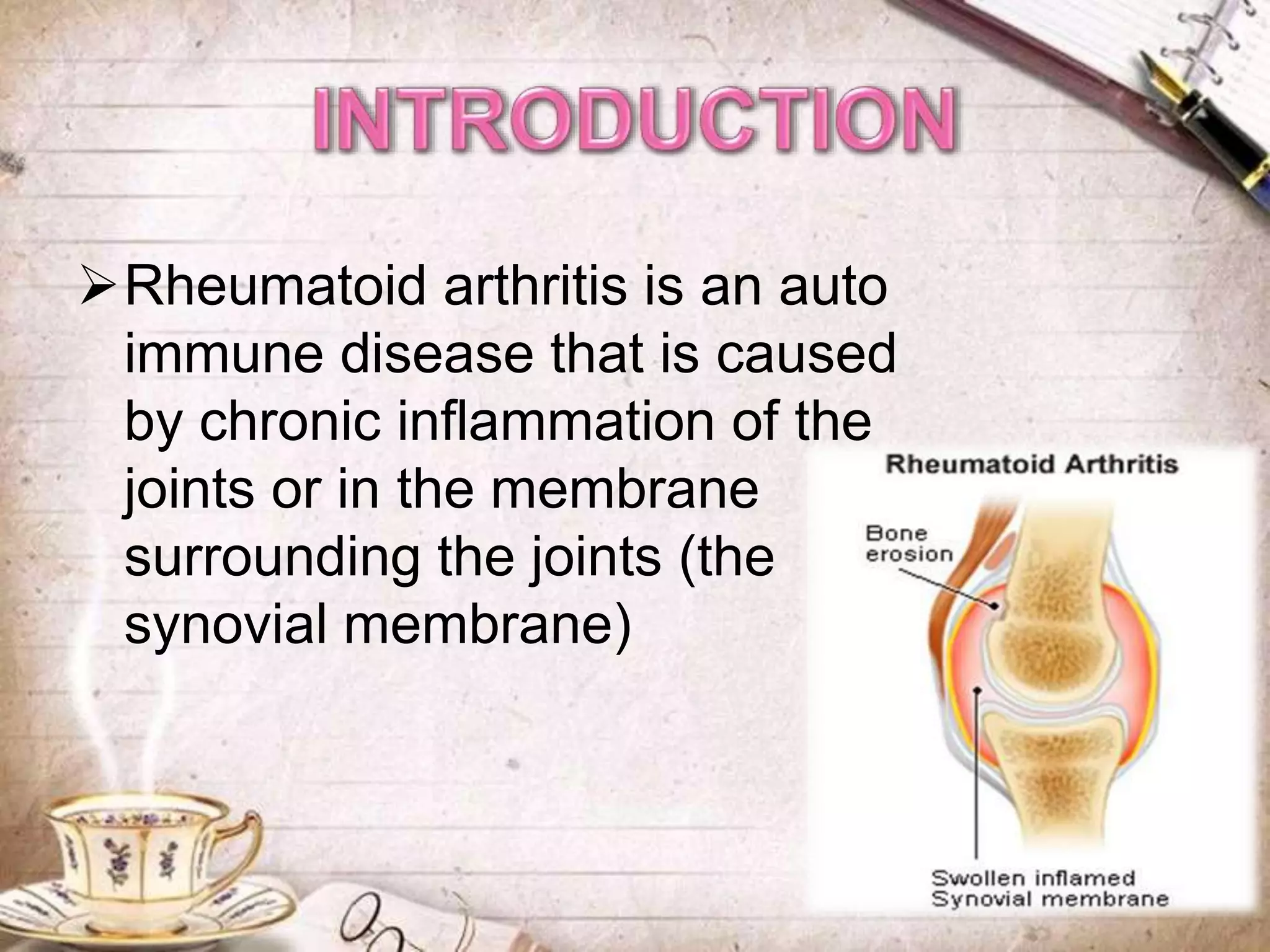
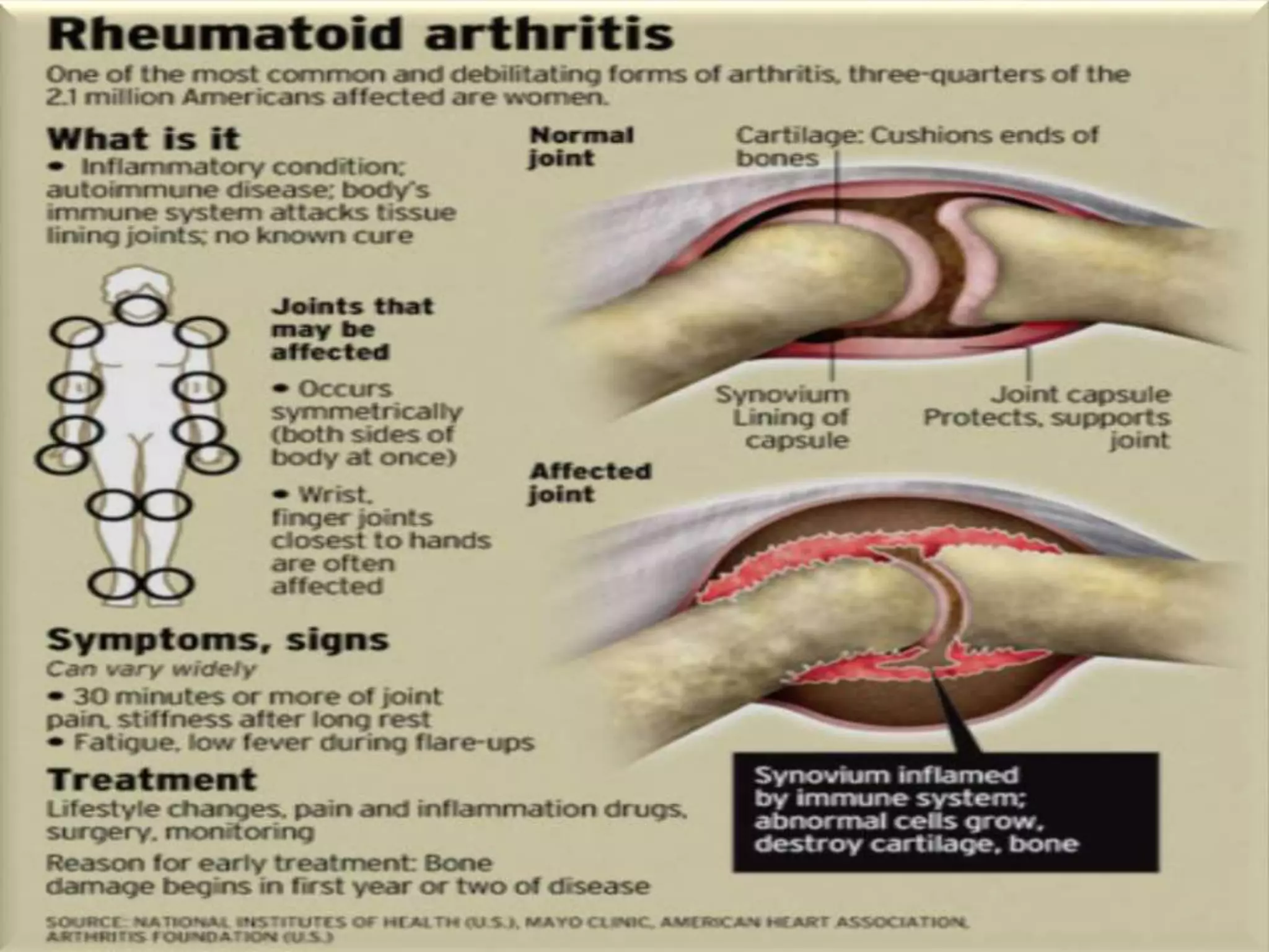
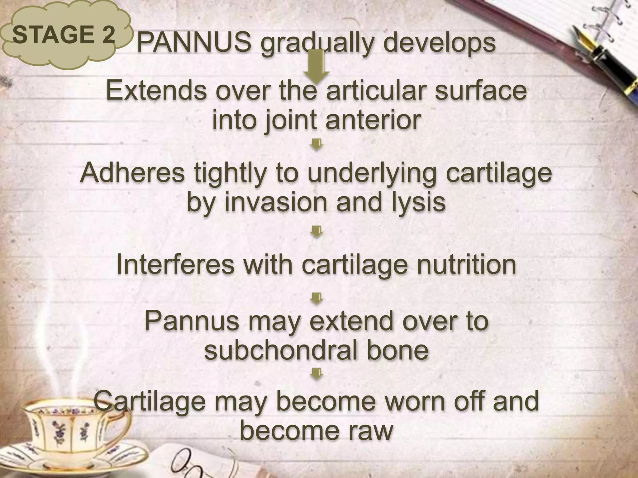
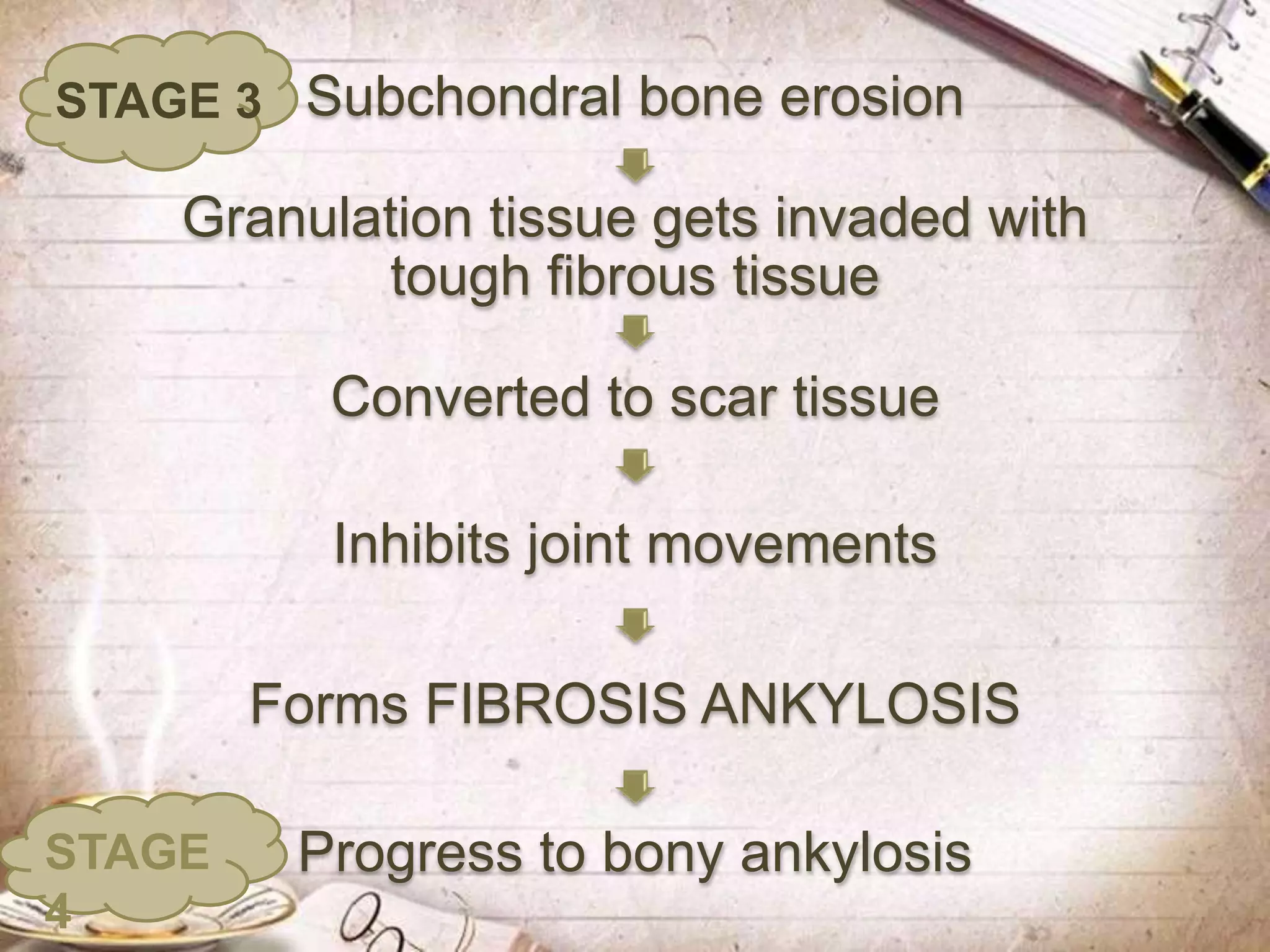
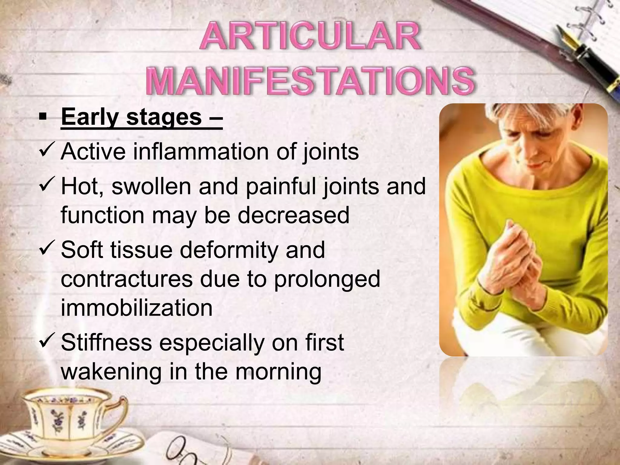
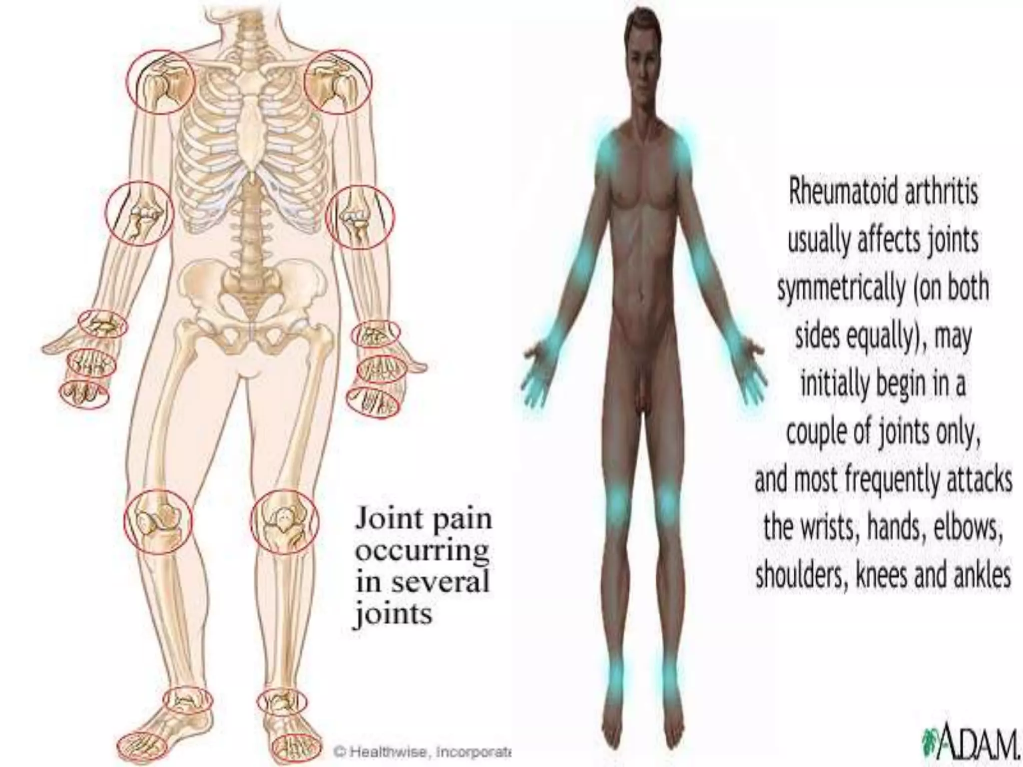
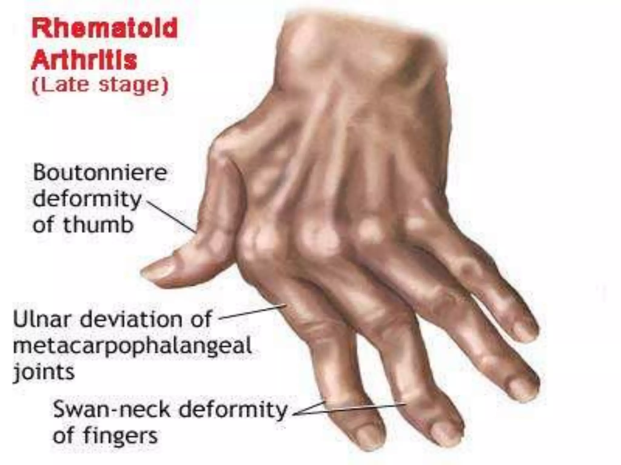
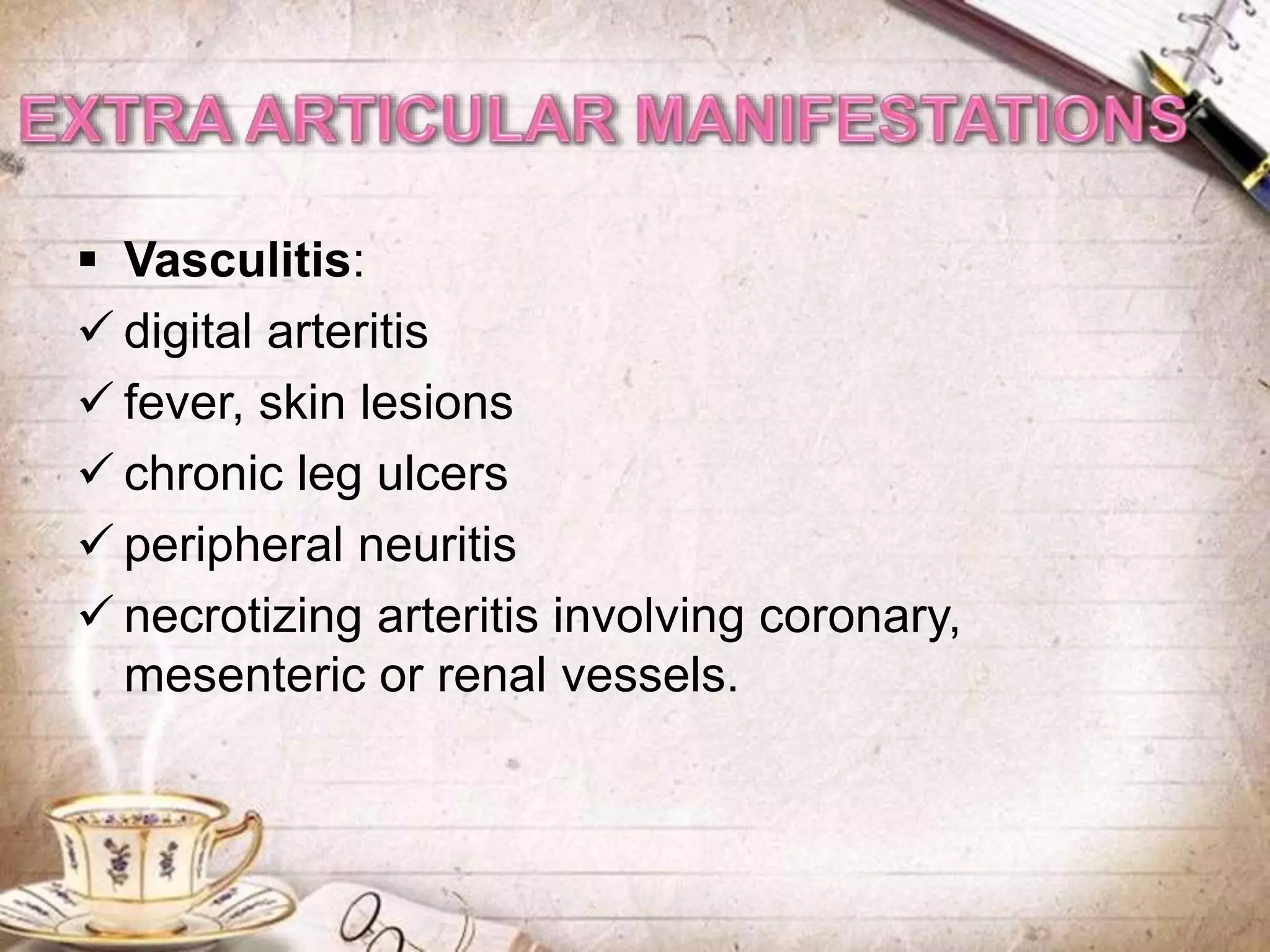
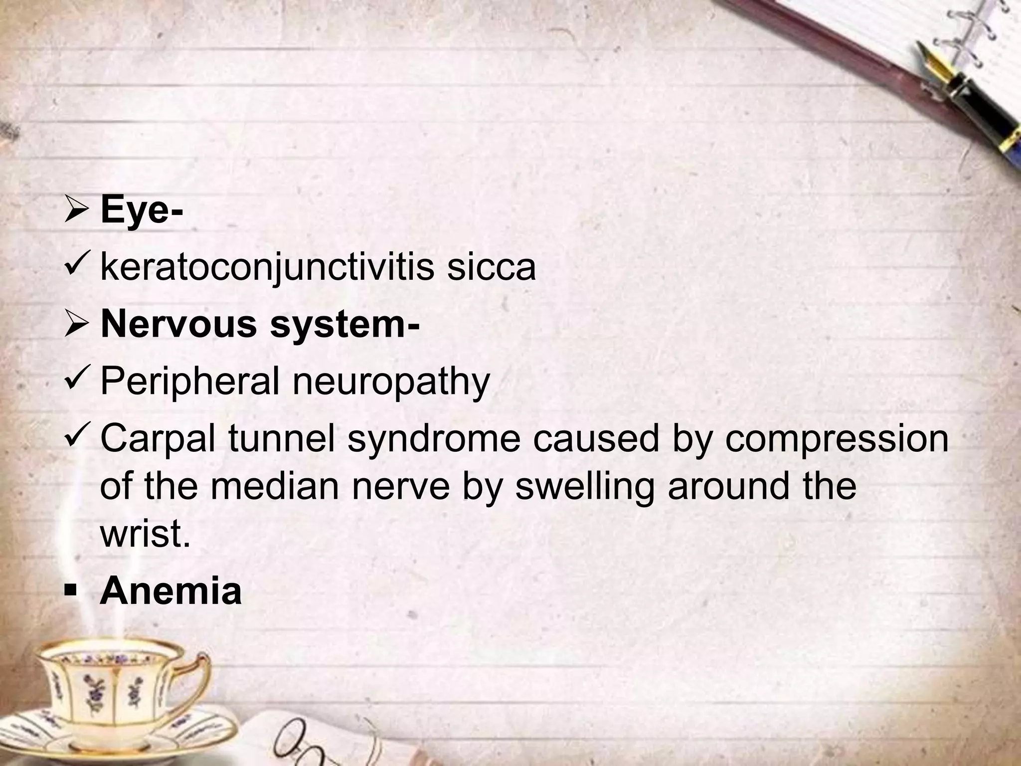
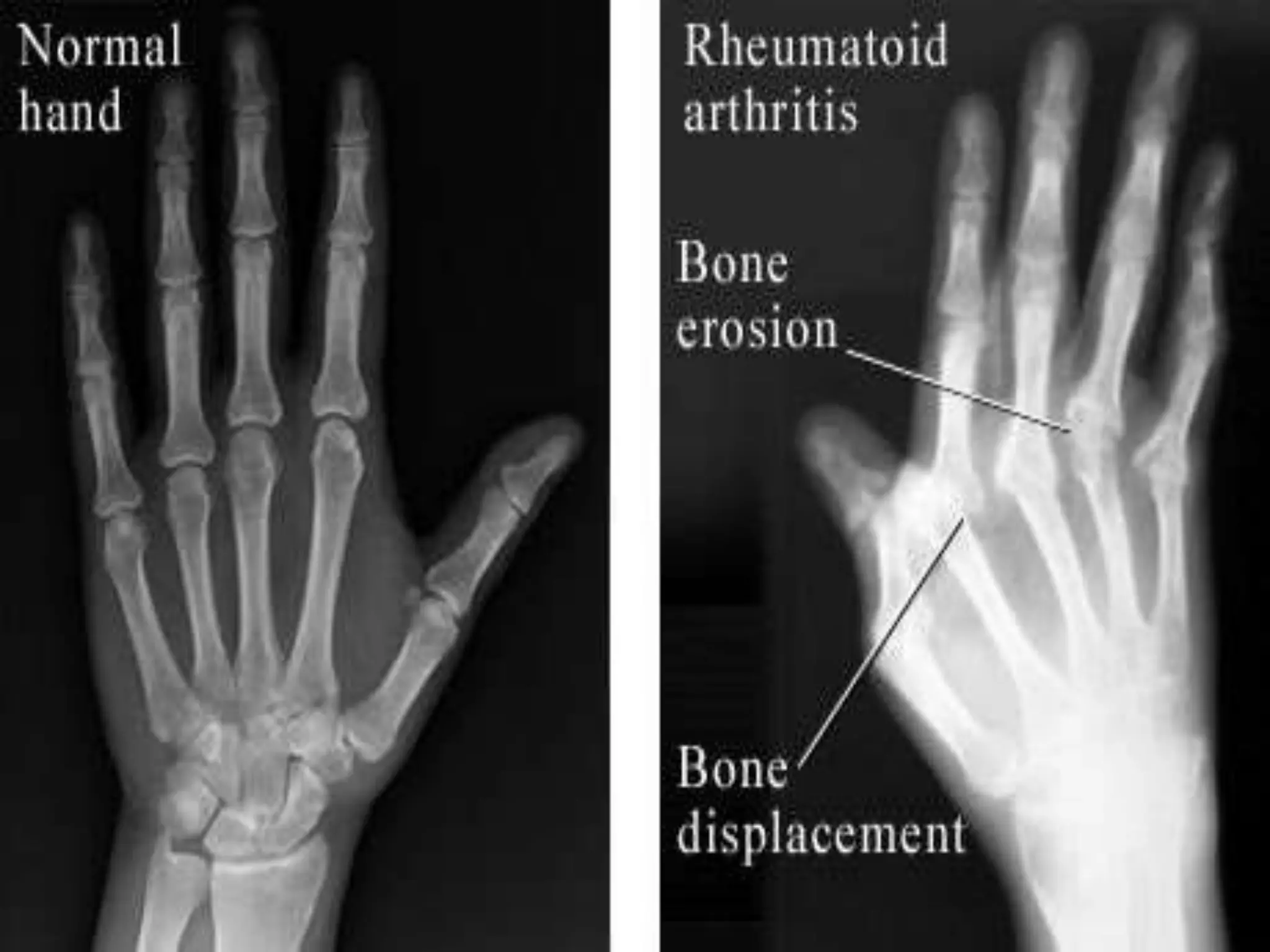
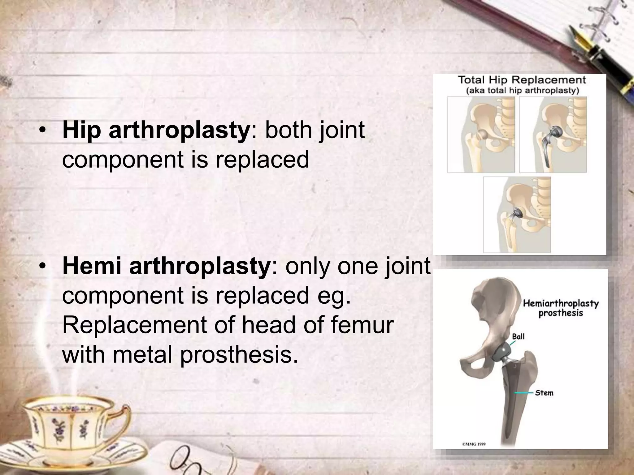
Rheumatoid arthritis is an auto
immune disease that is caused
by chronic inflammation of the
joints or in the membrane
surrounding the joints (the
synovial membrane)
 Rheumatoid arthritis (RA) is a chronic, systemic
autoimmune disorder whose major distinctive
feature is chronic, symmetrical and erosive
inflammation of the synovial tissue of the joints.
 The severity of the joint disease may fluctuate
over time, but progressive development of
various degrees of joint destruction, deformity
and disability is the most common outcome of
established disease.
• About 1% of the world’s population is afflicted by
rheumatoid arthritis.
• Onset is most frequent between the ages of 20
and 40, but people of any age can be affected.
• Women are three times more likely to get RA
than men, however women who are taking or
have taken oral contraceptives are less likely to
develop RA.
• Rheumatoid factor (RF), an
autoantibody directed against
immunoglobulin G (IgG)
• Antibodies against collagen,
Epstein-Barr virus, encoded
nuclear antigen
• Antikeratin antibody (AKA) &
anti-perinuclear factor (APF)
Genetic factors- People with RA are more likely to
have a gene called HLA- DR4, located on
chromosome 6.
Environmental factors- Infectious agents that may
induce RA are mycoplasma organisms, Epstein-
Barr virus, rubella virus and Streptococcus.
Smoking is also a major environmental risk factor.
Hormonal factors- During pregnancy, women
often experience a symptom improvement of RA,
due to increase in estrogen and progesterone
levels.
• The pathologic process involved in RA are
type 3( immune complex) and type 4( cell
mediated) reactions. If unarrested,
pathologic changes in RA pass through
four stages:
1. Synovitis
2. Pannus formation
3. Fibrous ankylosis
4. Bony ankylosis
Presentation of antigen to T cell
T and B cell proliferation
Activation of complement system
Activation of complex system &
release of lysosymes from
leukocytes
Proliferative inflammation
localized in joint capsule
Tissue thickens with edema
and congestion
SYNOVITIS
STAGE 1
PANNUS gradually develops
Extends over the articular surface
into joint anterior
Adheres tightly to underlying cartilage
by invasion and lysis
Interferes with cartilage nutrition
Pannus may extend over to
subchondral bone
Cartilage may become worn off and
become raw
STAGE 2
Subchondral bone erosion
Granulation tissue gets invaded with
tough fibrous tissue
Converted to scar tissue
Inhibits joint movements
Forms FIBROSIS ANKYLOSIS
Progress to bony ankylosis
STAGE 3
STAGE
4
1.Articular Manifestations
2.Extra Articular
Manifestations
 Early stages –
 Active inflammation of joints
 Hot, swollen and painful joints and
function may be decreased
 Soft tissue deformity and
contractures due to prolonged
immobilization
 Stiffness especially on first
wakening in the morning
 Later stages-
Deformity of hands and feet due
to misalignment resulting from
swelling, progressive joint
destruction or partial dislocation.
Muscle spasm and
weakness
 Others –
 Fever
 Rheumatoid nodules at joint capsules
 Fatigue
 Weakness
 Anorexia
 Weight loss
 Generalized aching
Rheumatoid nodules •Subcutaneous
•few millimetres to
a few centimetres
in diameter
•usually found over
bony prominences
ULNAR DRIFT
• Deviation of the MCP
(metacarpophalangeal) joints
of the fingers and medial
deviation of the wrist.
• Most marked in the little
finger and involves each
successive finger to a
lesser degree.
SWAN NECK DEFORMITY
• Hyperextension of PIP
(proximal interphalangeal) joint
with flexion of the MCP and
DIP (distal interphalangeal) joints.
• Results from contractures of
the intrinsic muscles and
tendons.
THE BOUTONNIERE
DEFORMITY
• Flexion of the PIP joints and
hyperextension of the DIP
joints.
• Due to rupture of the extensor
tendons over the fingers.
 Vasculitis:
 digital arteritis
 fever, skin lesions
 chronic leg ulcers
 peripheral neuritis
 necrotizing arteritis involving coronary,
mesenteric or renal vessels.
 Serositis:
 Lungs-
 Fibrosis of the lungs
 Pleural effusions
 Heart-
 Prone to atherosclerosis,
 Risk of myocardial infarction & stroke
 Others: pericarditis, endocarditis, left ventricular
failure, valvulitis and fibrosis.
 Eye-
 keratoconjunctivitis sicca
 Nervous system-
 Peripheral neuropathy
 Carpal tunnel syndrome caused by compression
of the median nerve by swelling around the
wrist.
 Anemia
Radiologic examination-
• This consists of X rays of both hands and of the
affected joints. The following features may be
present:
• Reduced joint space
• Erosion articular margins
• Subchondral cysts
• Soft tissue swelling
Blood tests-
• Elevated ESR
• Decreased Hb value
• Positive test for C reactive protein
• ANA (antinuclear antibodies) – seen in
15-20% clients
• Rheumatoid factor (RF)- It can be
detected in serum by following tests:
• Latex fixation test- positive in titre 1:20
• Rose- waaler test – agglutination test
sheeps RBC are used as carrier.
Synovial fluid examination-
• Arthroscopic examination shows
synovial fluid is cloudy, milky or dark
yellow
• contains numerous inflammatory
components
• reduced viscosity
• WBC count is as high as
50,000/mm3
1. Morning stiffness of >1 hour most mornings
for at least 6 weeks.
2. Arthritis and soft-tissue swelling of >3 of 14
joints/joint groups, present for at least 6
weeks
3. Swelling of atleast 1 wrist, MCP, or PIP joint,
present for at least 6 weeks
4. Symmetric arthritis, present for at least 6
weeks
5. Subcutaneous nodules in specific places
6. Presence of Rheumatoid factor
7. Radiological changes suggestive of joint
erosion
 four or more of the seven criteria must be
met before the disease is classified as RA.
The aims are-
• The induction of remission and its
maintenance: the disease’s activity is
brought under control by drugs.
• The preservation of joint functions and
prevention of deformities: during the activity
of disease and thereafter by physiotherapy
and splinting.
• Repair of the joint damage which already
exists: it will relieve pain or facilitate
functions. It sometimes requires surgical
interventions eg. Synovectomy.
MEDICAL
MANAGEMENT
ORTHOPAEDIC
MANAGEMENT
It has 3 goals:
• Prevention of joint deformity
• Preservation of joint function
• Reduction of inflammation and pain.
It consists of
• Rest
• Physical therapy (isometric exercises)
• Hot and cold application (moist, dry heat, cold
packs)
• Diet
• Drugs
 Drugs like NSAID’s
 Disease Modifying Anti- Rheumatic
Drugs(DMARD’s)
 Steroids
Never massage acutely inflamed
joint.
• Aspirin / sodium salicylate:
 They are analgesic, anti-
inflammatory, relatively safe and
inexpensive.
 Analgesia is achieved in small
doses and large doses are
needed to reduce inflammation.
 Frequent doses (3 to 4 times)
are required, even when pain is
not present to keep blood
salicylate level high.
• NSAID’s: if aspirin is ineffective NSAID’s
are given.
They are anti – inflammatory, analgesics
and antipyretics e.g.
 Phenylbutazone
 Indomethacin
 Ibuprofen
* to be taken with histamine receptor
antagonist.
• Corticosteroids
 Wysolone
 Prednisolone
• Intra- articular injections of corticosteroids
• Immunosuppressive (cytotoxic) agents:
Methotrexate,acts as an immunosuppresent,
blocking the inflammatory process of RA.
• No particular dietary
modification is required.
• The client is encouraged to eat
a nutritious diet rich in vitamins,
proteins, iron.
• If client is overweight, they are
taught to loose weight to relieve
stress on affected joints.
• Tendon transfers and
osteotomy:
 Prevents progressive deformity
which would be caused by muscle
spasm.
 Nodules or bony tumors can be
surgically removed.
 Osteotomy may improve the
function of deformed joints or limbs.
• Synovectomy:
 It helps in maintaining joint
functions.
 Early surgical removal of the
synovium helps prevent
recurrent inflammation.
• Joint replacement:
eg. Arthrodesis or arthroplasty( with or
without replacement of joint parts with
prosthesis).
 Arthrodesis( fusion) sacrifices function
of the joint but relieves pain in
severely damaged joints.
 Fusion may be performed on the
wrists to fix affected part in a
functional position so it can be used
more effectively.
• Arthrodesis –
surgical fusion of joints
• Hip arthroplasty: both joint
component is replaced
• Hemi arthroplasty: only one joint
component is replaced eg.
Replacement of head of femur
with metal prosthesis.
Thus the surgerical management is divided
into:
• Preventive surgery: eg synovectomy
• Palliative surgery: for relief eg tendon transfer
• Reconstructive surgery: for rehabilitation eg.
Arthroplasty, total joint replacement
Rhematoid arthiritis

Rhematoid arthiritis

  • 2.
    Rheumatoid arthritis isan auto immune disease that is caused by chronic inflammation of the joints or in the membrane surrounding the joints (the synovial membrane)
  • 3.
     Rheumatoid arthritis(RA) is a chronic, systemic autoimmune disorder whose major distinctive feature is chronic, symmetrical and erosive inflammation of the synovial tissue of the joints.  The severity of the joint disease may fluctuate over time, but progressive development of various degrees of joint destruction, deformity and disability is the most common outcome of established disease.
  • 5.
    • About 1%of the world’s population is afflicted by rheumatoid arthritis. • Onset is most frequent between the ages of 20 and 40, but people of any age can be affected. • Women are three times more likely to get RA than men, however women who are taking or have taken oral contraceptives are less likely to develop RA.
  • 6.
    • Rheumatoid factor(RF), an autoantibody directed against immunoglobulin G (IgG) • Antibodies against collagen, Epstein-Barr virus, encoded nuclear antigen • Antikeratin antibody (AKA) & anti-perinuclear factor (APF)
  • 7.
    Genetic factors- Peoplewith RA are more likely to have a gene called HLA- DR4, located on chromosome 6. Environmental factors- Infectious agents that may induce RA are mycoplasma organisms, Epstein- Barr virus, rubella virus and Streptococcus. Smoking is also a major environmental risk factor. Hormonal factors- During pregnancy, women often experience a symptom improvement of RA, due to increase in estrogen and progesterone levels.
  • 8.
    • The pathologicprocess involved in RA are type 3( immune complex) and type 4( cell mediated) reactions. If unarrested, pathologic changes in RA pass through four stages: 1. Synovitis 2. Pannus formation 3. Fibrous ankylosis 4. Bony ankylosis
  • 9.
    Presentation of antigento T cell T and B cell proliferation Activation of complement system Activation of complex system & release of lysosymes from leukocytes Proliferative inflammation localized in joint capsule Tissue thickens with edema and congestion SYNOVITIS STAGE 1
  • 10.
    PANNUS gradually develops Extendsover the articular surface into joint anterior Adheres tightly to underlying cartilage by invasion and lysis Interferes with cartilage nutrition Pannus may extend over to subchondral bone Cartilage may become worn off and become raw STAGE 2
  • 11.
    Subchondral bone erosion Granulationtissue gets invaded with tough fibrous tissue Converted to scar tissue Inhibits joint movements Forms FIBROSIS ANKYLOSIS Progress to bony ankylosis STAGE 3 STAGE 4
  • 12.
  • 13.
     Early stages–  Active inflammation of joints  Hot, swollen and painful joints and function may be decreased  Soft tissue deformity and contractures due to prolonged immobilization  Stiffness especially on first wakening in the morning
  • 15.
     Later stages- Deformityof hands and feet due to misalignment resulting from swelling, progressive joint destruction or partial dislocation. Muscle spasm and weakness
  • 16.
     Others – Fever  Rheumatoid nodules at joint capsules  Fatigue  Weakness  Anorexia  Weight loss  Generalized aching
  • 17.
    Rheumatoid nodules •Subcutaneous •fewmillimetres to a few centimetres in diameter •usually found over bony prominences
  • 18.
    ULNAR DRIFT • Deviationof the MCP (metacarpophalangeal) joints of the fingers and medial deviation of the wrist. • Most marked in the little finger and involves each successive finger to a lesser degree.
  • 19.
    SWAN NECK DEFORMITY •Hyperextension of PIP (proximal interphalangeal) joint with flexion of the MCP and DIP (distal interphalangeal) joints. • Results from contractures of the intrinsic muscles and tendons.
  • 20.
    THE BOUTONNIERE DEFORMITY • Flexionof the PIP joints and hyperextension of the DIP joints. • Due to rupture of the extensor tendons over the fingers.
  • 22.
     Vasculitis:  digitalarteritis  fever, skin lesions  chronic leg ulcers  peripheral neuritis  necrotizing arteritis involving coronary, mesenteric or renal vessels.
  • 23.
     Serositis:  Lungs- Fibrosis of the lungs  Pleural effusions  Heart-  Prone to atherosclerosis,  Risk of myocardial infarction & stroke  Others: pericarditis, endocarditis, left ventricular failure, valvulitis and fibrosis.
  • 24.
     Eye-  keratoconjunctivitissicca  Nervous system-  Peripheral neuropathy  Carpal tunnel syndrome caused by compression of the median nerve by swelling around the wrist.  Anemia
  • 25.
    Radiologic examination- • Thisconsists of X rays of both hands and of the affected joints. The following features may be present: • Reduced joint space • Erosion articular margins • Subchondral cysts • Soft tissue swelling
  • 27.
    Blood tests- • ElevatedESR • Decreased Hb value • Positive test for C reactive protein • ANA (antinuclear antibodies) – seen in 15-20% clients • Rheumatoid factor (RF)- It can be detected in serum by following tests: • Latex fixation test- positive in titre 1:20 • Rose- waaler test – agglutination test sheeps RBC are used as carrier.
  • 28.
    Synovial fluid examination- •Arthroscopic examination shows synovial fluid is cloudy, milky or dark yellow • contains numerous inflammatory components • reduced viscosity • WBC count is as high as 50,000/mm3
  • 29.
    1. Morning stiffnessof >1 hour most mornings for at least 6 weeks. 2. Arthritis and soft-tissue swelling of >3 of 14 joints/joint groups, present for at least 6 weeks 3. Swelling of atleast 1 wrist, MCP, or PIP joint, present for at least 6 weeks 4. Symmetric arthritis, present for at least 6 weeks
  • 30.
    5. Subcutaneous nodulesin specific places 6. Presence of Rheumatoid factor 7. Radiological changes suggestive of joint erosion  four or more of the seven criteria must be met before the disease is classified as RA.
  • 32.
    The aims are- •The induction of remission and its maintenance: the disease’s activity is brought under control by drugs. • The preservation of joint functions and prevention of deformities: during the activity of disease and thereafter by physiotherapy and splinting. • Repair of the joint damage which already exists: it will relieve pain or facilitate functions. It sometimes requires surgical interventions eg. Synovectomy.
  • 33.
  • 34.
    It has 3goals: • Prevention of joint deformity • Preservation of joint function • Reduction of inflammation and pain.
  • 35.
    It consists of •Rest • Physical therapy (isometric exercises) • Hot and cold application (moist, dry heat, cold packs) • Diet
  • 36.
    • Drugs  Drugslike NSAID’s  Disease Modifying Anti- Rheumatic Drugs(DMARD’s)  Steroids Never massage acutely inflamed joint.
  • 37.
    • Aspirin /sodium salicylate:  They are analgesic, anti- inflammatory, relatively safe and inexpensive.  Analgesia is achieved in small doses and large doses are needed to reduce inflammation.  Frequent doses (3 to 4 times) are required, even when pain is not present to keep blood salicylate level high.
  • 38.
    • NSAID’s: ifaspirin is ineffective NSAID’s are given. They are anti – inflammatory, analgesics and antipyretics e.g.  Phenylbutazone  Indomethacin  Ibuprofen * to be taken with histamine receptor antagonist.
  • 39.
    • Corticosteroids  Wysolone Prednisolone • Intra- articular injections of corticosteroids • Immunosuppressive (cytotoxic) agents: Methotrexate,acts as an immunosuppresent, blocking the inflammatory process of RA.
  • 40.
    • No particulardietary modification is required. • The client is encouraged to eat a nutritious diet rich in vitamins, proteins, iron. • If client is overweight, they are taught to loose weight to relieve stress on affected joints.
  • 41.
    • Tendon transfersand osteotomy:  Prevents progressive deformity which would be caused by muscle spasm.  Nodules or bony tumors can be surgically removed.  Osteotomy may improve the function of deformed joints or limbs.
  • 42.
    • Synovectomy:  Ithelps in maintaining joint functions.  Early surgical removal of the synovium helps prevent recurrent inflammation.
  • 43.
    • Joint replacement: eg.Arthrodesis or arthroplasty( with or without replacement of joint parts with prosthesis).  Arthrodesis( fusion) sacrifices function of the joint but relieves pain in severely damaged joints.  Fusion may be performed on the wrists to fix affected part in a functional position so it can be used more effectively.
  • 44.
  • 45.
    • Hip arthroplasty:both joint component is replaced • Hemi arthroplasty: only one joint component is replaced eg. Replacement of head of femur with metal prosthesis.
  • 46.
    Thus the surgericalmanagement is divided into: • Preventive surgery: eg synovectomy • Palliative surgery: for relief eg tendon transfer • Reconstructive surgery: for rehabilitation eg. Arthroplasty, total joint replacement