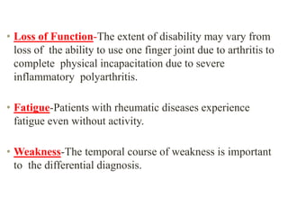
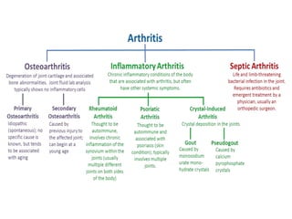
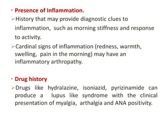
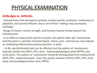
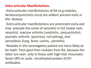
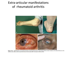
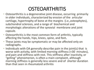
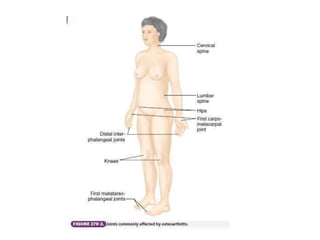
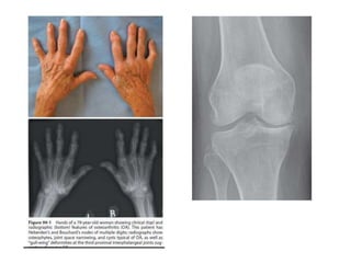
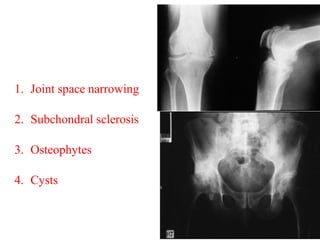
This document provides an overview of arthritis, including:
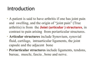
- Definitions of true arthritis versus periarticular joint pain
- Common symptoms of arthritis like pain, swelling, stiffness, limitation of motion
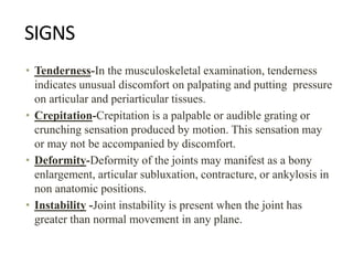
- Classification of arthritis based on number of joints involved and duration
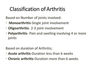
- Approaches to evaluating monoarthritis and polyarthritis through history, exam, labs, imaging
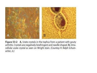
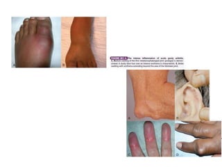
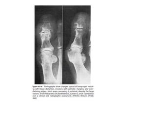
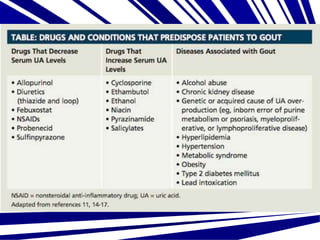
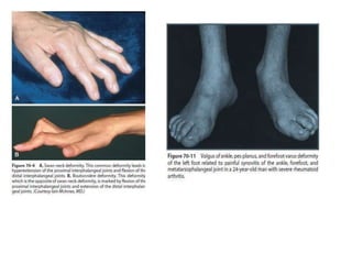
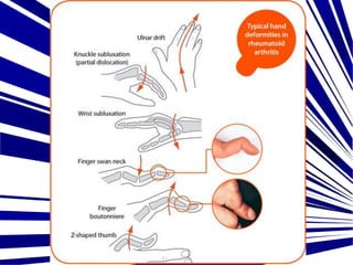
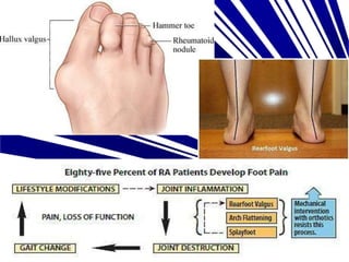
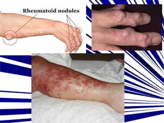
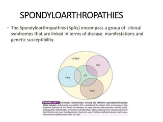
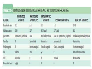
- Details on specific types of arthritis like gout, pseudogout, osteoarthritis
This document provides guidance on evaluating a patient presenting with arthritis. It describes how arthritis is defined and classified based on the number and duration of involved joints. Key components of the history include symptoms, physical exam findings, classification, differential diagnoses, and initial investigations for monoarthritis and polyarthritis. Initial workup may include blood tests, imaging, and synovial fluid analysis to help identify conditions like gout, pseudogout, septic arthritis, trauma, and rheumatic diseases.
The document discusses the approach to diagnosing arthritis. It covers the main pathophysiologic types of joint disease including synovitis, enthesitis, crystal deposition, infection, and structural/mechanical derangements. For each type, it describes the characteristic pathologic features. In evaluating a patient's joint pain, the history and physical exam aim to differentiate inflammatory from noninflammatory arthritis and determine the specific pathophysiologic process involved based on features like onset, duration, distribution and symmetry of joint involvement, and extra-articular manifestations. Signs on physical exam of inflammatory arthritis include swelling, pain with motion, erythema, warmth, and limited range of motion.
This document discusses arthritis and related diseases. It defines arthritis as inflammation of a joint and describes its main causes and types. Rheumatoid arthritis is discussed in detail, including its pathology, clinical features, diagnostic criteria, and treatments. Ankylosing spondylitis and osteoarthritis are also summarized. The document provides overviews of the classifications, presentations, investigations, and management approaches for different forms of arthritis.
Osteoarthritis and rheumatoid arthritis are chronic joint disorders. Osteoarthritis involves the progressive breakdown of articular cartilage in a joint. It is associated with aging and risk factors like obesity, joint injury, and genetics. Rheumatoid arthritis is an autoimmune disease where the immune system attacks the joints, causing pain, stiffness, and swelling. It can eventually damage cartilage and bone within joints and may affect other organs. Both diseases are diagnosed based on symptoms, physical exam, x-rays, and blood tests. Treatment focuses on reducing pain and inflammation, maintaining joint mobility, and may include medications, weight loss, or joint replacement surgery.
This document discusses bone and joint infections. It begins by classifying infections as either pyogenic (bacterial), tuberculous, or other causes. Osteomyelitis is defined as a bone infection that can be caused by bacteria, fungi, parasites or viruses. Symptoms of osteomyelitis can be acute, subacute, or chronic. Common sites of bone infection in children are the metaphysis around the knee. Imaging plays an important role in diagnosis, with plain radiography, CT, MRI, bone scans, and ultrasound all discussed. Biopsy may be needed to confirm infection and identify the organism. Brodie's abscess, a characteristic subacute pyogenic bone infection, is also mentioned.
This document provides an overview of inflammatory joint conditions and childhood arthropathies. It defines synovial joints and describes the structure and functions of synovial fluid. Rheumatoid arthritis is characterized as an autoimmune disease that causes inflammation in the joints, commonly affecting the hands and feet. The stages of rheumatoid arthritis progression and its clinical features are outlined. Childhood arthropathies like juvenile idiopathic arthritis can affect multiple joint types and have different subtypes depending on symptoms. Diagnosis and treatment of both conditions involve examinations of synovial fluid, blood tests, imaging and medications.
Arthritis is inflammation of one or more joints that can be caused by infection, trauma, or other causes. The main types discussed are osteoarthritis, rheumatoid arthritis, septic arthritis, gout, and psoriatic arthritis. Common signs include pain, stiffness, and limited movement of the affected joints. Management focuses on relieving pain and modifying the immune system. Complications can include permanent joint deformity, fistula formation, and development of nodules.
1. Key physical exam findings that help differentiate joint diseases include the presence of swelling, erythema, warmth, tenderness, range of motion, pain characteristics, and duration of symptoms.
2. The major arthritides like osteoarthritis, rheumatoid arthritis, and gout/pseudogout can be compared based on features such as onset, pathology, number and type of joints involved, and associated extra-articular findings.
3. Osteoarthritis is the most common type of joint disease and diagnosis is based on clinical and radiographic evidence. Treatment focuses on medications, lifestyle changes, and sometimes surgery. Rheumatoid arthritis is an autoimmune disease treated initially with disease-mod
This document provides information on Charcot foot, including:
- A history of Charcot foot first being described in the 1700s and studied in more detail in the 1800s.
- Charcot foot is defined as a non-infective, destructive condition affecting bones and joints caused by neuropathy leading to fractures and joint destruction.
- Risk factors include diabetes, alcoholism, leprosy and other neurological conditions.
- It presents clinically with foot swelling, warmth, pain and loss of function and can be classified into stages based on radiographic findings.
- Management involves offloading with casting or bracing, surgery for deformities, and sometimes amputation for severe cases.
This document discusses different types of arthritis, including osteoarthritis, rheumatoid arthritis, and gouty arthritis. It describes the causes, symptoms, risk factors, diagnosis, and treatment options for each type. Osteoarthritis is caused by wear and tear on joints and commonly affects the hips and knees. Rheumatoid arthritis is an autoimmune disorder that causes inflammation of the joints and can affect other parts of the body. Gouty arthritis occurs when uric acid crystals accumulate in a joint, causing inflammation. Treatment involves medications, physical therapy, bracing, and sometimes surgery.
This document discusses various types of arthritis including osteoarthritis, rheumatoid arthritis, and gouty arthritis. It describes the causes, symptoms, risk factors, and treatments for each type. The major treatments discussed are medications, physical therapy, orthotic devices, and surgery. Orthotic devices like braces, insoles, and toe pads can help reduce stress on joints and relieve pain caused by different forms of arthritis. While orthotic treatments cannot cure arthritis, they can help slow its progression and make daily activities less painful.
Arthritis and arthroplasty- dr. Mahmoud Abdel KareemAhmed-shedeed
This document provides information about osteoarthritis (OA), including its definition, prevalence, risk factors, pathology, diagnosis, natural history, differential diagnosis, and treatment. It notes that OA is the most common form of arthritis, affecting over 20 million people in the US. Risk factors include age, obesity, family history, and previous joint injury or disorder. Diagnosis is typically based on symptoms like pain and stiffness, physical exam findings, and x-ray evidence of cartilage loss, bone spurs, and bone changes. Treatment includes conservative options like medications, exercise, and weight loss, as well as intra-articular injections or surgery for advanced cases.
Arthritis can involve either the joints (articular) or the areas around the joints (periarticular). It has many potential causes including trauma, infection, or autoimmune issues. A thorough history and physical exam can help determine if the arthritis is acute or chronic, monoarticular or polyarticular, and point to possible diagnoses like gout, osteoarthritis, rheumatoid arthritis, or rheumatic fever. Blood tests, imaging, joint fluid analysis, and other diagnostic tests may be used to confirm the diagnosis. Treatment depends on the underlying cause but may include medications like NSAIDs, steroids, DMARDs, or biologics as well as lifestyle changes.
The document discusses seronegative spondyloarthropathies, a group of disorders that share clinical features like inflammatory axial arthritis and enthesitis. It focuses on ankylosing spondylitis (AS), describing its pathology, clinical manifestations including stiffness and fusion of the spine, extra-articular involvement like uveitis, and treatments including NSAIDs and TNF inhibitors. Surgical treatments for severe AS spinal deformities like osteotomies and joint replacement are also summarized.
This document discusses various types of crystals found in rheumatology, including mono-sodium urate (gout), calcium pyrophosphate dehydrate (CPPD), basic calcium phosphate (BCP), cholesterol crystal, lipid crystal, and corticosteroid crystal. It presents several case studies demonstrating the clinical presentation and investigations for patients with these various crystal-related arthritides. Key points include the importance of identifying crystals in synovial fluid to confirm diagnoses, characteristic radiographic and ultrasound findings, and treatment approaches for acute flares and long-term management.
This document provides information about gout and hyperuricemia. It discusses uric acid metabolism and the causes of elevated uric acid levels. Gout is characterized by recurrent attacks of acute inflammatory arthritis caused by deposition of urate crystals in the joints. Risk factors include genetics, sex, adrenal dysfunction, electrolyte imbalances, and vascular changes. Treatment involves medications to reduce uric acid levels such as allopurinol and febuxostat, in addition to symptomatic treatments for acute flares like NSAIDs, colchicine, and corticosteroids. Chronic tophaceous gout can lead to joint damage and other complications if uric acid levels are not well controlled.
1. Seronegative spondyloarthritis is a group of inflammatory diseases characterized by inflammatory back pain, spinal and peripheral joint involvement, and enthesitis with a negative rheumatoid factor.
2. This family includes ankylosing spondylitis, psoriatic arthritis, enteropathic arthritis, reactive arthritis, and undifferentiated spondyloarthropathy.
3. Psoriatic arthritis is characterized by seronegative polysynovitis, erosive arthritis, enthesitis, dactylitis, and sacroiliitis or spondylitis. It commonly presents with distal interphalangeal joint involvement and nail changes.
This document provides information about various types of arthritis from an expert in rheumatology. It begins with an introduction to arthritis and how it can originate from the joint or surrounding tissues. It then discusses the diagnostic approach and evaluation of a patient with arthritis. The rest of the document discusses specific types of arthritis in more detail, including septic arthritis, gout, osteoarthritis, and rheumatoid arthritis. It provides information on clinical features, investigations, diagnosis, and management for each type.
This presentation focuses on different types of arthritis/joint disorders. It provides stepwise approach to evaluation and diagnoses and it's truly wonderful to have a broad overview of many joint conditions in one presentation - ranging from osteoarthritis, gout, rheumatoid arthritis, septic arthritis, to ankylosing spondilitis, and many others, including fibromyaligia.
Osteomyelitis has been recognized since ancient times. It is an infection of bone that can be caused by bacteria, fungi, or other microbes entering through the bloodstream or directly through trauma. The most common type is acute hematogenous osteomyelitis, which typically affects children under 15 and presents as a rapidly destructive infection of the metaphysis of long bones. Staphylococcus aureus is the primary causative organism. Diagnosis involves blood tests, imaging like x-rays, ultrasound, bone scans or MRI, and tissue sampling. Treatment involves antibiotics along with surgical drainage and debridement if abscesses form or the infection fails to respond to antibiotics alone. Chronic osteomyelitis can develop if not properly
Synovial biopsy provides tissue that can be used to better understand the pathophysiological mechanisms of arthritis through techniques like immunohistochemistry, electron microscopy, and molecular biology. It is not normally required for routine diagnosis but can help evaluate new treatment approaches. There are different types of synovial biopsies including needle biopsy, arthroscopic biopsy, and open surgical biopsy. Needle biopsy is the most common technique and samples are obtained from joints like the knee using a 14-gauge needle. Synovial biopsy helps diagnose conditions like infectious arthritis, autoimmune diseases like rheumatoid arthritis, and crystal-induced arthritides.
Osteoarthritis is a degenerative joint disease that involves the breakdown of articular cartilage and underlying bone. It most commonly affects weight-bearing joints like the knees and hips. Risk factors include age over 45, female sex, joint injuries, obesity, and hereditary factors. Symptoms include pain, stiffness, swelling, and loss of function. Diagnosis is made based on symptoms and confirmed with x-rays showing cartilage loss, bone spurs, and bone changes. Treatment focuses on pain management, physical therapy, braces, and surgery like joint replacement for severe cases.
This document provides an overview of musculoskeletal disorders, focusing on arthritis. It defines arthritis as inflammation of one or more joints. The three most common types of arthritis are osteoarthritis, rheumatoid arthritis, and gouty arthritis. Osteoarthritis is a non-inflammatory degenerative joint condition characterized by cartilage breakdown and new bone growth. Rheumatoid arthritis is a chronic autoimmune disorder causing symmetrical polyarthritis. Gouty arthritis results from uric acid crystal deposition in joints and tissues. The document discusses the causes, symptoms, investigations, and management of these three arthritis types.
Spontaneous Osteonecrosis of the Knee.pptxcheryl712552
Spontaneous osteonecrosis of the knee (SONK) is a type of osteonecrosis where there is cell death of bone and bone marrow due to interrupted blood supply, typically affecting the medial femoral condyle. It most commonly affects active females over 50 and can cause sudden severe knee pain. Diagnosis involves X-ray, MRI, and bone scan. Conservative treatment is usually tried initially but surgery may be needed if the lesion is large or has collapsed. Surgical options include arthroscopy, core decompression, osteotomy, or knee replacement depending on the stage of disease.
Approach to a patient with arthritis by Dr Imtiaz.pptxDRIMTIAZ3
This document provides guidance on approaching a patient presenting with arthritis. It details the types of information that should be gathered through history taking (including symptoms, disease characteristics, medical history) and clinical examination (inspection, palpation, range of motion testing and assessment of extra-articular signs). Laboratory investigations and imaging may then help classify the arthritis and identify underlying causes, which could include conditions like rheumatoid arthritis, gout, psoriatic arthritis and more. A thorough evaluation is required to diagnose the specific cause of a patient's arthritis.
This document discusses various types of acquired heart disease, including ischemic/hypoxic, hypertensive, infectious, inflammatory, metabolic, nutritional, and traumatic causes. It provides details on specific conditions like Kawasaki disease, pericarditis, myocarditis, infective endocarditis, and rheumatic heart disease. Kawasaki disease causes inflammation in blood vessels and can lead to coronary artery aneurysms if untreated. Pericarditis results in inflammation of the pericardium, while myocarditis is an infection and inflammation of the myocardium. Infective endocarditis is a microbial infection of the heart valves or walls. Rheumatic fever is an autoimmune response to a streptococcal infection that
This document discusses various cardiomyopathies including dilated, hypertrophic, and restrictive cardiomyopathies. It provides details on dilated cardiomyopathy including causes, clinical presentation, workup, and management. Hypertrophic cardiomyopathy etiology, presentation, and treatment are summarized. The document also reviews pericarditis including presentation, ECG and imaging findings, differential diagnosis, and treatment. Common pediatric heart murmurs and features that suggest an innocent murmur versus potential cardiac pathology are presented.
Respiratory disorders are the second leading cause of emergency room visits in children. The pediatric airway is smaller in diameter than an adult's and more susceptible to obstruction. Common respiratory emergencies in children include croup, epiglottitis, foreign body aspiration, and asthma. It is important to properly assess a child's respiratory status using the ABCDE method, treat life-threatening issues immediately, and be prepared for their condition to deteriorate rapidly. Maintaining a patent airway and providing supplemental oxygen are often critical in pediatric respiratory emergencies.
Anatomy of Bone and Fracture Healing.pptesicOrtho1
Bone anatomy and fracture healing is discussed. Bones are composed of organic and inorganic materials and have a microscopic structure including haversian systems. Fractures heal through stages including hematoma formation, granulation tissue development, callus formation, consolidation, and remodeling. Many factors can influence fracture healing including the injury characteristics, patient health, bone properties, and treatment approach. Rigid fixation, proper alignment of fragments, and limiting motion are important for optimal healing.
The document discusses general principles of fracture management. It defines fractures and classifies them based on whether the skin is broken (open vs closed) and the mechanism of injury. Treatment approaches are also summarized, including splinting, definitive treatments like casting, plating or external fixation, and rehabilitation. Open fractures require additional initial management steps like antibiotics and wound care before definitive treatment.
Osteoarthritis is a degenerative joint disease characterized by cartilage loss, bone changes, and loss of joint function. Symptoms include pain, swelling, and stiffness. Conservative treatment focuses on pain control, preventing further cartilage damage, and restoring function. This involves medications like NSAIDs, supplements to support cartilage, physical therapy including modalities like ultrasound-guided injections, laser therapy, and Pilates-based exercises. Viscosupplementation therapy and platelet-rich plasma therapy may also help manage osteoarthritis symptoms.
This document discusses various elbow injuries. It begins by describing elbow anatomy and development of the elbow bones in children. It then discusses common elbow fractures in children such as supracondylar fractures, lateral condyle fractures, and radial neck fractures. Treatment options for displaced and non-displaced fractures are provided. The document also discusses complications of fractures and injuries commonly seen in adults such as olecranon fractures, radial head fractures, and elbow dislocations. Surgical treatment techniques like tension band wiring and plating are described.
These lecture slides, by Dr Sidra Arshad, offer a simplified look into the mechanisms involved in the regulation of respiration:
Learning objectives:
1. Describe the organisation of respiratory center
2. Describe the nervous control of inspiration and respiratory rhythm
3. Describe the functions of the dorsal and respiratory groups of neurons
4. Describe the influences of the Pneumotaxic and Apneustic centers
5. Explain the role of Hering-Breur inflation reflex in regulation of inspiration
6. Explain the role of central chemoreceptors in regulation of respiration
7. Explain the role of peripheral chemoreceptors in regulation of respiration
8. Explain the regulation of respiration during exercise
9. Integrate the respiratory regulatory mechanisms
10. Describe the Cheyne-Stokes breathing
Study Resources:
1. Chapter 42, Guyton and Hall Textbook of Medical Physiology, 14th edition
2. Chapter 36, Ganong’s Review of Medical Physiology, 26th edition
3. Chapter 13, Human Physiology by Lauralee Sherwood, 9th edition
Clinic ^%[+27633867063*Abortion Pills For Sale In Tembisa Central19various
Clinic ^%[+27633867063*Abortion Pills For Sale In Tembisa Central Clinic ^%[+27633867063*Abortion Pills For Sale In Tembisa CentralClinic ^%[+27633867063*Abortion Pills For Sale In Tembisa CentralClinic ^%[+27633867063*Abortion Pills For Sale In Tembisa CentralClinic ^%[+27633867063*Abortion Pills For Sale In Tembisa Central
Rasamanikya is a excellent preparation in the field of Rasashastra, it is used in various Kushtha Roga, Shwasa, Vicharchika, Bhagandara, Vatarakta, and Phiranga Roga. In this article Preparation& Comparative analytical profile for both Formulationon i.e Rasamanikya prepared by Kushmanda swarasa & Churnodhaka Shodita Haratala. The study aims to provide insights into the comparative efficacy and analytical aspects of these formulations for enhanced therapeutic outcomes.
Promoting Wellbeing - Applied Social Psychology - Psychology SuperNotesPsychoTech Services
A proprietary approach developed by bringing together the best of learning theories from Psychology, design principles from the world of visualization, and pedagogical methods from over a decade of training experience, that enables you to: Learn better, faster!
- Video recording of this lecture in English language: https://youtu.be/kqbnxVAZs-0
- Video recording of this lecture in Arabic language: https://youtu.be/SINlygW1Mpc
- Link to download the book free: https://nephrotube.blogspot.com/p/nephrotube-nephrology-books.html
- Link to NephroTube website: www.NephroTube.com
- Link to NephroTube social media accounts: https://nephrotube.blogspot.com/p/join-nephrotube-on-social-media.html
Does Over-Masturbation Contribute to Chronic Prostatitis.pptxwalterHu5
In some case, your chronic prostatitis may be related to over-masturbation. Generally, natural medicine Diuretic and Anti-inflammatory Pill can help mee get a cure.
Adhd Medication Shortage Uk - trinexpharmacy.comreignlana06
The UK is currently facing a Adhd Medication Shortage Uk, which has left many patients and their families grappling with uncertainty and frustration. ADHD, or Attention Deficit Hyperactivity Disorder, is a chronic condition that requires consistent medication to manage effectively. This shortage has highlighted the critical role these medications play in the daily lives of those affected by ADHD. Contact : +1 (747) 209 – 3649 E-mail : sales@trinexpharmacy.com
Local Advanced Lung Cancer: Artificial Intelligence, Synergetics, Complex Sys...Oleg Kshivets
Overall life span (LS) was 1671.7±1721.6 days and cumulative 5YS reached 62.4%, 10 years – 50.4%, 20 years – 44.6%. 94 LCP lived more than 5 years without cancer (LS=2958.6±1723.6 days), 22 – more than 10 years (LS=5571±1841.8 days). 67 LCP died because of LC (LS=471.9±344 days). AT significantly improved 5YS (68% vs. 53.7%) (P=0.028 by log-rank test). Cox modeling displayed that 5YS of LCP significantly depended on: N0-N12, T3-4, blood cell circuit, cell ratio factors (ratio between cancer cells-CC and blood cells subpopulations), LC cell dynamics, recalcification time, heparin tolerance, prothrombin index, protein, AT, procedure type (P=0.000-0.031). Neural networks, genetic algorithm selection and bootstrap simulation revealed relationships between 5YS and N0-12 (rank=1), thrombocytes/CC (rank=2), segmented neutrophils/CC (3), eosinophils/CC (4), erythrocytes/CC (5), healthy cells/CC (6), lymphocytes/CC (7), stick neutrophils/CC (8), leucocytes/CC (9), monocytes/CC (10). Correct prediction of 5YS was 100% by neural networks computing (error=0.000; area under ROC curve=1.0).
Muktapishti is a traditional Ayurvedic preparation made from Shoditha Mukta (Purified Pearl), is believed to help regulate thyroid function and reduce symptoms of hyperthyroidism due to its cooling and balancing properties. Clinical evidence on its efficacy remains limited, necessitating further research to validate its therapeutic benefits.
share - Lions, tigers, AI and health misinformation, oh my!.pptxTina Purnat
• Pitfalls and pivots needed to use AI effectively in public health
• Evidence-based strategies to address health misinformation effectively
• Building trust with communities online and offline
• Equipping health professionals to address questions, concerns and health misinformation
• Assessing risk and mitigating harm from adverse health narratives in communities, health workforce and health system
TEST BANK For Community Health Nursing A Canadian Perspective, 5th Edition by...Donc Test
TEST BANK For Community Health Nursing A Canadian Perspective, 5th Edition by Stamler, Verified Chapters 1 - 33, Complete Newest Version Community Health Nursing A Canadian Perspective, 5th Edition by Stamler, Verified Chapters 1 - 33, Complete Newest Version Community Health Nursing A Canadian Perspective, 5th Edition by Stamler Community Health Nursing A Canadian Perspective, 5th Edition TEST BANK by Stamler Test Bank For Community Health Nursing A Canadian Perspective, 5th Edition Pdf Chapters Download Test Bank For Community Health Nursing A Canadian Perspective, 5th Edition Pdf Download Stuvia Test Bank For Community Health Nursing A Canadian Perspective, 5th Edition Study Guide Test Bank For Community Health Nursing A Canadian Perspective, 5th Edition Ebook Download Stuvia Test Bank For Community Health Nursing A Canadian Perspective, 5th Edition Questions and Answers Quizlet Test Bank For Community Health Nursing A Canadian Perspective, 5th Edition Studocu Test Bank For Community Health Nursing A Canadian Perspective, 5th Edition Quizlet Test Bank For Community Health Nursing A Canadian Perspective, 5th Edition Stuvia Community Health Nursing A Canadian Perspective, 5th Edition Pdf Chapters Download Community Health Nursing A Canadian Perspective, 5th Edition Pdf Download Course Hero Community Health Nursing A Canadian Perspective, 5th Edition Answers Quizlet Community Health Nursing A Canadian Perspective, 5th Edition Ebook Download Course hero Community Health Nursing A Canadian Perspective, 5th Edition Questions and Answers Community Health Nursing A Canadian Perspective, 5th Edition Studocu Community Health Nursing A Canadian Perspective, 5th Edition Quizlet Community Health Nursing A Canadian Perspective, 5th Edition Stuvia Community Health Nursing A Canadian Perspective, 5th Edition Test Bank Pdf Chapters Download Community Health Nursing A Canadian Perspective, 5th Edition Test Bank Pdf Download Stuvia Community Health Nursing A Canadian Perspective, 5th Edition Test Bank Study Guide Questions and Answers Community Health Nursing A Canadian Perspective, 5th Edition Test Bank Ebook Download Stuvia Community Health Nursing A Canadian Perspective, 5th Edition Test Bank Questions Quizlet Community Health Nursing A Canadian Perspective, 5th Edition Test Bank Studocu Community Health Nursing A Canadian Perspective, 5th Edition Test Bank Quizlet Community Health Nursing A Canadian Perspective, 5th Edition Test Bank Stuvia
Histololgy of Female Reproductive System.pptxAyeshaZaid1
Dive into an in-depth exploration of the histological structure of female reproductive system with this comprehensive lecture. Presented by Dr. Ayesha Irfan, Assistant Professor of Anatomy, this presentation covers the Gross anatomy and functional histology of the female reproductive organs. Ideal for students, educators, and anyone interested in medical science, this lecture provides clear explanations, detailed diagrams, and valuable insights into female reproductive system. Enhance your knowledge and understanding of this essential aspect of human biology.
2. Introduction
• A patient is said to have arthritis if one has joint pain
and swelling, and the origin of “joint pain” (True
arthritis) is from the Joint (articular ) structures, in
contrast to pain arising from periarticular structures.
• Articular structures include Synovium, synovial
fluid, cartilage, intraarticular ligaments, the joint
capsule and the adjacent bone
• Periarticular structures include ligaments, tendons,
bursae, muscle, fascia , bone and nerve.
4. SYMPTOMS
• Pain-Pain is a subjective hurting sensation or experience
described in various terms, often of actual or perceived
physical damage.
• Swelling-Patients with inflammatory arthritis may describe
swelling of joints in a distribution typical of a specific
disease—symmetric swelling of the metacarpophalangeal
joints and wrists in rheumatoid arthritis, or swelling of several
toes and a knee in psoriatic arthritis
• Stiffness-Discomfort and limitation on attempted movement
of joints after a period of inactivity.
• Limitation of Motion-Duration of restriction and determining
the rapidity.
5. • Loss of Function-The extent of disability may vary from
loss of the ability to use one finger joint due to arthritis to
complete physical incapacitation due to severe
inflammatory polyarthritis.
• Fatigue-Patients with rheumatic diseases experience
fatigue even without activity.
• Weakness-The temporal course of weakness is important
to the differential diagnosis.
6. SIGNS
• Tenderness-In the musculoskeletal examination, tenderness
indicates unusual discomfort on palpating and putting pressure
on articular and periarticular tissues.
• Crepitation-Crepitation is a palpable or audible grating or
crunching sensation produced by motion. This sensation may
or may not be accompanied by discomfort.
• Deformity-Deformity of the joints may manifest as a bony
enlargement, articular subluxation, contracture, or ankylosis in
non anatomic positions.
• Instability -Joint instability is present when the joint has
greater than normal movement in any plane.
7. Classification ofArthritis
Based on Number of joints involved:
• Monoarthritis-Single joint involvement
• Oligoarthritis- 2-3 joint involvement
• Polyarthritis- Pain and swelling involving 4 or more
joints
Based on duration of Arthritis;
• Acute arthritis-Duration less than 6 weeks
• Chronic arthritis-Duration more than 6 weeks
11. HISTORY
Age-Gout usually occurs in age group of 30-60 years,
Degenervative disorders associated with old age
Occupation- Increased prevalence of OA of the elbows, knees,
and spine in miners, farmers, firefighters, mill workers, unskilled
manual laborers
Drug history- Gout precipitated by Loop and Thiazide diuretics,
Ethambutol, Pyrizinamide.
Sexual history- Gonococcal arthritis, Reactive arthritis
12. • Pain and joint stiffness, its diurnal variation, and
aggravating and relieving factors should be sought,
and a history of swelling should be established.
• Specific features of relevance include trauma, joint
locking.
• Presence of systemic symptoms (fevers ,sweats, rigors,
and weight loss)-suggestive of septic arthritis
• Inquiring about ocular, oral, respiratory,
gastrointestinal, or skin symptoms can facilitate the
diagnosis.
13.
14. EXAMINATION
• Look, feel, and move joints to assess function—comparing the
affected joint and normal side
• In any acutely swollen joint, examination should include looking
for local signs of inflammation (pain, erythema, swelling, heat, and
loss of function).
• Any patient with preceding arthritis should be assessed for chronic
changes to joint structure and disability. Local synovial swelling
and/or effusion, joint instability, limited movement, and deformity
of any single joint necessitate detailed investigation.
• Local monoarticular tenderness without swelling could indicate
enthesitis, tendinitis, bursitis, or bone disease.
• A complete examination should look specifically for ocular signs,
skin rashes,ulcers, and nodules
15. INVESTIGATIONS
1)BLOOD
•Inflammatory, septic, and crystal arthrits cause
elevated (ESR), C-reactive protein (CRP), and white
cell count (WCC), often associated with anemia.
•Systemic disease involvement is assessed by renal,
liver, muscle, or bone biochemical screening and
protein electrophoresis.
•Raised uric acid levels suggest a diagnosis of gout.
16. • In acute hemarthrosis, a platelet count, INR, and
clotting studies are warranted.
• Blood cultures are mandatory in patients with
suspected septic arthritis and should precede
antibiotic prescription
• Viral screening (IgG and IgM antibodies),
antistreptolysin-O test (ASOT), and Lyme serology
can be diagnostic in relevant situations.
17. 2)URINE
•The urinary tract can be a source of gram-negative
bacteria in septic arthritis in the elderly.
•Significant proteinuria and or hematuria and red
cell casts indicate renal damage in SLE, vasculitis, or
subacute bacterial endocarditis
18. IMAGING STUDIES
• A range of imaging studies assist in diagnosis of acute
monoarthritis.
1.Plain radiographs identify soft tissue swelling, calcium in
periarticular tissues, fractures, local bone disease, and loose
bodies, as well as destructive changes in long-standing
arthritis
2.Computed tomography (CT): CT scanning better identifies
fractures, bone diseases, and intra-abdominal and chest
pathology. It is useful when magnetic resonance imaging
(MRI) is contraindicated. In acute arthritis, CT scan can show
osteomyelitis over and above acute inflammation.
3.Musculoskeletal ultrasound (US): In acute monoarthritis US
can show loculated synovial fluid to better target aspiration
and injection and power Doppler views can demonstrate
increased blood flow in active synovitis.
19. 4.MRI: Best technique for soft tissueimaging,MRI can diagnose
internal ligament damage and tendon enthesitis and is most
effective in identifying avascular necrosis of bone. MRI is also useful
when identifying the extent of inflammation in acute monoarthritis
and subclinical joint involvement.
5.Arthrography: Imaging internal joint structure after injection of
radiopaque solutions in association with CT scanning is useful for
hip cartilage tears and in situations when MRI is not feasible.
6.Radionuclide scans.
•Bone scintigraphy is helpful when excluding bone and joint
disorders in patients with chronic pain syndromes.
•Bone scans show differences in the pattern of joint involvement
between inflammatory conditions and osteoarthritis.
• Labeled white cell scans can identify areas of infection, especially
when the source of infection is uncertain in patients with septic
arthritis
20. SYNOVIAL FLUIDANALYSIS
The most useful test in acute monoarthritis is examination
of synovial fluid, which should be analyzed for:
•Color and cloudiness
•Predominant cell type
•Gram stain to detect bacteria
•Polarized light analysis to identify uric acid or calcium
pyrophosphate dehydrate (CPPD) crystals.
•Synovial fluid culture can provide results even when a
Gram stain is negative.
21.
22. SYNOVIAL OR BONEBIOPSY
Arthroscopic synovial biopsy is necessary in
• Tuberculosis
• Sarcoidosis,
• Amyloidosis
• Pigmented villonodular synovitis
• Foreign body synovitis
23. GOUT
• The three stages of gout are asymptomatic hyperuricemia, acute and
intercritical gout, and chronic gouty arthritis.
• Podagra is the classic monoarthritis of the first metatarsophalangeal joint, but
other lower limb joints can be affected.
• Patients tend to be obese males, aged 40 to 50 with hypertension,
and consumers of excess alcohol.
• Increasingly, postmenopausal females with low estrogens . Many drugs raise
serum urate levels and predispose to gout attacks, especially diuretics
,ethambutol ,pyrizinamide
• Tophi are indicative of the diagnosis.
• Blood Investigations reveal increase in White cell count, ESR,and CRP are raised
• Serum uric acid may be raised, but levels are low in 33% during
acute attacks.
• Renal and liver function should be assessed.
24. • Needle-shaped uric acid crystals (that are negatively
birefringent under polarized light) are present in synovial fluid
or tophi aspirate and confirm the diagnosis.
• Routine radiographs frequently show no bony abnormalities
but may identify erosions after repeated or prolonged attacks.
• Diagnostic ultrasound and MRI can identify synovial fluid for
aspiration, tophi, and erosive disease
25.
26.
27.
28.
29. PSEUDOGOUT
• Patients with pseudogout or pyrophosphate (CPPD) arthropathy present
with similar symptoms, usually in the knee or wrist in older females often
concurrent with osteoarthritis.
• Acute attacks often occur following a trigger such as infection, trauma, or
surgery.
• Calcinosis can be seen cartilage, and periarticular tissues can be seen on a
radiograph.
• Ultrasound may assist in the diagnosis of pyrophosphate arthritis because
crystal deposits can be seen.
• Synovial fluid microscopy demonstrates rhomboid-shaped crystals .
• Culture should exclude coexistent septic arthritis.
• Hemarthrosis necessitates review for an occult fracture.
• Repeat imaging (e.g., using MRI) may be necessary to formally exclude
bone injury if clinical suspicion exists.
30.
31. Acute Calcific Periarthritis
•Calcium deposition in periarticular tissues is common adjacent to upper
and lower limb large joints.
•Many patients are asymptomatic, but an acute shoulder
monoarthropathy with loss of function is well recognized.
•Calcium crystals can be associated with subacromial bursitis, identified on
routine radiographs, ultrasound, or MRI.
•Hypercalcemia necessitates that hyperparathyroidism should be excluded.
Calcium Phosphate Crystal Arthritis
•Intra-articular deposits of basic calcium phosphate (BCP) are rare but
present in older female patients with osteoarthritis, with a destructive
shoulder arthropathy usually on the dominant side (Milwaukee shoulder)
as an acute on chronic monoarthritis.
•Synovial fluid can be viscous and blood-stained and may contain calcium
aggregates and cartilage fragments.
•Plain radiographs show upward shoulder dislocation, and MRI exhibits
characteristic features.
32. APPROACH TOPOLYARTHRITIS
1)HISTORY:
•Demographics. Age, sex, and family background may
provide clues to the type of arthritis. Gout is more
common in men ; osteoarthritis affects older patients
more often than younger counterparts, and in
spondyloarthritides familial association
•Symptom Onset. If patients present with abrupt onset
of symptoms, consider infection, gout, pseudogout, or
trauma, whereas if symptoms were present for
months/years, rheumatoid arthritis (RA), psoriatic
arthritis (PsA), chronic infection (e.g., syphilis, hepatitis,
human immunodeficiency virus [HIV]) and OA are
differentials.
33. • Pattern of Joint Involvement.
Patients who present with involvement of distal
interphalangeal joints (DIPs)may indicate osteoarthritis
or psoriatic arthritis
Symptoms in proximal interphalangeal joints (PIPs) and
metacarpophalangeal joints(MCPs) favour rheumatoid
arthritis
Involvement of large joints such as hips and shoulders
may suggest polymyalgia rheumatica or a
spondyloarthritis.
34. • Presence of Inflammation.
History that may provide diagnostic clues to
inflammation, such as morning stiffness and response
to activity.
Cardinal signs of inflammation (redness, warmth,
swelling, pain in the morning) may have an
inflammatory arthropathy.
• Drug history
Drugs like hydralazine, isoniazid, pyrizinamide can
produce a lupus like syndrome with the clinical
presentation of myalgia, arthalgia and ANA positivity.
35. PHYSICALEXAMINATION
Arthralgia vs. Arthritis.
•Characteristics that distinguish synovitis include warmth, erythema, tenderness to
palpation, and synovial effusion. Any or all of these findings may accompany
arthralgia.
•Range of motion, muscle strength, and function may be limited around the
inflamed joint.
•In an effort to reduce joint volume and pain, the patient often will involuntarily
hold the joint in a position of partial flexion. Hence, joint contractures may indicate
an underlying inflammatory process (present or past)
•. In RA, any diarthrodial joint can be affected, but the pattern of involvement
typically involves the MCPs, PIPs, wrist, metatarsophalangeal joints (MTPs), and
ankle joints. This pattern of involvement should be distinguished from osteoarthritis
(DIPs, PIPs, carpometacarpal , knee, hip ,spine), psoriatic arthritis (DIPs, PIPs, wrist,
toes), and pseudogout (knee, wrist, MTPs).
36. Extra-articular Manifestations.
•Extra-articular manifestations of RA (e.g.nodules,
keratoconjunctivitis sicca) are seldom present early in
the disease.
•Extra-articular manifestations are prominent early and
may precede the onset of synovitis in SLE (malar rash,
serositis) reactive arthritis (urethritis, conjunctivitis),
psoriatic arthritis (psoriasis, nail pitting), and
sarcoidosis (lung, fever, uveitis, parotitis).
•Nodules in the seronegative patient are more likely to
be tophi from gout than nodules from RA, because the
latter are seen only in those with high-titer rheumatic
factor (RF) or cyclic citrullinated protein (CCP)
antibodies.
38. LABORATORY TESTS AND RADIOLOGIC
STUDIES
• Laboratory investigation of polyarthritis is indicated with
chronicity (symptoms longer than 6 weeks), failure to respond to
initial therapy, and the presence of systemic (e.g., fever, rash) or
neurologic symptoms.
Acute Phase Reactants
• Elevations in acute phase reactants such as erythrocyte
sedimentation rate (ESR) and C-reactive protein (CRP) provide a
surrogate measure of inflammation; both ESR and CRP have been
correlated with poor prognosis and worse radiologic outcomes.
• Some may have elevations in other acute phase reactants such
as ferritin, haptoglobin, ceruloplasmin, and complement levels.
• Anemia of chronic disease and elevated platelets and white cell
count
39. • When clinical suspicion for Vasculitis is high, i.e patient
has arthritis, proteinuria, and active sediments in urine,
suspicious nodules on chest radiograph, Anti-neutrophil
cytoplasmic antibody (ANCA) test should be involved.
• Baseline Liver function tests, Renal function tests,
Examination of urine for proteinuria and active
sediments, Plain chest radiograph ,ECG and
Echocardiogram should be done in all chronic
inflammatory polyarthritis
40. Serologies
• Serum rheumatoid factor (RF),autoantibody (typically
immunoglobulin [Ig]M) that binds to the Fc component of IgG
and may play a role in acute inflammatory arthritis.
• Approximately 20% of patients meeting American College of
Rheumatology(ACR) classification criteria for RA are seronegative.
• The presence of the RF has been found to be predictive of
persistent disease and progression with radiologic damage in
patients with inflammatory arthritis.
• Other serologic markers that have been evaluated for use in early
diagnosis of RA include the Anticitrullinated protein antibodies
(ACPAs or CCP Ab), which are nearly as sensitive as RF but are far
more specific for RA.
• If the CCP Ab test is combined with the RF, one can expect a
sensitivity of 58% with a specificity of 100%; positive and
negative predictive values are 100% and 88%, respectively.
41. Genetic Markers
• Associations have been observed between regions of
the Major histocompatibility complex (MHC) on
chromosome 6 and rheumatic diseases.
• There is a strong association between HLA-B27 and the
Seronegative spondyloarthritides.
• HFE gene found in patients with hereditary
hemochromatosis (HHC). HHC is an autosomal
recessive disorder of iron metabolism; joint pain is the
most common complaint
42. Synovial Fluid Analysis
• In patients with oligoarthritis or in those who present
with joint effusions, arthrocentesis may be useful in
diagnosing patients and relieving symptoms.
• Synovial fluid from an inflamed joint is typically yellow
and turbulent from inflammatory cells. White cell
counts are typically greater than 10,000 cells/mm
(range, 5000 to 50,000 cells/mm), with a
predominance in neutrophils.
• Prompt evaluation under polarized microscopy will
maximize the yield for identifying crystals.
43. Imaging:
Indications for radiography include
(1)History of trauma or injury (to exclude fracture)
(2)Persistence of joint pain and swelling longer
than 6 weeks
(3)Suspicion of septic or gouty arthritis
(4)As a baseline evaluation for a newly diagnosed
polyarticular condition
44. X-RAY
Characteristic findings on radiographs of Inflammatory
arthritis
may include:
•Soft tissue swelling,
•Chondrocalcinosis
•Joint effusion
•Juxta-articular osteopenia
•Symmetric loss of articular cartilage with joint space
narrowing
•Bony erosions- important markers of progressive
damage
45.
46. USG ANDMRI
Helps in detection of synovitis when clinical examination
and
conventional radiographs have failed. Advantages of
these
devices include
Ability to detect subtle synovitis and soft tissue
abnormalities as tendon rupture or tenosynovitis
To permit more accurate placement of the needle in
diagnostic arthrocentesis and therapeutic
Injections.
47.
48.
49. RHEUMATOIDARTHRITIS
• Rheumatoid arthritis (RA) is a complex disease involving numerous
cell types, including macrophages, T cells, B cells, fibroblasts,
chondrocytes, neutrophils, mast cells, and dendritic cells
• The ratio of female-to-male patients is 2 : 1 to 3 : 1
50.
51.
52.
53.
54.
55.
56.
57. Early treatment with a disease modifying drug is
standard of care
Non-disease modifying
–NSAIDs
–Prednisone
Disease modifying
–Methotrexate – most common first line, usually around 15-
20mg/week with daily folate 1mg/day
–Sulfasalazine, leflunomide also effective
–Biological agents such as TNF-alpha blockers, abatacept,
rituximab, and tocilizumab are all second or third line
Treatment
58. Goal of treatment is clinical remission if
possible
Control of disease prevents bone erosions
and subsequent deformity and loss of function
All disease modifying drugs are
immunosuppressive, non-biologics have risk
of GI intolerance and hair loss, TNF blockers
are associated with re-activation of
tuberculosis and rarely an MS-like disease,
other biologics are not currently in wide use
Treatment
59. OSTEOARTHRITIS
• Osteoarthritis is a degenerative joint disease, occurring primarily
in older individuals, characterized by erosion of the articular
cartilage, hypertrophy of bone at the margins (i.e.,osteophytes),
subchondral sclerosis, and a range of biochemical and
morphologic alterations of the synovial membrane and joint
capsule.
• Osteoarthritis is the most common form of arthritis, typically
affecting the hands, hips, knees, spine, and feet.
• These joints may be symptomatic or may be affected only on
radiographs.
• Individuals with OA generally describe pain in the joint(s) that is
worse with activity, with limited morning stiffness (<30 minutes),
and pain and stiffness with rest. This stiffness after inactivity, or
“gelling” phenomenon, is often a main complaint, although
morning stiffness is generally less severe and of shorter duration
than that seen in rheumatoid arthritis
63. OSTEOARTHRITIS
TREATMENT
1.Protection of affected joints from overloading Weight
loss
Use of walking stick
2.Exercise of supporting muscles around joints to avoid
wasting.
3.Supportive measures such as pain relief by analgesics or
NSAIDs.
4.Hyaluronic acid injections.
5.Glucosamine & chondroitin
6.Surgical treatment
68. ANKYLOSINGSPONDYLITIS
• AS is the most common inflammatory disorder of the axial
skeleton.
• The following is a useful rule of thumb: AS occurs in 0.2% of the
general population, in 2% of the B27-positive population, and in
20% of B27-positive individuals with an affected family member.
• There is a male preponderance in the disease, with the male-
to-female ratio ranging from 2.5 : 1 to 5 : 1.
• AS typically begins in young adulthood with an increasing
prevalence of radiographic sacroiliitis. At the other end of the
age spectrum, a small number of patients with late-onset AS
may have sacroiliitis and oligoarthritis.
• The classic manifestation of AS is the onset of low back pain that
persists for more than 3 months, is accompanied by early-
morning stiffness, and is typically improved by exercise.
69.
70. PSORIATICARTHRITIS
• PsA develops in 5 to 7% of patients with psoriasis.Most cases arise in
patients with established cutaneous disease, The age at onset can
range from 30 to 55 years, with an equal predilection for PsA in women
and men. Psoriatic spondylitis has a slight male preponderance.
• The most common form, is an Asymmetrical oligoarthritis that may
involve both large and small joints Dactylitis, arising as sausage digits
• In the second subset there is selective targeting of the Distal
interphalangeal joints, seen in of patients. These changes are strongly
associated with nail dystrophy, of which the features are onycholysis,
subungual keratosis, pitting, and oil drop–like staining.
• The third subset has a Symmetrical polyarthritis that mimics RA in
many ways, except for the absence of rheumatoid nodules and
rheumatoid factor.
• The fourth clinical variant is Psoriatic spondylitis, 50% of such patients
are B27 positive.
• Finally, Arthritis Mutilans (5% of patients) is a destructive, erosive
arthritis that affects large and small joints and can be associated with
marked deformities and significant disability.
71. Radiographic changes in PsA to the appearance of asymmetrical
sacroiliitis with syndesmophytes that are bulky, asymmetrical,
and nonmarginal. The classic “pencil-in-cup” deformity may be
seen in patients with distal interphalangeal joint disease or
arthritis mutilans.
72. ENTEROPATHICARTHRITIS
• EA refers to the arthritis associated with Crohn’s disease (CD)
or ulcerative colitis (UC).
• It is typically an inflammatory, nonerosive polyarthritis, predominantly
of large joints In general, the clinical activity of the peripheral arthritis
parallels the disease activity