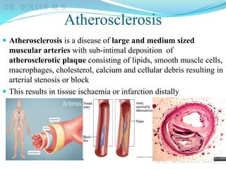
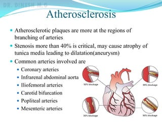
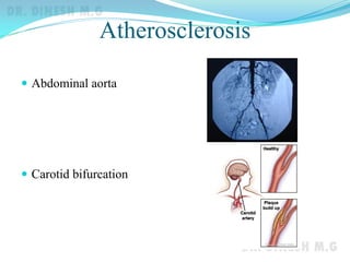
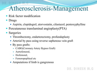
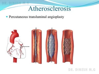
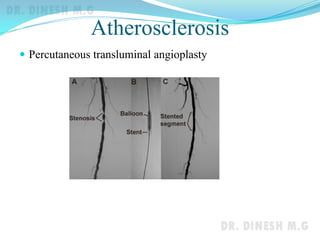
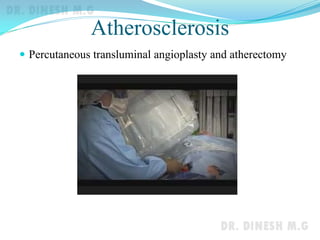
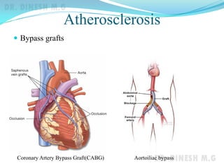
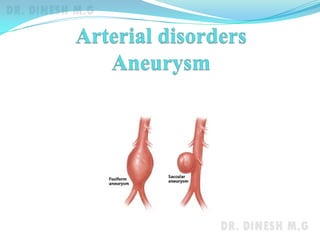
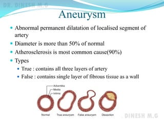
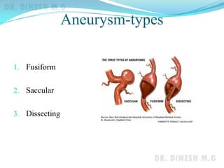
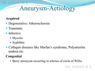
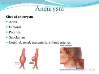
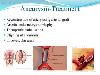
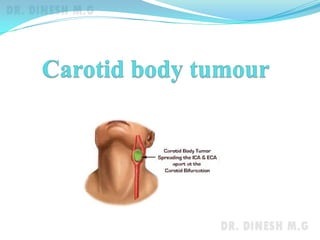
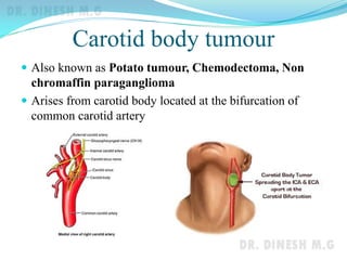
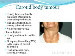
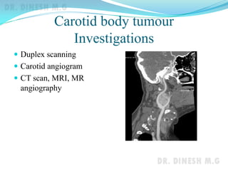
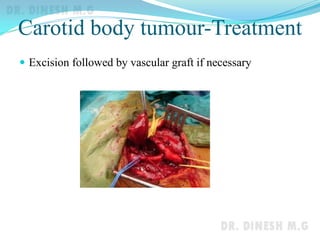
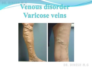
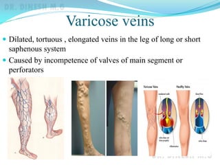
Dr. Dinesh. M.G
Professor of Surgery
J.J.M.M.C.
Davangere
Atherosclerosis
 Atherosclerosis is a disease of large and medium sized
muscular arteries with sub-intimal deposition of
atherosclerotic plaque consisting of lipids, smooth muscle cells,
macrophages, cholesterol, calcium and cellular debris resulting in
arterial stenosis or block
 This results in tissue ischaemia or infarction distally
Atherosclerosis
 Atherosclerotic plaques are more at the regions of
branching of arteries
 Stenosis more than 40% is critical, may cause atrophy of
tunica media leading to dilatation(aneurysm)
 Common arteries involved are
 Coronary arteries
 Infrarenal abdominal aorta
 Iliofemoral arteries
 Carotid bifurcation
 Popliteal arteries
 Mesenteric arteries
Atherosclerosis
Atherosclerosis
 Abdominal aorta
 Carotid bifurcation
Atherosclerosis-Risk factors
Definitive
• Hypercholesterolaemia and
hyperlipidaemia
• Cholesterol > 200mg%
• High LDL > 100mg%
• Low HDL < 35mg%
• Smoking
• Hypertension
• Diabetes mellitus
Relative
• Age-elderly
• Males
• Hypertriglyceridaemia
• Sedentary life, obesity
• Family history
Atherosclerosis-clinical features
 Common after 50 years of age
 Males>Females, Positive family history for
atherosclerosis
 Smoking, hypertension, diabetes and raised cholesterol
levels
 Arterial wall thickening
 Thrill or bruit over carotid, femoral, renal arteries
 Features of ischaemia in the limb
 Claudication
 Ulceration or gangrene of parts of limb
 Feeble or absent pulsations
 Transient ischaemic attacks, chest pain, sudden black out,
post prandial abdominal pain or altered renal function
Atherosclerosis-Investigations
 Blood sugar
 Fasting lipid profile
 ECG
 Echocardiogram
 Tread mill test
 Ultrasound abdomen
 Duplex scanning of suspected arteries
 Angiogram showing site, extent & percentage of block and
collaterals
 DSA
 CT angiogram
 MR angiogram
Atherosclerosis-Management
 Risk factor modification
 Drugs
 Aspirin, clopidogrel, atorvostatin, cilastazol, pentoxyphylline
 Percutaneous transluminal angioplasty(PTA)
 Surgeries
 Thrombectomy, endarterectomy, profundaplasty
 Arterial by pass using reverse saphenous vein graft
 By pass grafts:
 CABG(Coronary Artery Bypass Graft)
 Aortofemoral,
 Iliofemoral,
 Femoropopliteal etc
 Amputations if limb is gangrenous
Atherosclerosis
 Percutaneous transluminal angioplasty
Atherosclerosis
 Percutaneous transluminal angioplasty
Atherosclerosis
 Percutaneous transluminal angioplasty and atherectomy
Atherosclerosis
 Bypass grafts
Coronary Artery Bypass Graft(CABG) Aortoiliac bypass
Aneurysm
 Abnormal permanent dilatation of localised segment of
artery
 Diameter is more than 50% of normal
 Atherosclerosis is most common cause(90%)
 Types
 True : contains all three layers of artery
 False : contains single layer of fibrous tissue as a wall
Aneurysm-types
1. Fusiform
2. Saccular
3. Dissecting
Aneurysm-Aetiology
Acquired
 Degenerative: Atherosclerosis
 Traumatic
 Infective
 Mycotic
 Syphilitic
 Collagen diseases like Marfan’s syndrome, Polyarteritis
nodosa etc.
Congenital
 Berry aneurysm occurring in arteries of circle of Willis
Aneurysm
Sites of aneurysm
 Aorta
 Femoral
 Popliteal
 Subclavian
 Cerebral, renal, mesenteric, splenic arteries
Aneurysm
Complications of aneurysm
 Thrombosis and distal ischaemia
 Embolism and acute arterial occlusion
 Pressure effects
 On bone(erosion)
 Skin
 Veins (oedema)
 Nerves( pain, paraesthesia)
 Stomach(erosion leading to haematemesis)
 Oesophagus (dysphagia)
 Rupture and haemorrhage
 Infection of aneurysm
Aneurysm-clinical features
 Pulsatile swelling
 Thrill & bruit
 Distal oedema due to venous compression
 Altered sensation due to compression of nerves
 Erosion into bones, joints, trachea or oesophagus
 Embolism resulting in gangrene of toes, digits
Investigations
 Duplex scanning
 Angiogram( CT, MRI, DSA)
 Blood sugar, lipid profile, echocardiography
Aneurysm-Treatment
 Reconstruction of artery using arterial graft
 Arterial endoaneurysmorrhaphy
 Therapeutic embolisation
 Clipping of aneurysm
 Endovascular graft
Aneurysm
Open & Endovascular repair
Carotid body tumour
 Also known as Potato tumour, Chemodectoma, Non
chromaffin paraganglioma
 Arises from carotid body located at the bifurcation of
common carotid artery
Carotid body tumour
 Usually benign or locally
malignant. Occasionally
lymphatic spread occurs
 Well encapsulated, hard
creamy yellowish tumour
 Not hormonally active
Clinical features
 Usually unilateral in middle
age
 Firm pulsatile swelling(75%)
in the region of carotid
bifurcation
 Head ache, neck pain,
syncope, dysphagia
Carotid body tumour
Investigations
 Duplex scanning
 Carotid angiogram
 CT scan, MRI, MR
angiography
Carotid body tumour-Treatment
 Excision followed by vascular graft if necessary
Varicose veins
 Dilated, tortuous , elongated veins in the leg of long or short
saphenous system
 Caused by incompetence of valves of main segment or
perforators
Varicose veins
Varicose veins
Aetiology
 Primary
 Congenital weakness of wall of veins or absence of valves
 Prolonged standing
 Congenital AV malformation
 Secondary
 Pregnancy
 Pelvic tumours
 Traumatic arteriovenous fistula
Varicose veins-clinical features
 Dull ache in calf in the
evening
 Bleeding
 Eczema
 Pigmentation
 Varicose ulcer
Varicose veins
Signs
 Brodie-Trendelenburg test for SFJ and perforator incompetence
 Triple tourniquet test
 Perthe’ test for patency of deep veins
Complications
 Haemorrhage
 Pigmentation, eczema and ulcer
 Periosteitis
 Equinus deformity
 Thrombophlebitis
Varicose veins
Investigations
 Duplex scanning of leg veins
Treatment
 Elastic stockings
 Injection sclerotherapy for
below knee varicosities
 Juxtafemoral ligation of great
saphenous
vein(Trendelenburg’s
operation)
 Stripping of veins
 Subfascial perforator ligation
 Endovenous laser ablation
Varicose veins
Endovascular laser treatment
Thank you

Arterial and venous disorders

  • 1.
    Dr. Dinesh. M.G Professorof Surgery J.J.M.M.C. Davangere
  • 2.
    Atherosclerosis  Atherosclerosis isa disease of large and medium sized muscular arteries with sub-intimal deposition of atherosclerotic plaque consisting of lipids, smooth muscle cells, macrophages, cholesterol, calcium and cellular debris resulting in arterial stenosis or block  This results in tissue ischaemia or infarction distally
  • 3.
    Atherosclerosis  Atherosclerotic plaquesare more at the regions of branching of arteries  Stenosis more than 40% is critical, may cause atrophy of tunica media leading to dilatation(aneurysm)  Common arteries involved are  Coronary arteries  Infrarenal abdominal aorta  Iliofemoral arteries  Carotid bifurcation  Popliteal arteries  Mesenteric arteries
  • 4.
  • 5.
  • 6.
    Atherosclerosis-Risk factors Definitive • Hypercholesterolaemiaand hyperlipidaemia • Cholesterol > 200mg% • High LDL > 100mg% • Low HDL < 35mg% • Smoking • Hypertension • Diabetes mellitus Relative • Age-elderly • Males • Hypertriglyceridaemia • Sedentary life, obesity • Family history
  • 7.
    Atherosclerosis-clinical features  Commonafter 50 years of age  Males>Females, Positive family history for atherosclerosis  Smoking, hypertension, diabetes and raised cholesterol levels  Arterial wall thickening  Thrill or bruit over carotid, femoral, renal arteries  Features of ischaemia in the limb  Claudication  Ulceration or gangrene of parts of limb  Feeble or absent pulsations  Transient ischaemic attacks, chest pain, sudden black out, post prandial abdominal pain or altered renal function
  • 8.
    Atherosclerosis-Investigations  Blood sugar Fasting lipid profile  ECG  Echocardiogram  Tread mill test  Ultrasound abdomen  Duplex scanning of suspected arteries  Angiogram showing site, extent & percentage of block and collaterals  DSA  CT angiogram  MR angiogram
  • 9.
    Atherosclerosis-Management  Risk factormodification  Drugs  Aspirin, clopidogrel, atorvostatin, cilastazol, pentoxyphylline  Percutaneous transluminal angioplasty(PTA)  Surgeries  Thrombectomy, endarterectomy, profundaplasty  Arterial by pass using reverse saphenous vein graft  By pass grafts:  CABG(Coronary Artery Bypass Graft)  Aortofemoral,  Iliofemoral,  Femoropopliteal etc  Amputations if limb is gangrenous
  • 10.
  • 11.
  • 12.
  • 13.
    Atherosclerosis  Bypass grafts CoronaryArtery Bypass Graft(CABG) Aortoiliac bypass
  • 15.
    Aneurysm  Abnormal permanentdilatation of localised segment of artery  Diameter is more than 50% of normal  Atherosclerosis is most common cause(90%)  Types  True : contains all three layers of artery  False : contains single layer of fibrous tissue as a wall
  • 16.
  • 17.
    Aneurysm-Aetiology Acquired  Degenerative: Atherosclerosis Traumatic  Infective  Mycotic  Syphilitic  Collagen diseases like Marfan’s syndrome, Polyarteritis nodosa etc. Congenital  Berry aneurysm occurring in arteries of circle of Willis
  • 18.
    Aneurysm Sites of aneurysm Aorta  Femoral  Popliteal  Subclavian  Cerebral, renal, mesenteric, splenic arteries
  • 19.
    Aneurysm Complications of aneurysm Thrombosis and distal ischaemia  Embolism and acute arterial occlusion  Pressure effects  On bone(erosion)  Skin  Veins (oedema)  Nerves( pain, paraesthesia)  Stomach(erosion leading to haematemesis)  Oesophagus (dysphagia)  Rupture and haemorrhage  Infection of aneurysm
  • 20.
    Aneurysm-clinical features  Pulsatileswelling  Thrill & bruit  Distal oedema due to venous compression  Altered sensation due to compression of nerves  Erosion into bones, joints, trachea or oesophagus  Embolism resulting in gangrene of toes, digits Investigations  Duplex scanning  Angiogram( CT, MRI, DSA)  Blood sugar, lipid profile, echocardiography
  • 21.
    Aneurysm-Treatment  Reconstruction ofartery using arterial graft  Arterial endoaneurysmorrhaphy  Therapeutic embolisation  Clipping of aneurysm  Endovascular graft
  • 22.
  • 24.
    Carotid body tumour Also known as Potato tumour, Chemodectoma, Non chromaffin paraganglioma  Arises from carotid body located at the bifurcation of common carotid artery
  • 25.
    Carotid body tumour Usually benign or locally malignant. Occasionally lymphatic spread occurs  Well encapsulated, hard creamy yellowish tumour  Not hormonally active Clinical features  Usually unilateral in middle age  Firm pulsatile swelling(75%) in the region of carotid bifurcation  Head ache, neck pain, syncope, dysphagia
  • 26.
    Carotid body tumour Investigations Duplex scanning  Carotid angiogram  CT scan, MRI, MR angiography
  • 27.
    Carotid body tumour-Treatment Excision followed by vascular graft if necessary
  • 29.
    Varicose veins  Dilated,tortuous , elongated veins in the leg of long or short saphenous system  Caused by incompetence of valves of main segment or perforators
  • 30.
  • 31.
    Varicose veins Aetiology  Primary Congenital weakness of wall of veins or absence of valves  Prolonged standing  Congenital AV malformation  Secondary  Pregnancy  Pelvic tumours  Traumatic arteriovenous fistula
  • 32.
    Varicose veins-clinical features Dull ache in calf in the evening  Bleeding  Eczema  Pigmentation  Varicose ulcer
  • 33.
    Varicose veins Signs  Brodie-Trendelenburgtest for SFJ and perforator incompetence  Triple tourniquet test  Perthe’ test for patency of deep veins Complications  Haemorrhage  Pigmentation, eczema and ulcer  Periosteitis  Equinus deformity  Thrombophlebitis
  • 34.
    Varicose veins Investigations  Duplexscanning of leg veins Treatment  Elastic stockings  Injection sclerotherapy for below knee varicosities  Juxtafemoral ligation of great saphenous vein(Trendelenburg’s operation)  Stripping of veins  Subfascial perforator ligation  Endovenous laser ablation
  • 35.
  • 36.