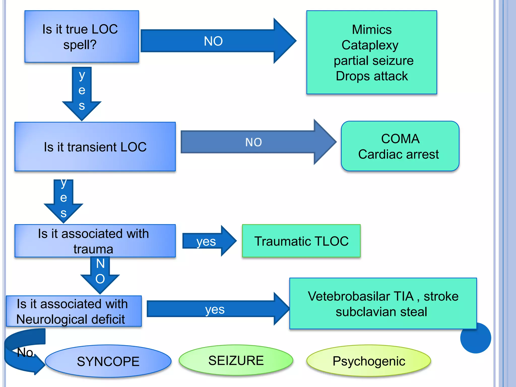
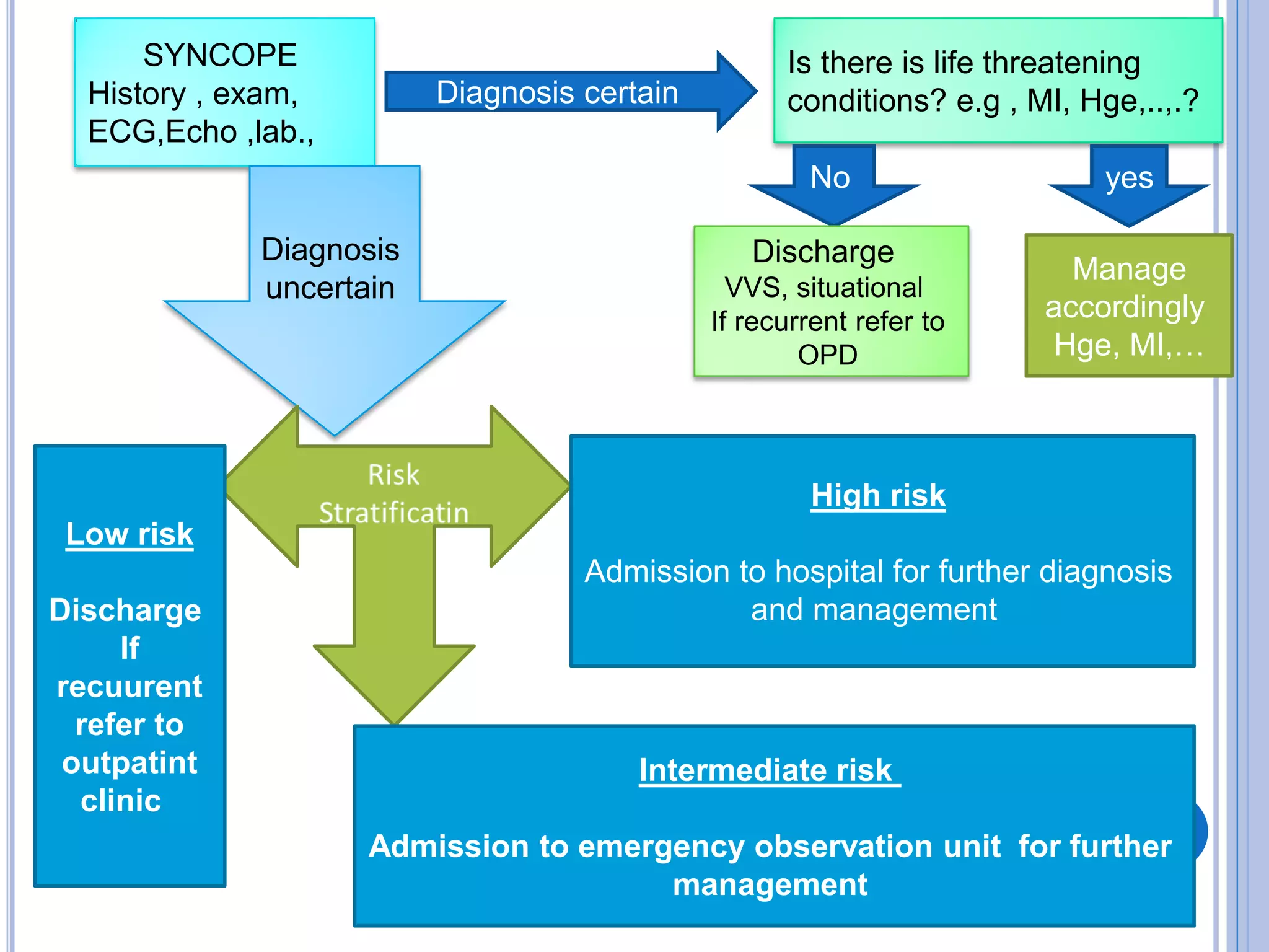
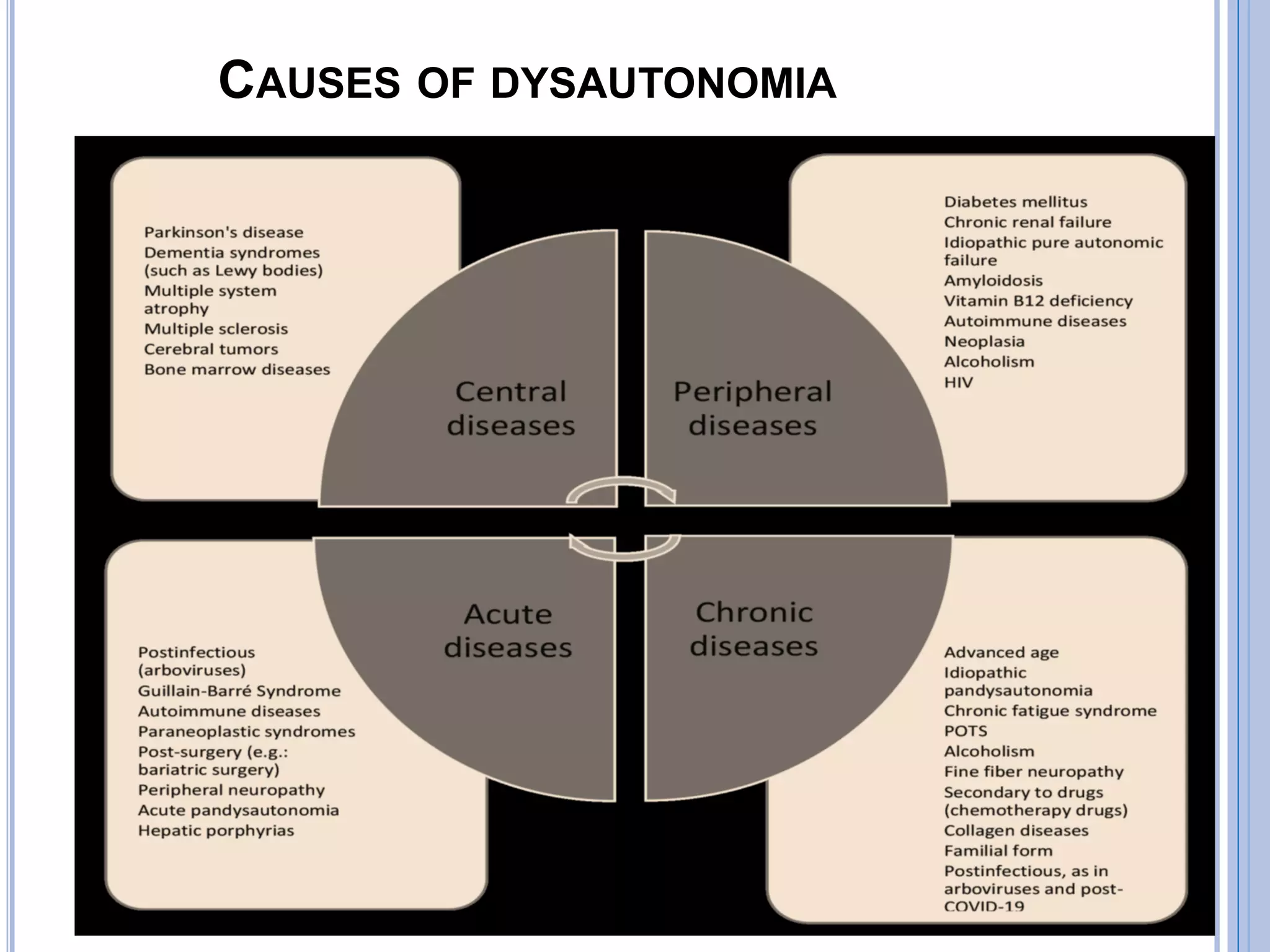
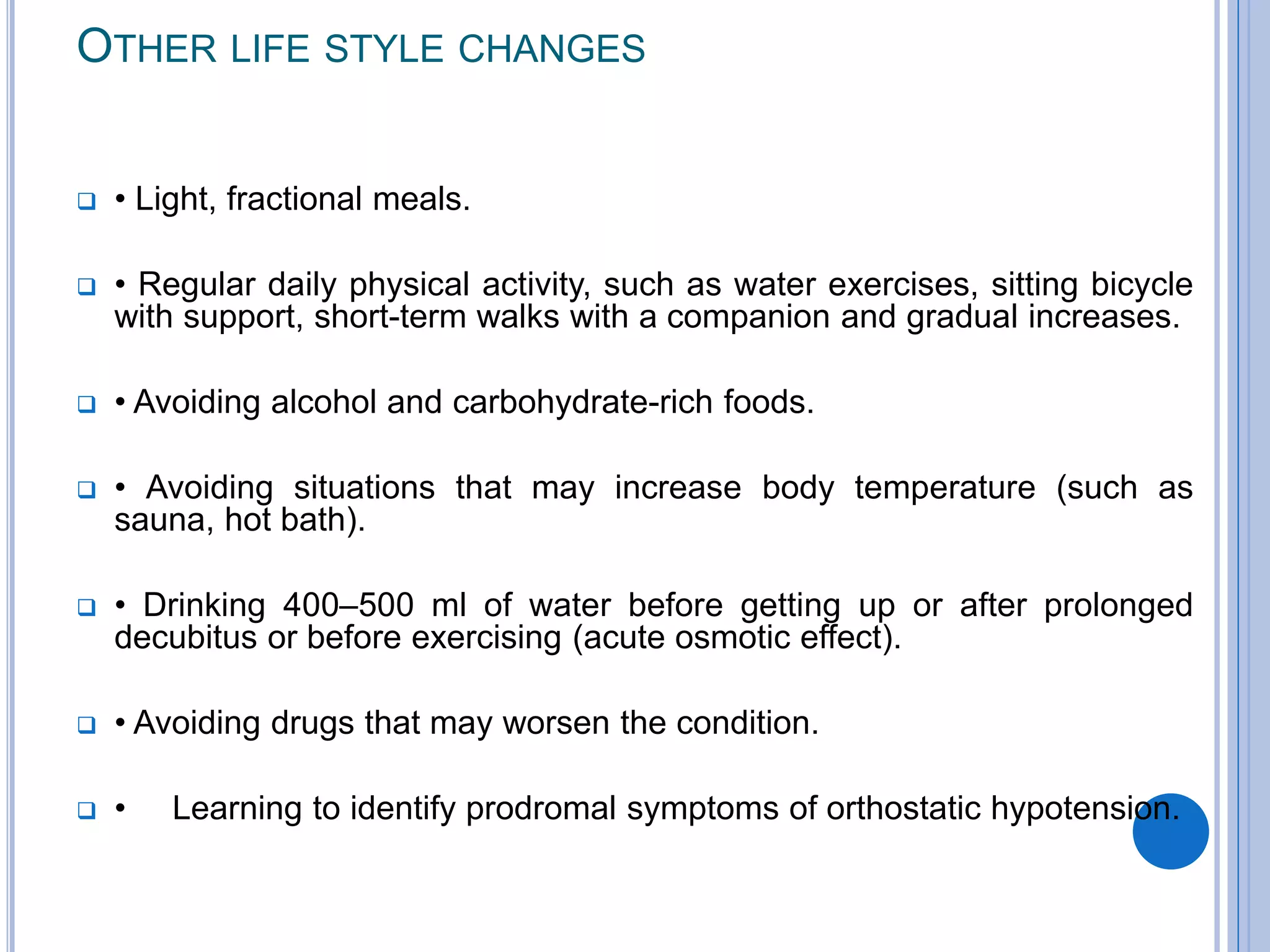
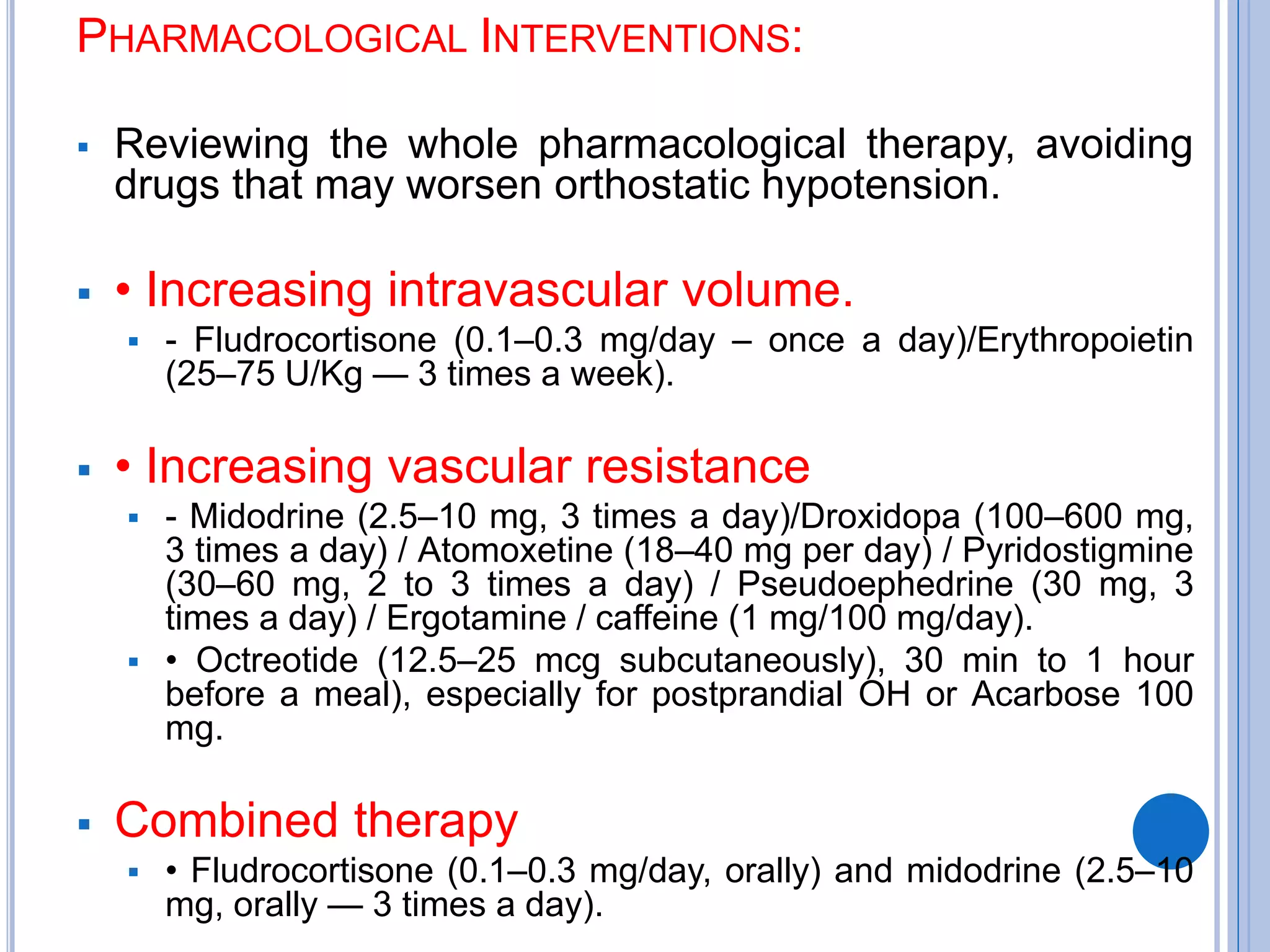
APPROACH TO (TLOC) TRANSIENT LOSS OF
CONSCIOUSNESS
SYNCOPE AND SEIZURE
Dr. Mohamed Habeel
Lecturer of Neurology
 Consciousness
 has two domains, wakefulness (arousal) and awareness. Arousal is
governed by the integrated function of different parts of the central
nervous system including the reticular activating system (RAS)
located within the upper and posterior part of brainstem, thalamus
and the major neuronal projections to bilateral cerebral hemisphere.
 LOC results from dysfunction of either upper brain stem or bilateral
cerebral hemisphere and thalamus. It could be transient (which is the
focus of this discussion) or more prolonged (e.g. coma).
 The two main groups of TLOC are ‗TLOC due to head trauma‘ and
‗non-traumatic TLOC‘ (which is covered in this discussion).
 The non-traumatic group of TLOC is :
 Syncope: is sudden cessation cerebral perfusion.
 Epileptic seizures: Abnormal excessive brain activity.
 Psychogenic TLOC: Psychological process of conversion.
Is it true LOC
spell? NO
Mimics
Cataplexy
partial seizure
Drops attack
y
e
s
Is it transient LOC
COMA
Cardiac arrest
y
e
s
Is it associated with
trauma
Traumatic TLOC
N
O
Is it associated with
Neurological deficit
yes
yes
Vetebrobasilar TIA , stroke
subclavian steal
SYNCOPE SEIZURE Psychogenic
No
 Seizures
 are abnormal, paroxysmal synchronous discharges of cortical
neurons resulting in abrupt neurologic manifestations that depend on
the area of the brain involved.
Seizures are classified as :
 generalized when they involve the entire brain, producing an abrupt
alteration in consciousness with or without motor manifestations.
 Focal seizures involve a single brain region, causing limited
dysfunction, which may include motor manifestations or non motor
manifestations such as sensory disturbances, behavioral/cognitive
changes, automatisms, or abnormal speech.
 Focal seizures are further subdivided into those with preserved
consciousness and those without.
 Status epilepticus is
 defined as 5 minutes or more of continuous clinical and/or
electrographic seizure activity, or repeated seizures between which
the patient fails to return to baseline.
 Syncope
 is defined as a symptom that present with an abrupt TLOC associated with
inability to maintain postural tone, with rapid and spontaneous recovery and
return to baseline neurologic function within seconds .
 There should not be clinical features of other non syncope causes of loss of
consciousness, such as seizure, antecedent head trauma, or apparent LOC (ie,
pseudo syncope)
 Relevant terms and definitions :
 TLOC:
 is defined as a state of real or apparent LOC with loss of awareness,
characterized by amnesia for the period of unconsciousness, abnormal motor
control, loss of responsiveness, and a short duration (self-limited).
 Presyncope:
 The symptoms before syncope. These symptoms could include extreme
lightheadedness; visual sensations, such as ―tunnel vision‖ or ―graying out‖; and
variable degrees of altered consciousness without complete loss of
consciousness. Presyncope could progress to syncope, or it could abort without
syncope.
 It should be considered a spectrum of the same symptom and approached in a
similar fashion .
 Psychogenic pseudosyncope:
 A syndrome of apparent but not true loss of consciousness that may occur in the
absence of identifiable cardiac, reflex, neurological, or metabolic causes.
BEFORE A SPELL
Syncope Seizure
Sweating, lightheadedness,
graying of vision and/or
nausea
Common Rare
Occurring out of sleep Rare Occasional
Aura (déjà vu or olfactory
hallucination, unilateral
symptoms)
Rare Common
Identifiable Trigger (change
in position, prolonged
standing, emotion, Valsalva,
exercise)
Common Rare
DURING A SPELL
syncope Seizure
Duration of loss of
consciousness
≤ 10 secs ≥60 secs
Movements A few rhythmic
jerks of the
limbs, lasting
<5-10seconds
Prolonged stiffening
of the limbs (tonic),
transitioning to
rhythmic jerking of
the limbs (clonic),
lasting ~30-90
seconds
Automatic behavior
(lip smacking,
picking, patting)
Occasional Common (focal
dyscognitive
seizures)
Tongue biting
(lateral)
Rare Occasional
Pallor Common Rare
Cyanosis Rare Common
AFTER A SPELL
syncope seizure
Frothing/ hypersalivation
/vomiting
Rare common
Confusion/
disorientation
Rare <30
seconds
common
Diffuse muscle pain Rare /brief common
Creatinine kinase (CK)
elevation
Rare common
Focal neurological
signs
Rare occasional
Incontinence Rare occasional
Headache Rare common
Amnesia for the event Rare /less common common
 Syncope is brief loss of consciousness caused by a
sudden reduction of cerebral blood flow.
 Presyncope refers to the sensation of impending loss of
consciousness, but the patient does not actually pass
out.
 Syncope is a cardiovascular symptom. Regardless of the
underlying causes of the syncope, the final common
pathway is interrupted blood flow for a short period of
time (e.g. 8-10 seconds) to both cerebral cortices or to
the brainstem ‗RAS‘ resulting in transient loss of
consciousness.
 Cerebral Autoregulation is the ability of the brain to
maintain cerebral blood flow relatively constant (40
to 60 ml/100 g/min) over a wide range (50 to 150
mm Hg) of arterial pressures.
 Syncope accounts for common ED visits and
hospital admission .
 Although most common causes of syncope are
benign, identification of life threatening causes
associated with syncope is sometimes challenging.
 The task is to confirm that the event was syncope
and to discover the cause of the syncopal attack.
 The pathophysiologic classification of the syncope (which
centers on a fall in systemic blood pressure and
decrease in global cerebral blood flow) defines syncope
in three classes :
 Reflex mediated syncope
 Orthostatic hypotension
 Cardiac syncope
Reflex mediated syncope
Secondary to pain ,
blood phobia, pain ,
prolonged standing in a
warm place,…
Vasovagal
Micturation , defecation ,
coughing ,….
Situational
Pressure over carotid
sinus , shaving , turning
head,..
Carotid sinus
hypersensitivity
Orthostatic (postural) hypotension
Volume depletion anemia, dehydration,…
Autonomic dysfunction Diabetic polyneuropathy,
parkinsonism, shy
drager,…
Drug induced Vasodilators, diuretic,
SSRI,…
MEDICATIONS THAT CAN CAUSE ORTHOSTATIC
HYPOTENSION
 Dopaminergic drugs >>>>>>>Levodopa, dopamine
agonists
 Tricyclic antidepressants >>>>>>Amitriptyline,
nortriptyline
 Anticholinergics>>>>>>> Atropine
 ↓ Pre-load>>>>>Diuretics, Nitrates
 Phosphodiesterase inhibitors>>>>Sildenafil, vardenafil
 Vasodilators >>>Alpha-1 adrenergic antagonists
 Ca++ blockers
 Other antihypertensives
Cardiac causes
Tachyarrhythmia's (ventricular
tachycardia, fibrillation)
Cardiac arrhythmias
Bradyarrhythmias (heart block,
sick sinus syndrome)
Pump failure (myocardial
infarction, tamponade )
Cardiac outflow failure
Obstruction to left ventricular
outflow (aortic stenosis,
hypertrophic cardiomyopathy)
Obstruction to pulmonary outflow
(pulmonary embolus)
SYNCOPE
History , exam,
ECG,Echo ,lab.,
Diagnosis certain
Is there is life threatening
conditions? e.g , MI, Hge,..,.?
yes
Manage
accordingly
Hge, MI,…
No
Discharge
VVS, situational
If recurrent refer to
OPD
Diagnosis
uncertain
High risk
Admission to hospital for further diagnosis
and management
Intermediate risk
Admission to emergency observation unit for further
management
Low risk
Discharge
If
recuurent
refer to
outpatint
clinic
DYSAUTONOMIA
 covers a range of clinical conditions with different
characteristics and prognoses. They are classified as:
 Cardiovascular Autonomic Neuropathy (CAN) .
 Postural Orthostatic Tachycardia Syndrome
(POTS).
 Chronic Fatigue Syndrome.
 Neurogenic Orthostatic Hypotension (nOH) .
 Carotid Sinus Hypersensitivity Syndrome.
VASOVAGAL SYNDROMES X DYSAUTONOMIA
Vasovagal syndromes are clinical situations that are
different from cardiovascular autonomic neuropathies.
As they do not represent intrinsic diseases in the
Autonomic Nervous System (ANS), resulting from reflex,
transient, benign mechanisms, therefore having a favorable
prognosis.
AUTONOMIC TESTING
 Sweat Test:
 Small plastic capsules are attached to the arms and legs. A machine is
used to stimulate the sweat glands and measure sweat production. You
may feel a slight local burning or tingling during this test.
 Heart Rate during Deep Breathing (HRDB) Test:
 This test measures your heart rate variation during deep breathing. You
will be asked to breathe deeply and steadily at a rate of 6 breaths per
minute guided by a moving green light.
 Valsalva Test:
 During this test you blow into a plastic tube for 15 seconds hard enough
to produce a pressure of 40mm/Hg as indicated by a timer that you will
be asked to observe. This test measures your blood pressure and heart
rate response to Valsalva maneuver. The Valsalva maneuver is
performed by attempting to forcibly exhale while keeping the mouth and
nose closed, usually done by closing one‘s mouth, pinching one‘s nose
shut while pressing out as if blowing up a balloon.
 The Tilt Table Test:
 After lying on a table for the first part of the test, you will be raised to a
nearly upright position. You will be secured to the table so you cannot
fall. The test will measure your blood pressure and pulse from laying to
upright.
CAUSES OF DYSAUTONOMIA
THERAPEUTIC APPROACH TO DYSAUTONOMIA
Neurogenic Orthostatic Hypotension
 Non Pharmacological Interventions:
 Reduced venous retention in lower limbs
• Physical counter-maneuvers (e.g.: crossing the legs, squatting,
moving the legs, hand compression); Slow change in position.
• Compressive clothing (elastic stockings, preferably waistline-high
— 30–40 mmHg and/or abdominal straps 20–30 mmHg).
 Increased central blood volume
• Increasing sodium intake (2-3 g/day or 5–7.5 g NaCl) or higher
doses, in the absence of supine hypertension, edema or heart
failure.
• Increasing water intake (2–3 liters/day).
• Head-up tilt sleeping (20-30 cm).
OTHER LIFE STYLE CHANGES
 • Light, fractional meals.
 • Regular daily physical activity, such as water exercises, sitting bicycle
with support, short-term walks with a companion and gradual increases.
 • Avoiding alcohol and carbohydrate-rich foods.
 • Avoiding situations that may increase body temperature (such as
sauna, hot bath).
 • Drinking 400–500 ml of water before getting up or after prolonged
decubitus or before exercising (acute osmotic effect).
 • Avoiding drugs that may worsen the condition.
 • Learning to identify prodromal symptoms of orthostatic hypotension.
PHARMACOLOGICAL INTERVENTIONS:
 Reviewing the whole pharmacological therapy, avoiding
drugs that may worsen orthostatic hypotension.
 • Increasing intravascular volume.
 - Fludrocortisone (0.1–0.3 mg/day – once a day)/Erythropoietin
(25–75 U/Kg — 3 times a week).
 • Increasing vascular resistance
 - Midodrine (2.5–10 mg, 3 times a day)/Droxidopa (100–600 mg,
3 times a day) / Atomoxetine (18–40 mg per day) / Pyridostigmine
(30–60 mg, 2 to 3 times a day) / Pseudoephedrine (30 mg, 3
times a day) / Ergotamine / caffeine (1 mg/100 mg/day).
 • Octreotide (12.5–25 mcg subcutaneously), 30 min to 1 hour
before a meal), especially for postprandial OH or Acarbose 100
mg.
 Combined therapy
 • Fludrocortisone (0.1–0.3 mg/day, orally) and midodrine (2.5–10
mg, orally — 3 times a day).
THANKS FOR ATTENTION

approach to transient loss of consciousness.pdf

  • 1.
    APPROACH TO (TLOC)TRANSIENT LOSS OF CONSCIOUSNESS SYNCOPE AND SEIZURE Dr. Mohamed Habeel Lecturer of Neurology
  • 3.
     Consciousness  hastwo domains, wakefulness (arousal) and awareness. Arousal is governed by the integrated function of different parts of the central nervous system including the reticular activating system (RAS) located within the upper and posterior part of brainstem, thalamus and the major neuronal projections to bilateral cerebral hemisphere.  LOC results from dysfunction of either upper brain stem or bilateral cerebral hemisphere and thalamus. It could be transient (which is the focus of this discussion) or more prolonged (e.g. coma).  The two main groups of TLOC are ‗TLOC due to head trauma‘ and ‗non-traumatic TLOC‘ (which is covered in this discussion).  The non-traumatic group of TLOC is :  Syncope: is sudden cessation cerebral perfusion.  Epileptic seizures: Abnormal excessive brain activity.  Psychogenic TLOC: Psychological process of conversion.
  • 4.
    Is it trueLOC spell? NO Mimics Cataplexy partial seizure Drops attack y e s Is it transient LOC COMA Cardiac arrest y e s Is it associated with trauma Traumatic TLOC N O Is it associated with Neurological deficit yes yes Vetebrobasilar TIA , stroke subclavian steal SYNCOPE SEIZURE Psychogenic No
  • 5.
     Seizures  areabnormal, paroxysmal synchronous discharges of cortical neurons resulting in abrupt neurologic manifestations that depend on the area of the brain involved. Seizures are classified as :  generalized when they involve the entire brain, producing an abrupt alteration in consciousness with or without motor manifestations.  Focal seizures involve a single brain region, causing limited dysfunction, which may include motor manifestations or non motor manifestations such as sensory disturbances, behavioral/cognitive changes, automatisms, or abnormal speech.  Focal seizures are further subdivided into those with preserved consciousness and those without.  Status epilepticus is  defined as 5 minutes or more of continuous clinical and/or electrographic seizure activity, or repeated seizures between which the patient fails to return to baseline.
  • 6.
     Syncope  isdefined as a symptom that present with an abrupt TLOC associated with inability to maintain postural tone, with rapid and spontaneous recovery and return to baseline neurologic function within seconds .  There should not be clinical features of other non syncope causes of loss of consciousness, such as seizure, antecedent head trauma, or apparent LOC (ie, pseudo syncope)  Relevant terms and definitions :  TLOC:  is defined as a state of real or apparent LOC with loss of awareness, characterized by amnesia for the period of unconsciousness, abnormal motor control, loss of responsiveness, and a short duration (self-limited).  Presyncope:  The symptoms before syncope. These symptoms could include extreme lightheadedness; visual sensations, such as ―tunnel vision‖ or ―graying out‖; and variable degrees of altered consciousness without complete loss of consciousness. Presyncope could progress to syncope, or it could abort without syncope.  It should be considered a spectrum of the same symptom and approached in a similar fashion .  Psychogenic pseudosyncope:  A syndrome of apparent but not true loss of consciousness that may occur in the absence of identifiable cardiac, reflex, neurological, or metabolic causes.
  • 7.
    BEFORE A SPELL SyncopeSeizure Sweating, lightheadedness, graying of vision and/or nausea Common Rare Occurring out of sleep Rare Occasional Aura (déjà vu or olfactory hallucination, unilateral symptoms) Rare Common Identifiable Trigger (change in position, prolonged standing, emotion, Valsalva, exercise) Common Rare
  • 8.
    DURING A SPELL syncopeSeizure Duration of loss of consciousness ≤ 10 secs ≥60 secs Movements A few rhythmic jerks of the limbs, lasting <5-10seconds Prolonged stiffening of the limbs (tonic), transitioning to rhythmic jerking of the limbs (clonic), lasting ~30-90 seconds Automatic behavior (lip smacking, picking, patting) Occasional Common (focal dyscognitive seizures) Tongue biting (lateral) Rare Occasional Pallor Common Rare Cyanosis Rare Common
  • 9.
    AFTER A SPELL syncopeseizure Frothing/ hypersalivation /vomiting Rare common Confusion/ disorientation Rare <30 seconds common Diffuse muscle pain Rare /brief common Creatinine kinase (CK) elevation Rare common Focal neurological signs Rare occasional Incontinence Rare occasional Headache Rare common Amnesia for the event Rare /less common common
  • 10.
     Syncope isbrief loss of consciousness caused by a sudden reduction of cerebral blood flow.  Presyncope refers to the sensation of impending loss of consciousness, but the patient does not actually pass out.  Syncope is a cardiovascular symptom. Regardless of the underlying causes of the syncope, the final common pathway is interrupted blood flow for a short period of time (e.g. 8-10 seconds) to both cerebral cortices or to the brainstem ‗RAS‘ resulting in transient loss of consciousness.
  • 11.
     Cerebral Autoregulationis the ability of the brain to maintain cerebral blood flow relatively constant (40 to 60 ml/100 g/min) over a wide range (50 to 150 mm Hg) of arterial pressures.
  • 12.
     Syncope accountsfor common ED visits and hospital admission .  Although most common causes of syncope are benign, identification of life threatening causes associated with syncope is sometimes challenging.  The task is to confirm that the event was syncope and to discover the cause of the syncopal attack.
  • 13.
     The pathophysiologicclassification of the syncope (which centers on a fall in systemic blood pressure and decrease in global cerebral blood flow) defines syncope in three classes :  Reflex mediated syncope  Orthostatic hypotension  Cardiac syncope
  • 14.
    Reflex mediated syncope Secondaryto pain , blood phobia, pain , prolonged standing in a warm place,… Vasovagal Micturation , defecation , coughing ,…. Situational Pressure over carotid sinus , shaving , turning head,.. Carotid sinus hypersensitivity
  • 15.
    Orthostatic (postural) hypotension Volumedepletion anemia, dehydration,… Autonomic dysfunction Diabetic polyneuropathy, parkinsonism, shy drager,… Drug induced Vasodilators, diuretic, SSRI,…
  • 16.
    MEDICATIONS THAT CANCAUSE ORTHOSTATIC HYPOTENSION  Dopaminergic drugs >>>>>>>Levodopa, dopamine agonists  Tricyclic antidepressants >>>>>>Amitriptyline, nortriptyline  Anticholinergics>>>>>>> Atropine  ↓ Pre-load>>>>>Diuretics, Nitrates  Phosphodiesterase inhibitors>>>>Sildenafil, vardenafil  Vasodilators >>>Alpha-1 adrenergic antagonists  Ca++ blockers  Other antihypertensives
  • 17.
    Cardiac causes Tachyarrhythmia's (ventricular tachycardia,fibrillation) Cardiac arrhythmias Bradyarrhythmias (heart block, sick sinus syndrome) Pump failure (myocardial infarction, tamponade ) Cardiac outflow failure Obstruction to left ventricular outflow (aortic stenosis, hypertrophic cardiomyopathy) Obstruction to pulmonary outflow (pulmonary embolus)
  • 18.
    SYNCOPE History , exam, ECG,Echo,lab., Diagnosis certain Is there is life threatening conditions? e.g , MI, Hge,..,.? yes Manage accordingly Hge, MI,… No Discharge VVS, situational If recurrent refer to OPD Diagnosis uncertain High risk Admission to hospital for further diagnosis and management Intermediate risk Admission to emergency observation unit for further management Low risk Discharge If recuurent refer to outpatint clinic
  • 21.
    DYSAUTONOMIA  covers arange of clinical conditions with different characteristics and prognoses. They are classified as:  Cardiovascular Autonomic Neuropathy (CAN) .  Postural Orthostatic Tachycardia Syndrome (POTS).  Chronic Fatigue Syndrome.  Neurogenic Orthostatic Hypotension (nOH) .  Carotid Sinus Hypersensitivity Syndrome.
  • 22.
    VASOVAGAL SYNDROMES XDYSAUTONOMIA Vasovagal syndromes are clinical situations that are different from cardiovascular autonomic neuropathies. As they do not represent intrinsic diseases in the Autonomic Nervous System (ANS), resulting from reflex, transient, benign mechanisms, therefore having a favorable prognosis.
  • 23.
    AUTONOMIC TESTING  SweatTest:  Small plastic capsules are attached to the arms and legs. A machine is used to stimulate the sweat glands and measure sweat production. You may feel a slight local burning or tingling during this test.  Heart Rate during Deep Breathing (HRDB) Test:  This test measures your heart rate variation during deep breathing. You will be asked to breathe deeply and steadily at a rate of 6 breaths per minute guided by a moving green light.  Valsalva Test:  During this test you blow into a plastic tube for 15 seconds hard enough to produce a pressure of 40mm/Hg as indicated by a timer that you will be asked to observe. This test measures your blood pressure and heart rate response to Valsalva maneuver. The Valsalva maneuver is performed by attempting to forcibly exhale while keeping the mouth and nose closed, usually done by closing one‘s mouth, pinching one‘s nose shut while pressing out as if blowing up a balloon.  The Tilt Table Test:  After lying on a table for the first part of the test, you will be raised to a nearly upright position. You will be secured to the table so you cannot fall. The test will measure your blood pressure and pulse from laying to upright.
  • 24.
  • 25.
    THERAPEUTIC APPROACH TODYSAUTONOMIA Neurogenic Orthostatic Hypotension  Non Pharmacological Interventions:  Reduced venous retention in lower limbs • Physical counter-maneuvers (e.g.: crossing the legs, squatting, moving the legs, hand compression); Slow change in position. • Compressive clothing (elastic stockings, preferably waistline-high — 30–40 mmHg and/or abdominal straps 20–30 mmHg).  Increased central blood volume • Increasing sodium intake (2-3 g/day or 5–7.5 g NaCl) or higher doses, in the absence of supine hypertension, edema or heart failure. • Increasing water intake (2–3 liters/day). • Head-up tilt sleeping (20-30 cm).
  • 26.
    OTHER LIFE STYLECHANGES  • Light, fractional meals.  • Regular daily physical activity, such as water exercises, sitting bicycle with support, short-term walks with a companion and gradual increases.  • Avoiding alcohol and carbohydrate-rich foods.  • Avoiding situations that may increase body temperature (such as sauna, hot bath).  • Drinking 400–500 ml of water before getting up or after prolonged decubitus or before exercising (acute osmotic effect).  • Avoiding drugs that may worsen the condition.  • Learning to identify prodromal symptoms of orthostatic hypotension.
  • 27.
    PHARMACOLOGICAL INTERVENTIONS:  Reviewingthe whole pharmacological therapy, avoiding drugs that may worsen orthostatic hypotension.  • Increasing intravascular volume.  - Fludrocortisone (0.1–0.3 mg/day – once a day)/Erythropoietin (25–75 U/Kg — 3 times a week).  • Increasing vascular resistance  - Midodrine (2.5–10 mg, 3 times a day)/Droxidopa (100–600 mg, 3 times a day) / Atomoxetine (18–40 mg per day) / Pyridostigmine (30–60 mg, 2 to 3 times a day) / Pseudoephedrine (30 mg, 3 times a day) / Ergotamine / caffeine (1 mg/100 mg/day).  • Octreotide (12.5–25 mcg subcutaneously), 30 min to 1 hour before a meal), especially for postprandial OH or Acarbose 100 mg.  Combined therapy  • Fludrocortisone (0.1–0.3 mg/day, orally) and midodrine (2.5–10 mg, orally — 3 times a day).
  • 28.