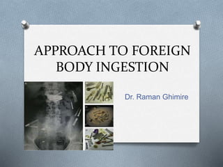
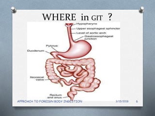
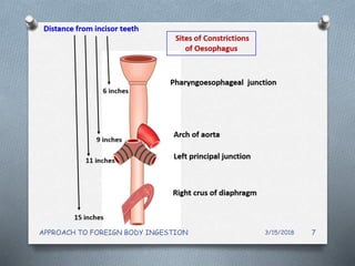
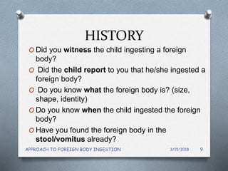
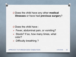
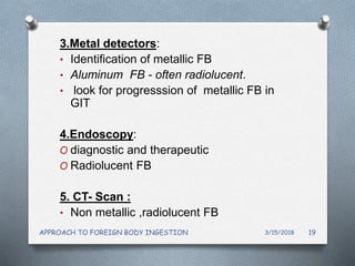
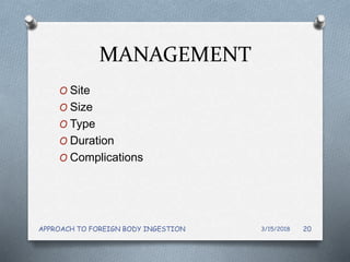
APPROACH TO FOREIGN
BODY INGESTION
Dr. Raman Ghimire
OVERVIEW
O Introduction
O What are FB ?
O Where FB are lodged?
O Who are at risk ?
O Clinical features
O Investigtions
O Management
O MCQs
O References
3/15/2018APPROACH TO FOREIGN BODY INGESTION 2
INTRODUCTION
O FBs of GIT are not so dangerous as air
way foreign bodies.
O very common problem among children
and elderly
O 80-90 % pass harmlessly
O 10–20% will require endoscopic
intervention
O < 1% will require surgery
3/15/2018APPROACH TO FOREIGN BODY INGESTION 3
WHAT ?
O Coin (45%)
O Fish bone (12%)
O Sharp (8%)
(nails, pins,blades )
O batteries. Keys, small toys
O Food bolus impactions
O Tooth brush : eating disorders
O Dentures partial , Teeth –artificial or natural
O Meat or chicken bone
3/15/2018APPROACH TO FOREIGN BODY INGESTION 4
WHO?
O Pediatric patients (80%)
AGE: 6 months - 4 years.
O Psychiatric patients
O Patients with underlying GI disorders
(malignancy,strictures,achalasia)
O Edentulous elderly patients
3/15/2018APPROACH TO FOREIGN BODY INGESTION 5
WHERE in GIT ?
3/15/2018APPROACH TO FOREIGN BODY INGESTION 6
3/15/2018APPROACH TO FOREIGN BODY INGESTION 7
70%
15%
15%
3/15/2018APPROACH TO FOREIGN BODY INGESTION 8
HISTORY
O Did you witness the child ingesting a foreign
body?
O Did the child report to you that he/she ingested a
foreign body?
O Do you know what the foreign body is? (size,
shape, identity)
O Do you know when the child ingested the foreign
body?
O Have you found the foreign body in the
stool/vomitus already?
3/15/2018APPROACH TO FOREIGN BODY INGESTION 9
O Does the child have any other medical
illnesses or have had previous surgery?
O Does the child have :
 Fever, abdominal pain, or vomiting?
 Stools? If so, how many times, what
color?
 Difficulty breathing ?
3/15/2018APPROACH TO FOREIGN BODY INGESTION 10
CLINICAL FEATURES
Esophageal foreign body symptoms
O Dysphagia
O Food refusal, weight loss
O Drooling, gagging
O Emesis/hematemesis
O Foreign body sensation
O Chest pain, sore throat
O Noisey breathing, difficulty breathing, cough,
O Unexplained fever
3/15/2018APPROACH TO FOREIGN BODY INGESTION 11
Stomach/lower GI tract foreign bodies
O Abdominal distention
O Abdominal pain
O vomiting
O Hematochezia
O Unexplained fever
3/15/2018APPROACH TO FOREIGN BODY INGESTION 12
PHYSICAL EXAMINATION
O Usually unremarkable
O Oral cavity/oropharynx/Neck :
• Drooling or pooling of secretions
• Impacted FB : hypopharynx
• Crepitus ,swelling : neck
O Per Abdomen: tenderness, rigidity,distention
(perforation/ obstruction)
O Chest : wheeze , stridor
3/15/2018APPROACH TO FOREIGN BODY INGESTION 13
INVESTIGATIONS
1. Lab investigations
• not necessary
• infections
• complications
3/15/2018APPROACH TO FOREIGN BODY INGESTION 14
2.Neck /Chest/Abdominal Radiography
O Initial Investigation of choice
O Most ingested foreign bodies are radiopaque
(60%)
Determines :
O Presence
O Type
O location of the foreign body.
O Identifying possible complications
3/15/2018APPROACH TO FOREIGN BODY INGESTION 15
3/15/2018APPROACH TO FOREIGN BODY INGESTION 16
3/15/2018
APPROACH TO FOREIGN BODY INGESTION
17
3/15/2018
APPROACH TO FOREIGN BODY INGESTION
18
3.Metal detectors:
• Identification of metallic FB
• Aluminum FB - often radiolucent.
• look for progresssion of metallic FB in
GIT
4.Endoscopy:
O diagnostic and therapeutic
O Radiolucent FB
5. CT- Scan :
• Non metallic ,radiolucent FB
3/15/2018APPROACH TO FOREIGN BODY INGESTION 19
MANAGEMENT
O Site
O Size
O Type
O Duration
O Complications
3/15/2018APPROACH TO FOREIGN BODY INGESTION 20
Removed immediately:
O Batteries in esophagus
O Length> 10cm
O Any object
Child: >1cm X 3cm
Adult: > 2cm X 5 cm
O Symptomatic, f/o perforation
O Esophageal FB (> 24 hrs at presentation)
O Sharp ,pointed FB
O Batteries remaining in the stomach (> 48 h )
3/15/2018APPROACH TO FOREIGN BODY INGESTION 21
Conservative management
(wait and watch )
O Coins in the esophagus : 12–24 Hrs
O Any asymptomatic blunt object : 1week
Child: <1cm X 3cm
Adult: < 2cm X 5 cm
O Asymptomatic Disk batteries and
cylindrical batteries in stomach : upto 48
Hrs.
O Progression ; serial xrays , metal detector
O laxatives to increase GI motility
3/15/2018APPROACH TO FOREIGN BODY INGESTION 22
Methods of removal
1.Endoscopy
O Procedure of choice
O Minimally invasive
O Success rate: 90-100%
O Can retrive FB up to 2nd part of duodenum
(FB beyond that often passes spontaneously)
3/15/2018APPROACH TO FOREIGN BODY INGESTION 23
2. Surgical
A. Cervical esophagostomy
B. Thoracotomy
C. laparotomy
INDICATIONS:
O Evidence of perforation, hemorrhage, fistula
formation, obstruction.
O FB fail to progress (lie beyond stomach)
O FB not retrieved endoscopically.
3/15/2018APPROACH TO FOREIGN BODY INGESTION 24
MCQs
1.Which is the most common FB in
esophagus
A. Button batteries
B. Coin
C. Fish bone
D. chicken bone
3/15/2018APPROACH TO FOREIGN BODY INGESTION 25
2. Where is the FB ?
AP: sagittal (end on) Lat: coronal (face )
oesophagus
3/15/2018APPROACH TO FOREIGN BODY INGESTION
26
REFERENCES
O Current opinion in pediatrics ;Foreign bodies in GIT
O African Journal of Emergency Medicine Volume 5,
Issue 4 December 2015, Pages 176-180;
Investigation and management of foreign body
ingestion in children at a major paediatric trauma unit
in South Africa
O Conners GP, Hadley JA. Esophageal coin with an
unusual radiographic appearance. Pediatric Emerg
Car. 2005;21:667-669.
O Raney LH, Losek JD. Child with esophageal coin and
atypical radiograph. J Emerg Med 2008;34:63-66
O Srilakshmi Narra, MD and Firas H. Al-Kawas MD;
The Importance of Preparation and Innovation in the
Endoscopic Management of Esophageal Foreign
Bodies
3/15/2018APPROACH TO FOREIGN BODY INGESTION 27
OTHANK YOU
3/15/2018APPROACH TO FOREIGN BODY INGESTION 28

Approach to foreign body ingestion

  • 1.
    APPROACH TO FOREIGN BODYINGESTION Dr. Raman Ghimire
  • 2.
    OVERVIEW O Introduction O Whatare FB ? O Where FB are lodged? O Who are at risk ? O Clinical features O Investigtions O Management O MCQs O References 3/15/2018APPROACH TO FOREIGN BODY INGESTION 2
  • 3.
    INTRODUCTION O FBs ofGIT are not so dangerous as air way foreign bodies. O very common problem among children and elderly O 80-90 % pass harmlessly O 10–20% will require endoscopic intervention O < 1% will require surgery 3/15/2018APPROACH TO FOREIGN BODY INGESTION 3
  • 4.
    WHAT ? O Coin(45%) O Fish bone (12%) O Sharp (8%) (nails, pins,blades ) O batteries. Keys, small toys O Food bolus impactions O Tooth brush : eating disorders O Dentures partial , Teeth –artificial or natural O Meat or chicken bone 3/15/2018APPROACH TO FOREIGN BODY INGESTION 4
  • 5.
    WHO? O Pediatric patients(80%) AGE: 6 months - 4 years. O Psychiatric patients O Patients with underlying GI disorders (malignancy,strictures,achalasia) O Edentulous elderly patients 3/15/2018APPROACH TO FOREIGN BODY INGESTION 5
  • 6.
    WHERE in GIT? 3/15/2018APPROACH TO FOREIGN BODY INGESTION 6
  • 7.
  • 8.
  • 9.
    HISTORY O Did youwitness the child ingesting a foreign body? O Did the child report to you that he/she ingested a foreign body? O Do you know what the foreign body is? (size, shape, identity) O Do you know when the child ingested the foreign body? O Have you found the foreign body in the stool/vomitus already? 3/15/2018APPROACH TO FOREIGN BODY INGESTION 9
  • 10.
    O Does thechild have any other medical illnesses or have had previous surgery? O Does the child have :  Fever, abdominal pain, or vomiting?  Stools? If so, how many times, what color?  Difficulty breathing ? 3/15/2018APPROACH TO FOREIGN BODY INGESTION 10
  • 11.
    CLINICAL FEATURES Esophageal foreignbody symptoms O Dysphagia O Food refusal, weight loss O Drooling, gagging O Emesis/hematemesis O Foreign body sensation O Chest pain, sore throat O Noisey breathing, difficulty breathing, cough, O Unexplained fever 3/15/2018APPROACH TO FOREIGN BODY INGESTION 11
  • 12.
    Stomach/lower GI tractforeign bodies O Abdominal distention O Abdominal pain O vomiting O Hematochezia O Unexplained fever 3/15/2018APPROACH TO FOREIGN BODY INGESTION 12
  • 13.
    PHYSICAL EXAMINATION O Usuallyunremarkable O Oral cavity/oropharynx/Neck : • Drooling or pooling of secretions • Impacted FB : hypopharynx • Crepitus ,swelling : neck O Per Abdomen: tenderness, rigidity,distention (perforation/ obstruction) O Chest : wheeze , stridor 3/15/2018APPROACH TO FOREIGN BODY INGESTION 13
  • 14.
    INVESTIGATIONS 1. Lab investigations •not necessary • infections • complications 3/15/2018APPROACH TO FOREIGN BODY INGESTION 14
  • 15.
    2.Neck /Chest/Abdominal Radiography OInitial Investigation of choice O Most ingested foreign bodies are radiopaque (60%) Determines : O Presence O Type O location of the foreign body. O Identifying possible complications 3/15/2018APPROACH TO FOREIGN BODY INGESTION 15
  • 16.
  • 17.
  • 18.
  • 19.
    3.Metal detectors: • Identificationof metallic FB • Aluminum FB - often radiolucent. • look for progresssion of metallic FB in GIT 4.Endoscopy: O diagnostic and therapeutic O Radiolucent FB 5. CT- Scan : • Non metallic ,radiolucent FB 3/15/2018APPROACH TO FOREIGN BODY INGESTION 19
  • 20.
    MANAGEMENT O Site O Size OType O Duration O Complications 3/15/2018APPROACH TO FOREIGN BODY INGESTION 20
  • 21.
    Removed immediately: O Batteriesin esophagus O Length> 10cm O Any object Child: >1cm X 3cm Adult: > 2cm X 5 cm O Symptomatic, f/o perforation O Esophageal FB (> 24 hrs at presentation) O Sharp ,pointed FB O Batteries remaining in the stomach (> 48 h ) 3/15/2018APPROACH TO FOREIGN BODY INGESTION 21
  • 22.
    Conservative management (wait andwatch ) O Coins in the esophagus : 12–24 Hrs O Any asymptomatic blunt object : 1week Child: <1cm X 3cm Adult: < 2cm X 5 cm O Asymptomatic Disk batteries and cylindrical batteries in stomach : upto 48 Hrs. O Progression ; serial xrays , metal detector O laxatives to increase GI motility 3/15/2018APPROACH TO FOREIGN BODY INGESTION 22
  • 23.
    Methods of removal 1.Endoscopy OProcedure of choice O Minimally invasive O Success rate: 90-100% O Can retrive FB up to 2nd part of duodenum (FB beyond that often passes spontaneously) 3/15/2018APPROACH TO FOREIGN BODY INGESTION 23
  • 24.
    2. Surgical A. Cervicalesophagostomy B. Thoracotomy C. laparotomy INDICATIONS: O Evidence of perforation, hemorrhage, fistula formation, obstruction. O FB fail to progress (lie beyond stomach) O FB not retrieved endoscopically. 3/15/2018APPROACH TO FOREIGN BODY INGESTION 24
  • 25.
    MCQs 1.Which is themost common FB in esophagus A. Button batteries B. Coin C. Fish bone D. chicken bone 3/15/2018APPROACH TO FOREIGN BODY INGESTION 25
  • 26.
    2. Where isthe FB ? AP: sagittal (end on) Lat: coronal (face ) oesophagus 3/15/2018APPROACH TO FOREIGN BODY INGESTION 26
  • 27.
    REFERENCES O Current opinionin pediatrics ;Foreign bodies in GIT O African Journal of Emergency Medicine Volume 5, Issue 4 December 2015, Pages 176-180; Investigation and management of foreign body ingestion in children at a major paediatric trauma unit in South Africa O Conners GP, Hadley JA. Esophageal coin with an unusual radiographic appearance. Pediatric Emerg Car. 2005;21:667-669. O Raney LH, Losek JD. Child with esophageal coin and atypical radiograph. J Emerg Med 2008;34:63-66 O Srilakshmi Narra, MD and Firas H. Al-Kawas MD; The Importance of Preparation and Innovation in the Endoscopic Management of Esophageal Foreign Bodies 3/15/2018APPROACH TO FOREIGN BODY INGESTION 27
  • 28.
    OTHANK YOU 3/15/2018APPROACH TOFOREIGN BODY INGESTION 28

Editor's Notes

  • #5 adults without psychiatric disorders, meat impaction is the most common cause of foreign body obstruction in the United States
  • #6 Ped pts risk of fb ingestion  tendency to put foreign bodies into their mouths and swallow some of them.may be fed by elder children Children with known GI tract abnormalities are more likely to encounter complications
  • #7 Can occur anywhere if GIT pathology +nt Pointed objs
  • #8 Ingested FB are commonly impacted in oesophagus d/t presence 4 Sites of natural constictions in oesophagus
  • #11 Ask to r/o complications : perforation, obstruction, respiratory compromise
  • #12 Respiratory symptoms due to esophageal foreign body is common in children because of their small and compressible tracheal lumen
  • #14 occasionally reveal an impacted foreign body in hypopharynx
  • #17 . There is a coin (a magnified US nickel) (white arrow) in the esophagus, impacted at the level of the aortic arch. The coin exceeds the diameter of the trachea (black arrows) so that it can not lie within the trachea.
  • #18 C- shaped cartilagenous ring ,defect posteriorly ,FB end on  Between swallows the esophagus is collapsed but the lumen can distend to approximately 2 cm in the anterior-posterior dimension and up to 3 cm laterally to accommodate a swallowed bolus. he cricopharyngeus (CP) muscle is a striated muscle attached to the cricoid cartilage. It forms a C-shaped muscular band that produces maximum tension in the anteroposterior direction and less tension in lateral direction.coin face en
  • #19 nce a disc battery moves past the esophagus and into stomach, systemic absorption is rare
  • #22 alkaline caustic material causing mucosal ulceration,mercury poisoning, lithium absorption
  • #25 blunt objects beyond the stomach that remain in the same location > 1week
  • #27 OESOPHAGUS However, on closer examination of both the lateral and A-P radiographs, it was obvious that the coin was aligned alongside and outside of the tracheal air column .