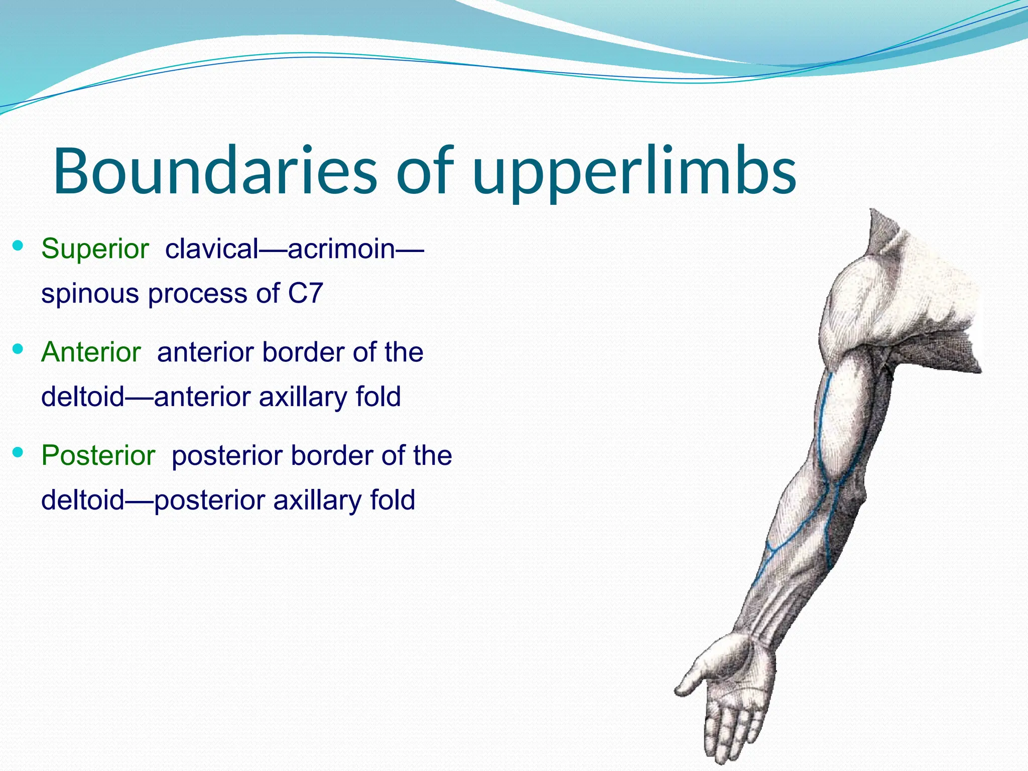
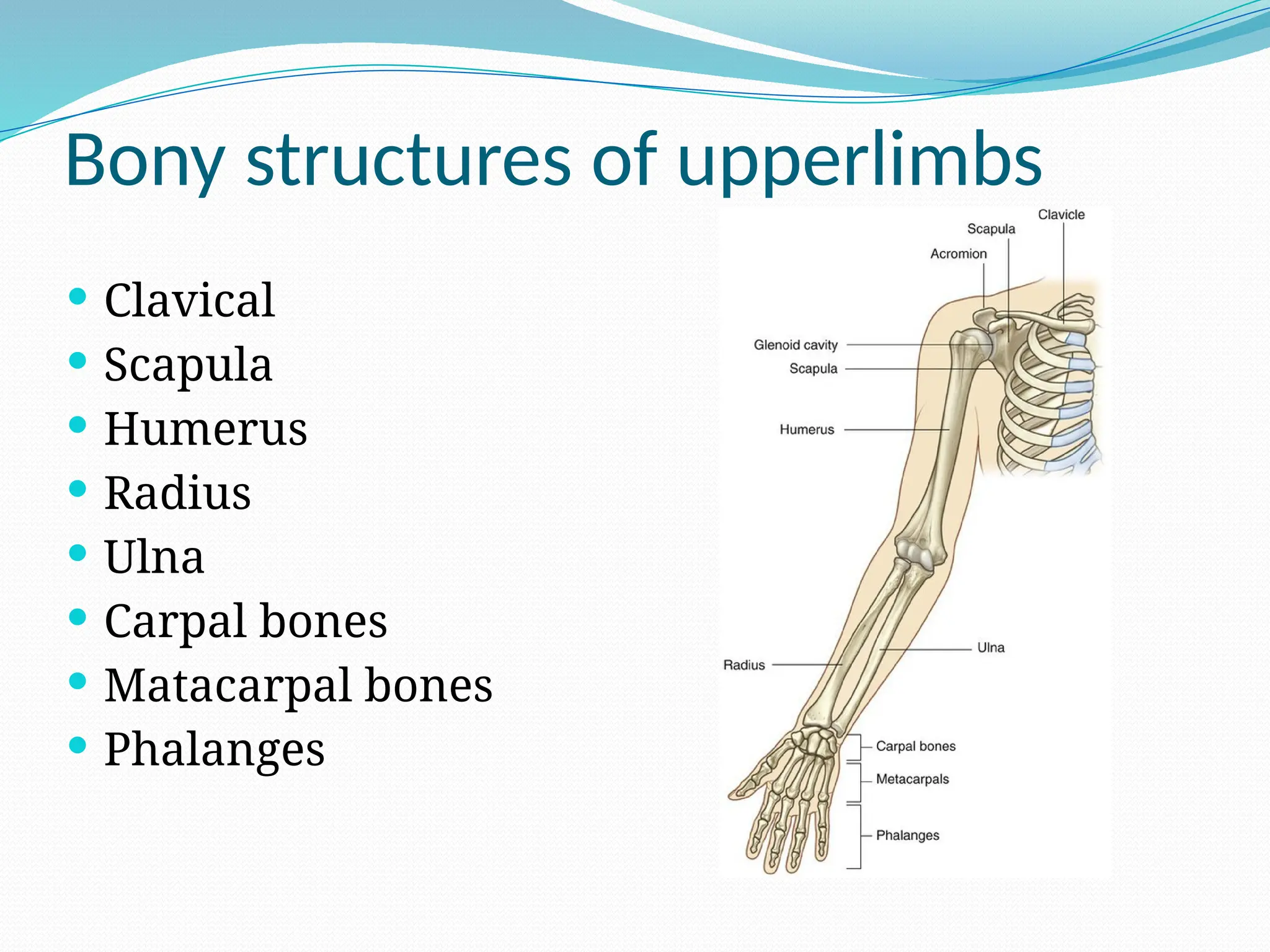
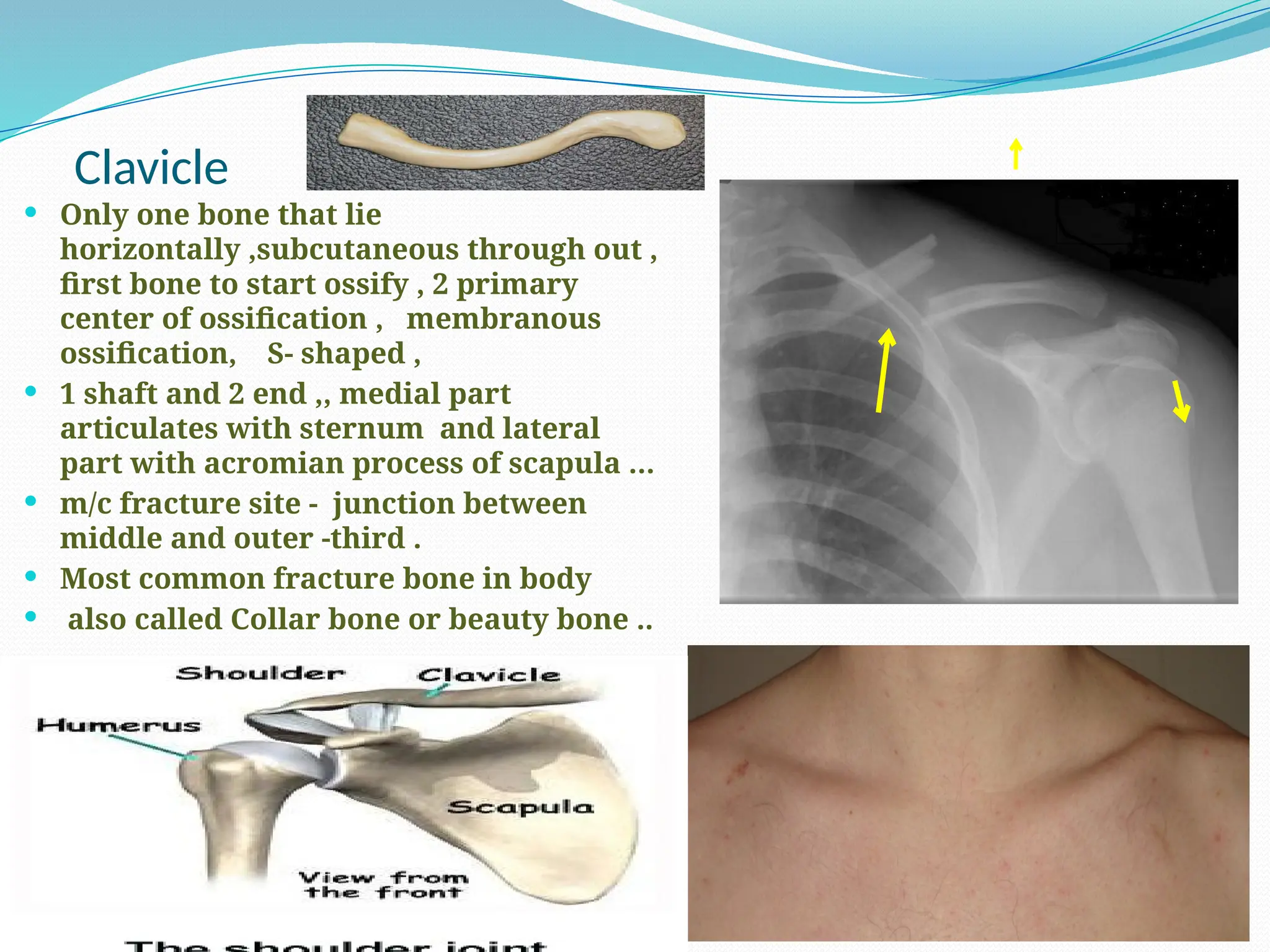
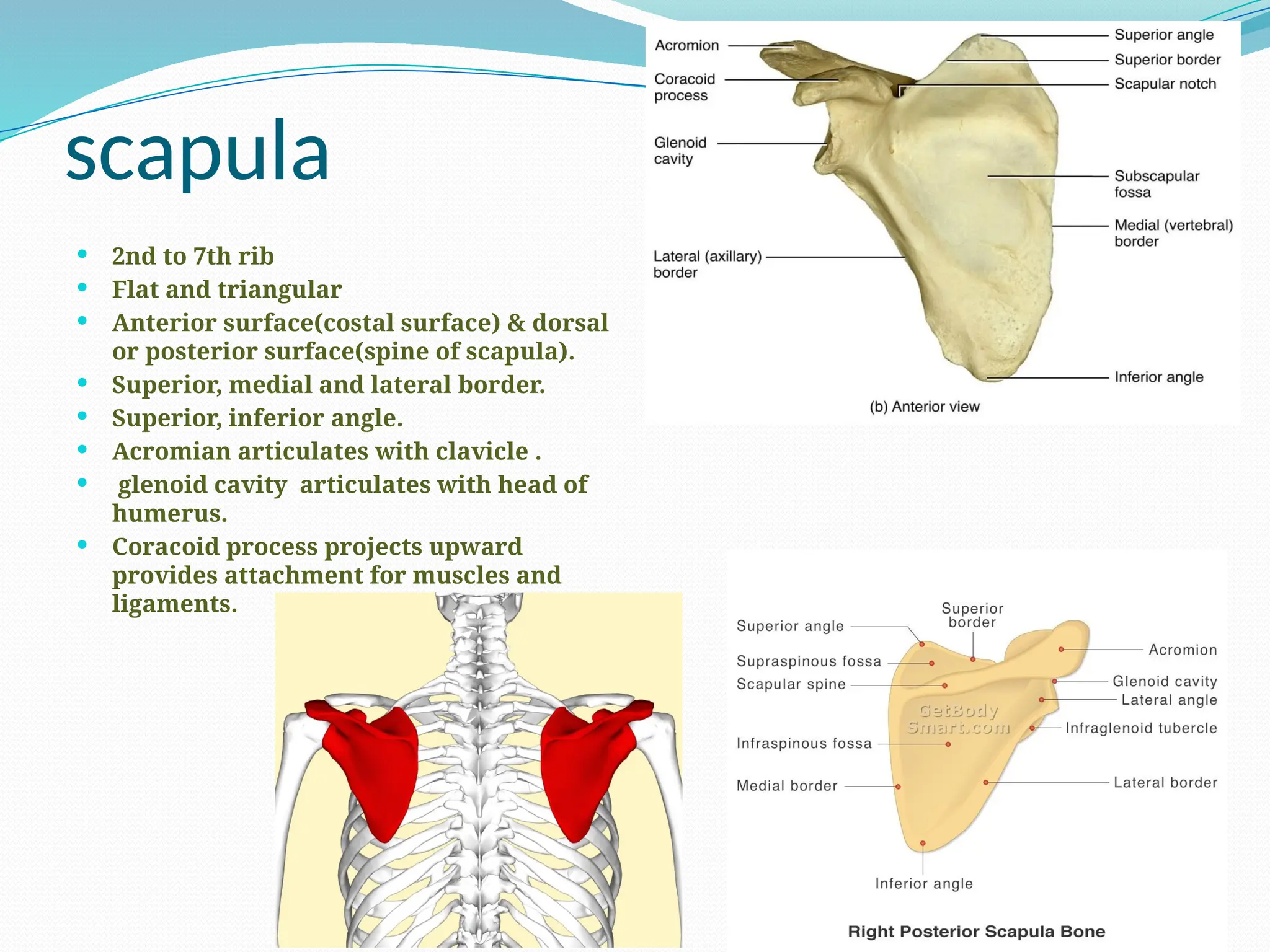
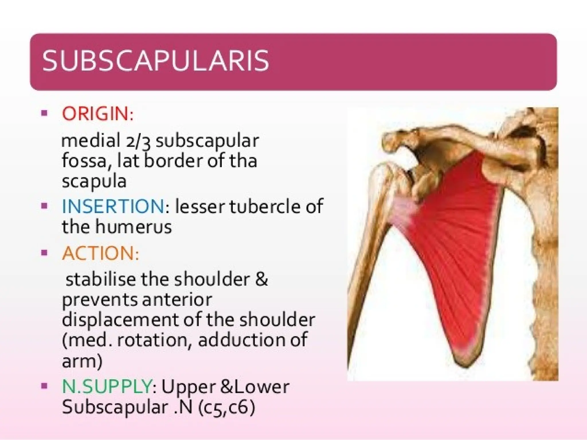
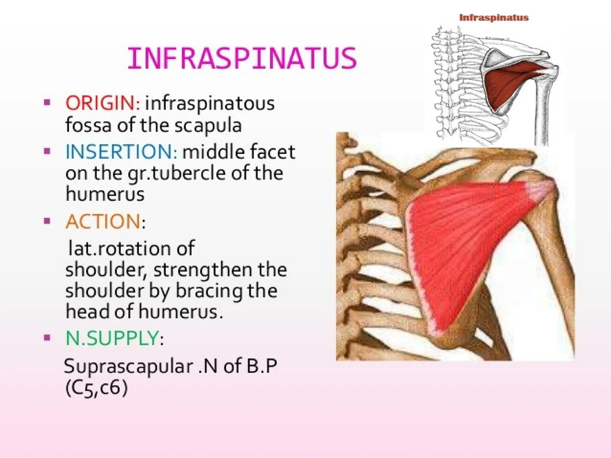
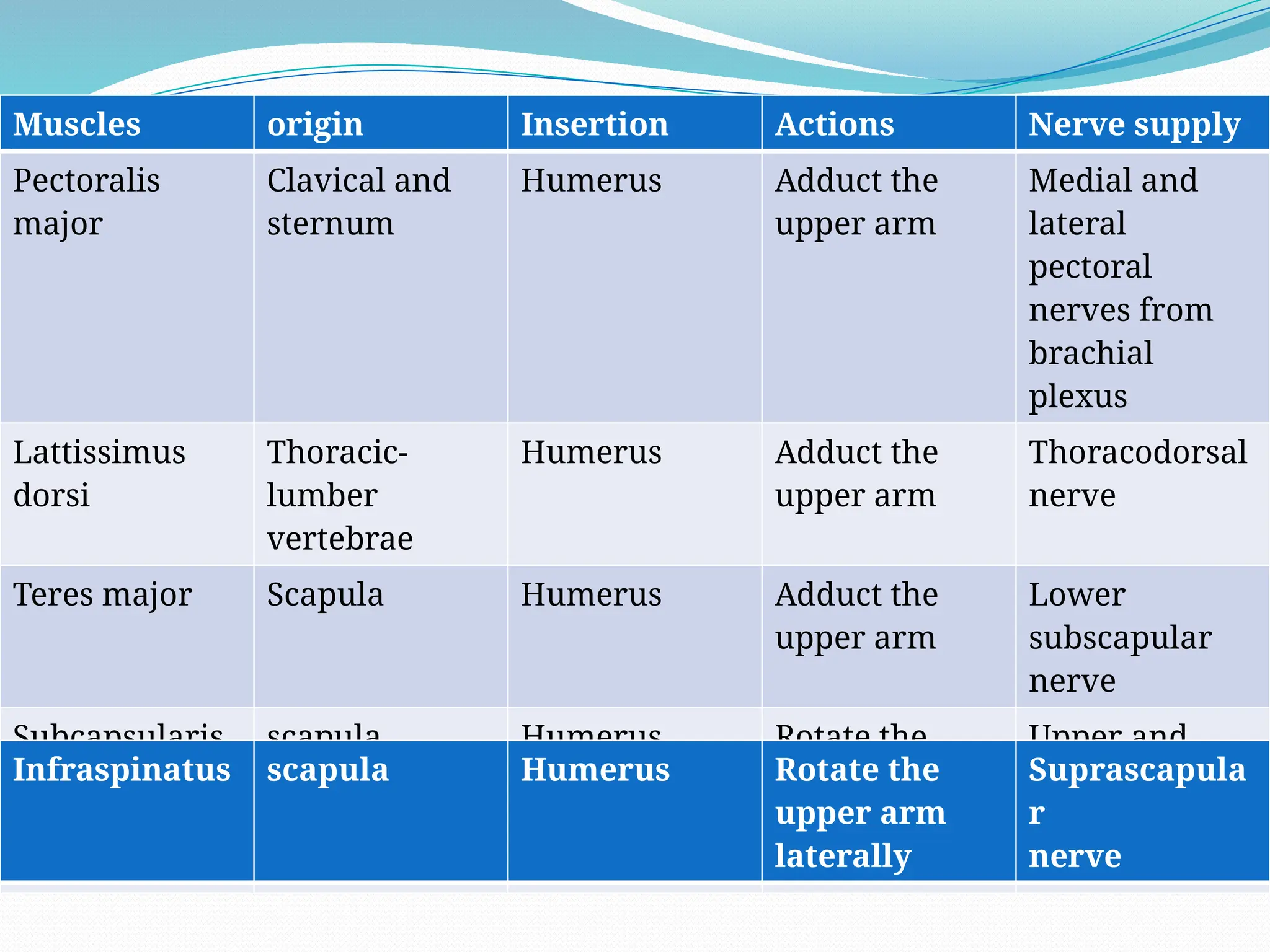
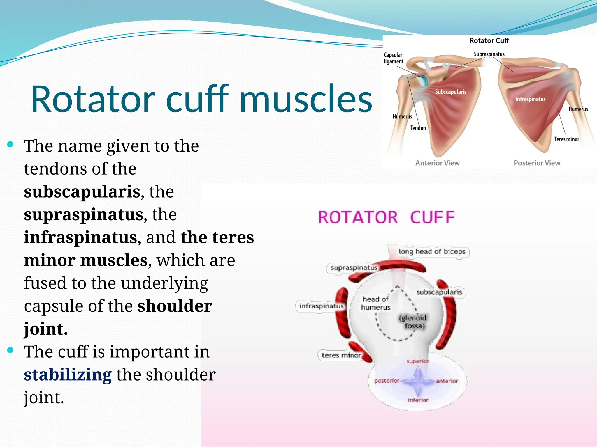
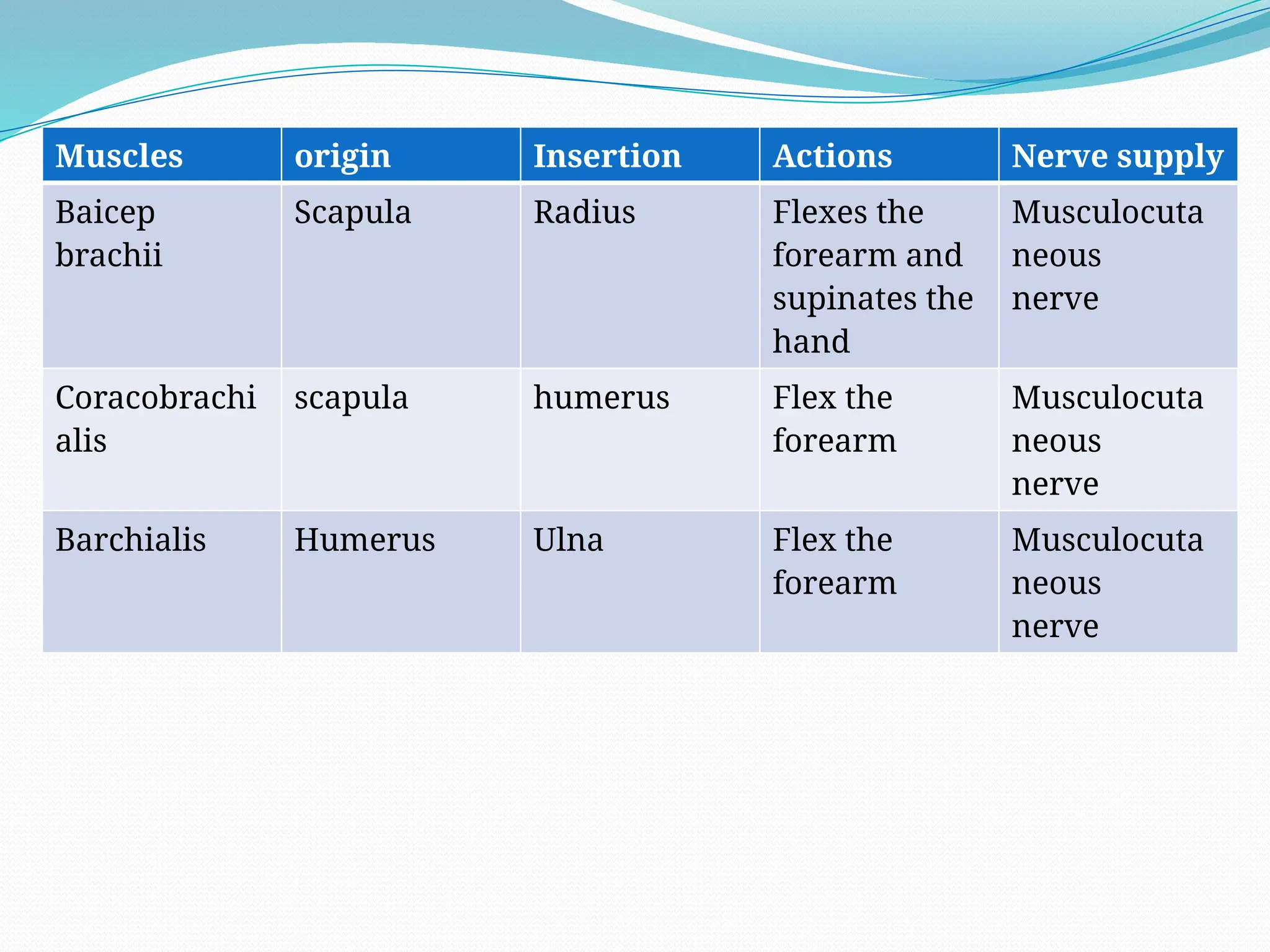
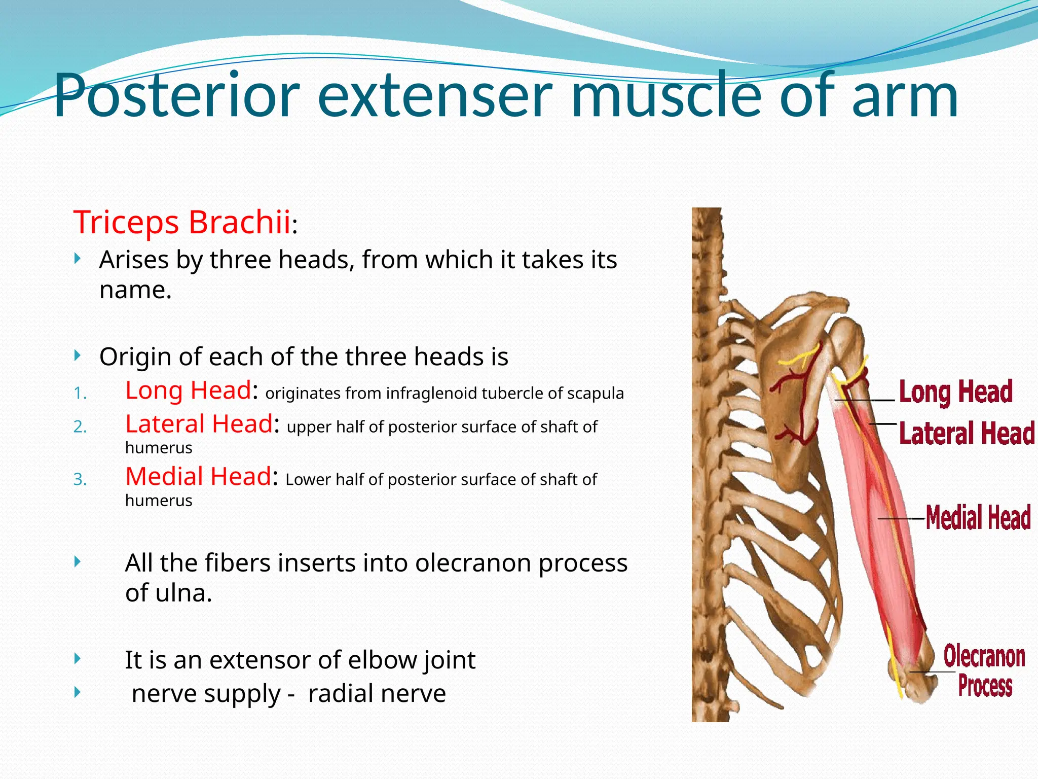
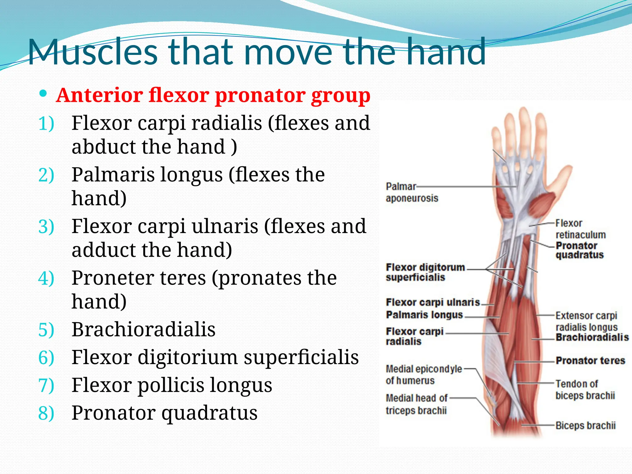
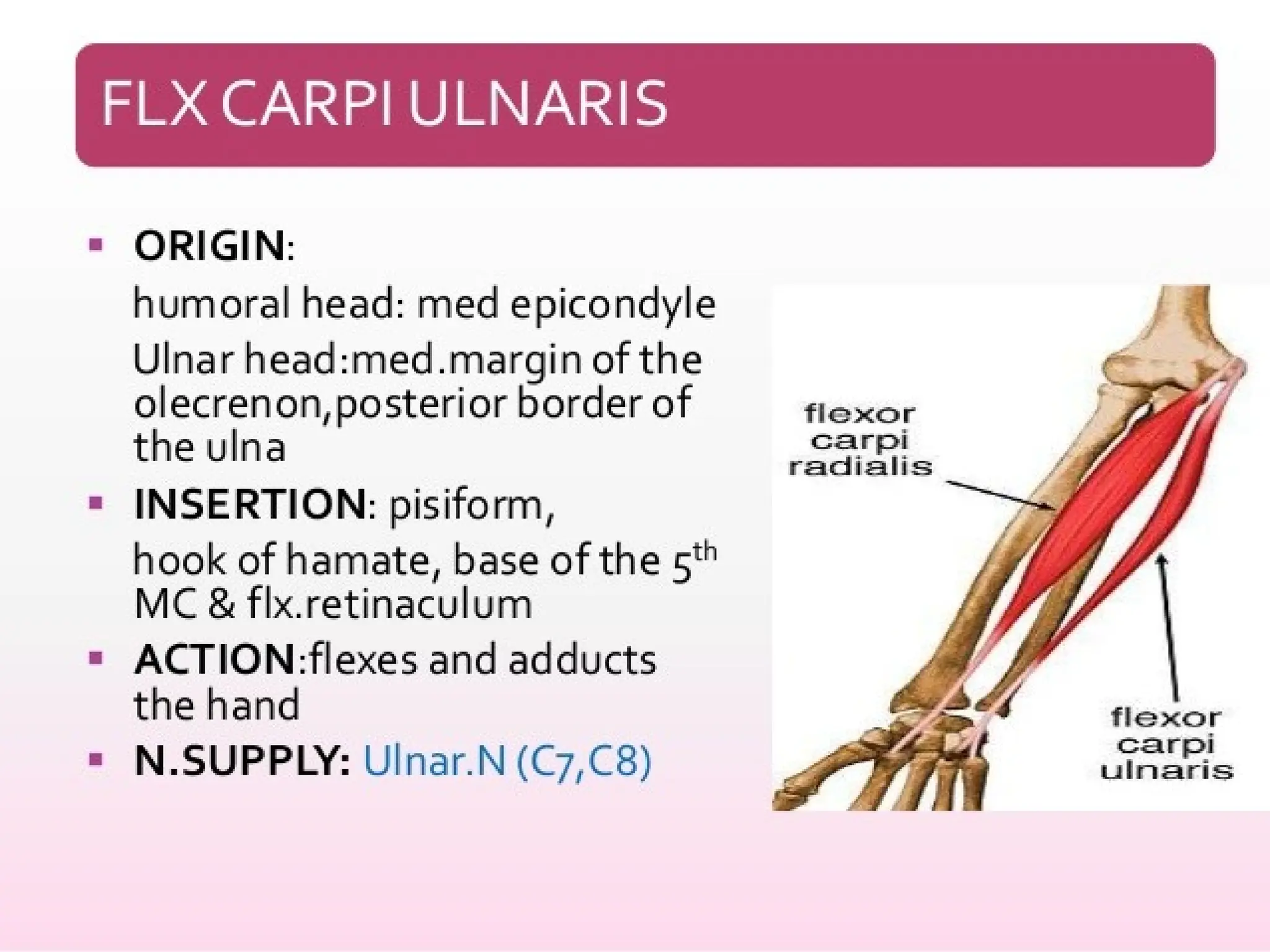
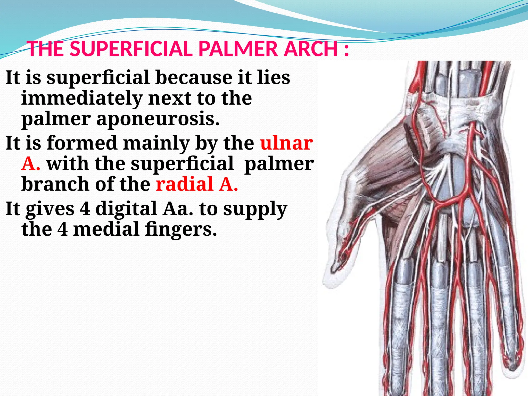
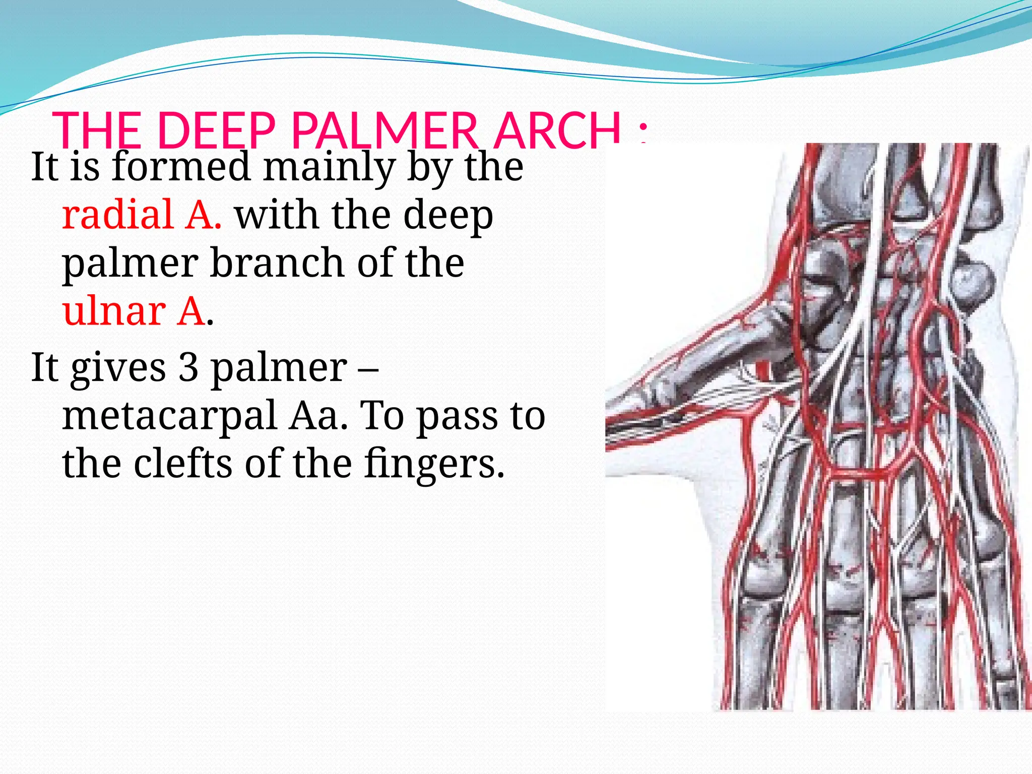
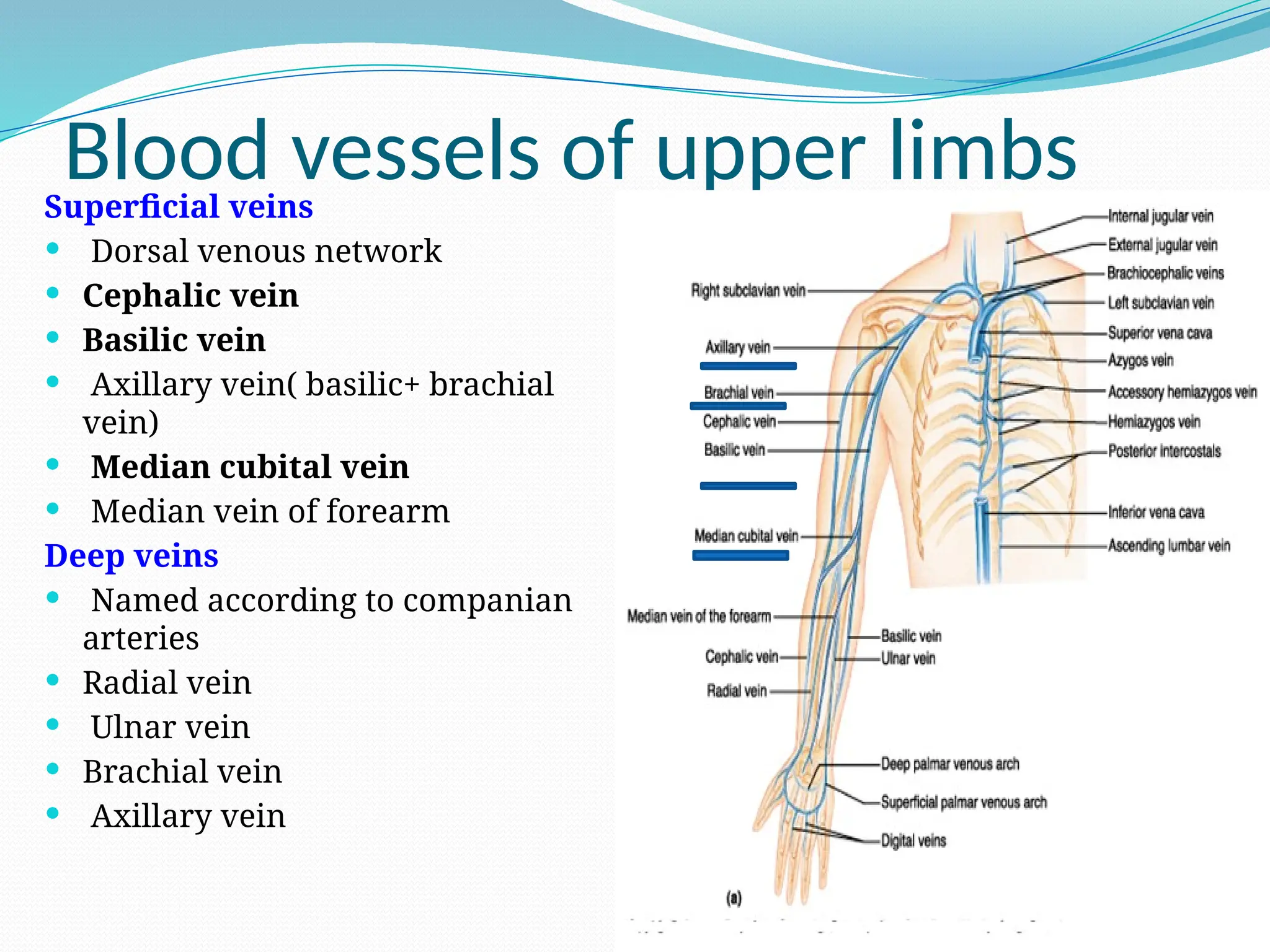
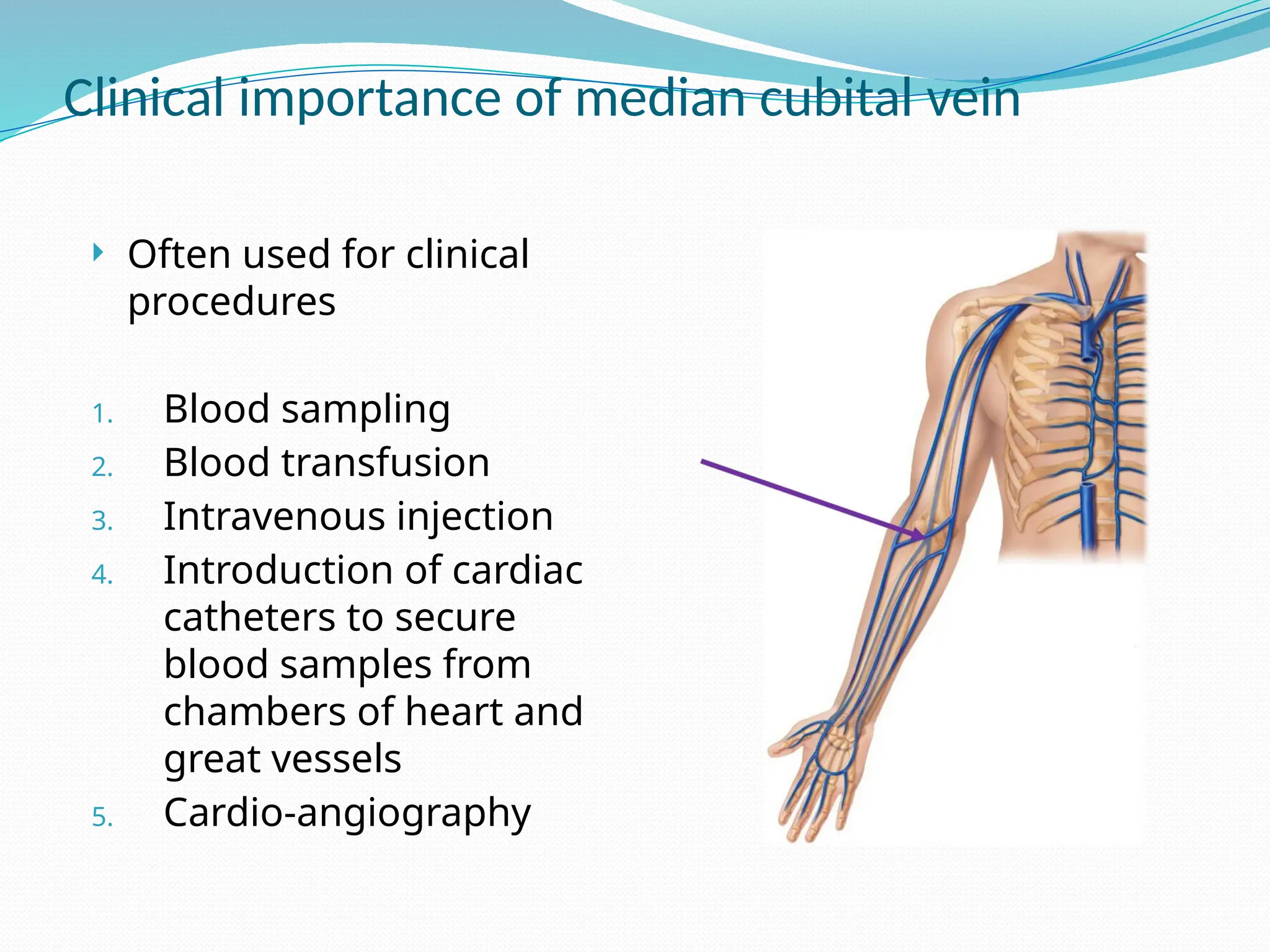
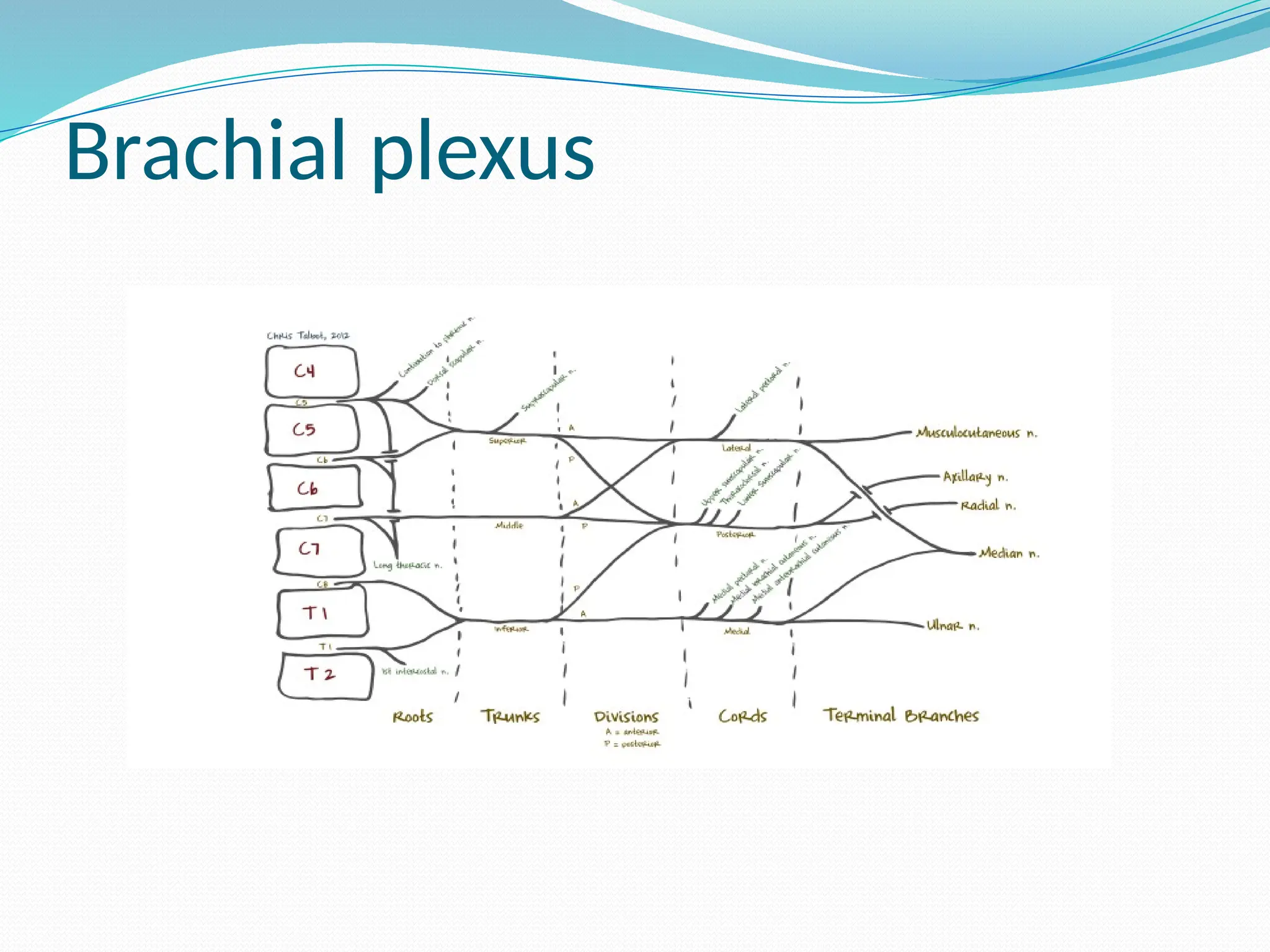
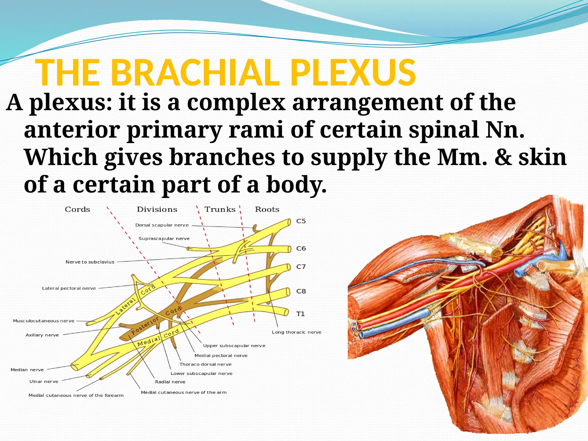
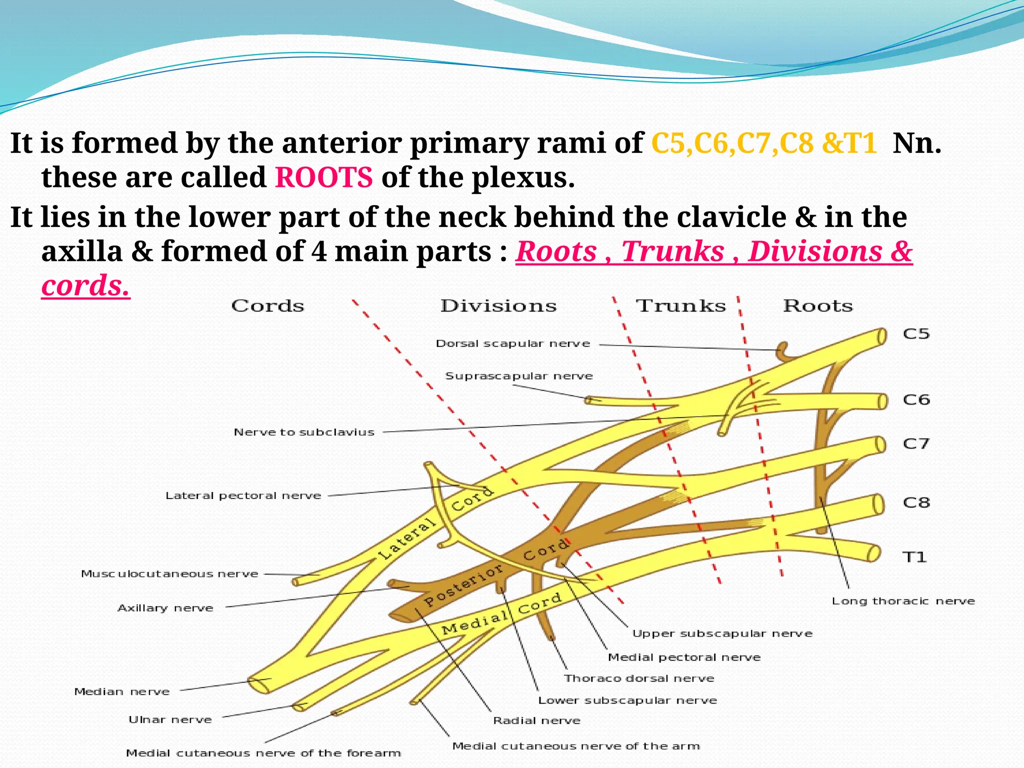
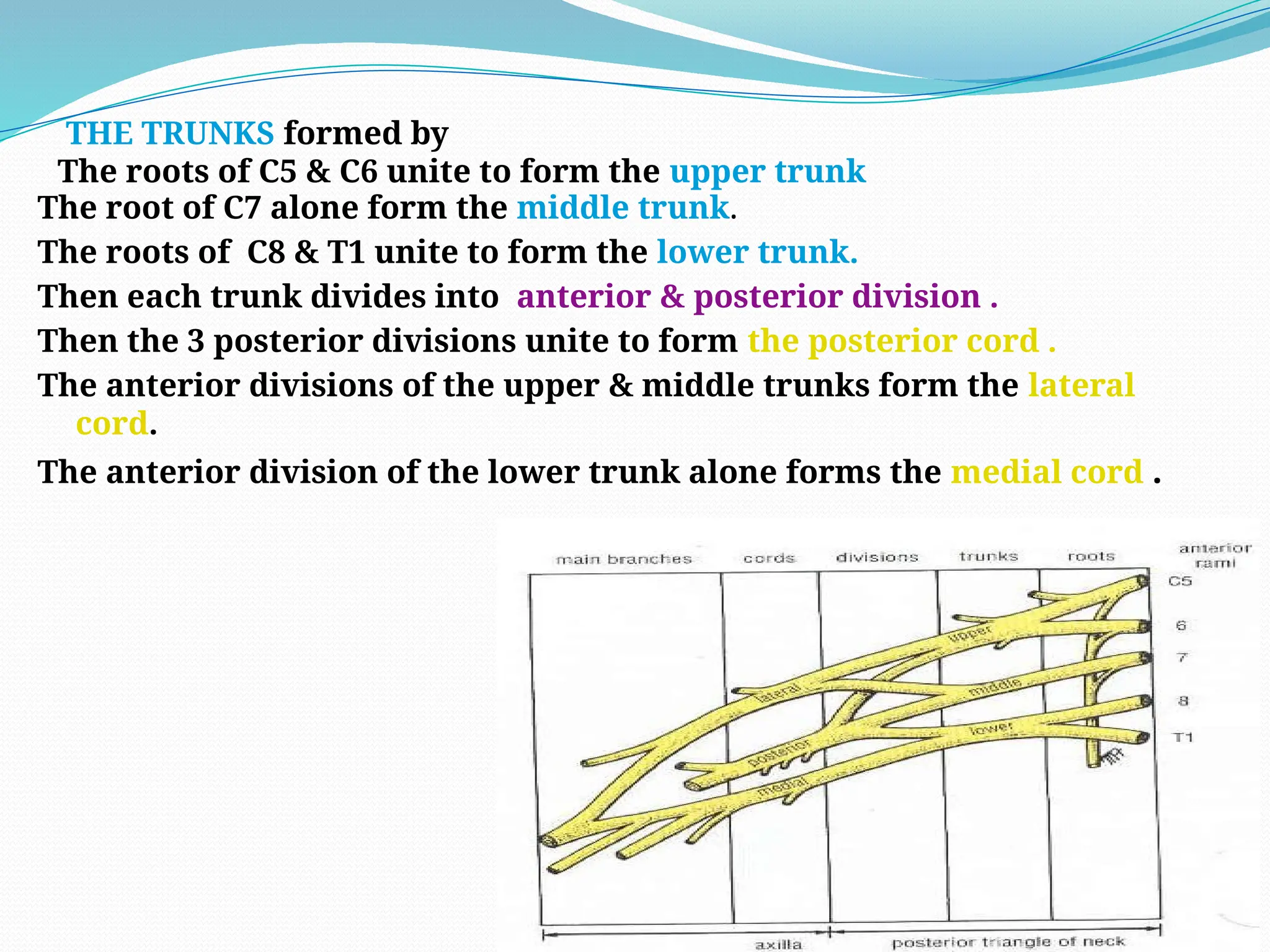
INTRODUCTION TO
UPPER LIMB
Dr. Ela kinra
Department of anatomy
Boundaries of upperlimbs
 Superior clavical—acrimoin—
spinous process of C7
 Anterior anterior border of the
deltoid—anterior axillary fold
 Posterior posterior border of the
deltoid—posterior axillary fold
Bony structures of upperlimbs
 Clavical
 Scapula
 Humerus
 Radius
 Ulna
 Carpal bones
 Matacarpal bones
 Phalanges
Clavicle
 Only one bone that lie
horizontally ,subcutaneous through out ,
first bone to start ossify , 2 primary
center of ossification , membranous
ossification, S- shaped ,
 1 shaft and 2 end ,, medial part
articulates with sternum and lateral
part with acromian process of scapula …
 m/c fracture site - junction between
middle and outer -third .
 Most common fracture bone in body
 also called Collar bone or beauty bone ..
scapula
 2nd to 7th rib
 Flat and triangular
 Anterior surface(costal surface) & dorsal
or posterior surface(spine of scapula).
 Superior, medial and lateral border.
 Superior, inferior angle.
 Acromian articulates with clavicle .
 glenoid cavity articulates with head of
humerus.
 Coracoid process projects upward
provides attachment for muscles and
ligaments.
humerus
 Longest and largest bone of upper limb
 Body and two ends
 Head, greater and lesser tubercle/
intertubecular sulcus or bicipital
groove.
 Anatomical neck
 Surgical neck=head meets the shaft
 Lower end is flat and two articular
surfaces
 Capitulum for head of radius
 Trochlea for articulation with trochlear
notch of ulna.
 Lateral and medial epicondyles for
muscles and ligament attachment
 Radial fossa for radial head when elbow
is flexed
 Coronoid fossa for coronoid process of
ulna during same movement.
 Olecranon fossa posteriorly for
olecranon process of ulna during
extension.
Humerus fractures and nerve injury
 surgical neck fracture - axillary
nerve injury .
 shaft #- radial nerve injury
 supracondylar # - median nerve
injury
 .
Supracondylar fracture
Radius and ulna
Radius
 Fracture of the distal end
(Colle’s fracture):
 It is due to a fall on the
outstretched hand in patients
over (50) years.
 The distal fragment of the radius
is pulled posteriorly and
superiorly
 The distal articular surface is
directed posteriorly.
 The posterior displacement
produces a posterior bump.
 The deformity is referred to as,
‘dinner-fork deformity’ because
the forearm and wrist resemble
the shape of a dinner fork.
 Smith’s fracture is a reversed
Cole’s as the distal segment is
displaced anteriorly
Carpal bones
8 in numbers
 proximal row (lateral to medial )
Scaphoid ( boat shaped )
Lunate( half –moon )
Triquetrum ( three
cornered
Pisiform ( pea Shaped )
Distal row
Trapezium ( four sided )
Trapezoid ( babys shoes
shaped )
Capitate ( head ,,, biggest )
Hamate ( hook)
Scaphoid fracture is most common .
Fracture of the Scaphoid Bone
 Common in young adults
 Fracture line passes through the
narrowest part of the bone
 If the fragments will not unite
properly, there will be permanent
pain and weakness at the wrist
 Deep tenderness in the anatomical
snuff box after a fall on an
outstretched hand in a young adult is
an indication of fracture of scaphoid
bone
Elbow Joint
 Dislocations are common
and most are posterior. Are
more common in children,
due to a fall on outstretched
hand. The distal end of
humerus is pushed
anteriorly through weak
part of the capsule
 Pulled Elbow: occurs in
children, when the child is
lifted by the upper limb.
The radial head is pulled out
of the annular ligament
Tennis Elbow
 Caused by partial tear or
degeneration of the origin
of superficial extensor
muscles attached to the
lateral epicondyle
 It results due to excessive
use of these muscles as in
tennis, violinists and
housewives.
 Results in pain and
tenderness over the lateral
epicondyle that radiates to
the lateral side of the
forearm
Golfer’s Elbow (Medial Epicondylitis)
 Caused by partial tear
or degeneration of the
origin of superficial
flexor muscles
attached to the medial
epicondyle
 It results due to
excessive use of these
muscles as in playing
golf
 Results in pain and
tenderness over the
medial epicondyle
that radiates to the
Bones of Hand
 Carpal bones
 Metacarpals
 Phalanges
From lateral to medial:
1- the proximal row : scaphoid
,lunate , triquetral &
pisiform .
2- the distal row : trapezium ,
trapezoid , capitate &
hamate
Deltoid
origin – from lateral 1/3rd
of clavicle , acromion
process and spine of scapula.
insertion – into the deltoid tuberosity of humerus .
Action- act as main muscle for abduction of shoulder
joint . Its anterior part is flexor and medial
rotator of humerus while posterior part is extensior
and lateral roatatior of humerus.
supraspinatus- deep to
trapezius, arises from supraspinatus
fossa of scapula. Fibers crosses above
the shoulder joint and is inserted into
greater tubercle of humerus. Stabilizes
shoulder joint and abducts the
humerus.
Muscles that abduct the upper arm
Muscles origin Insertion Actions Nerve supply
Deltiod Scapula and
clavical
Humerus Abduct the
upper arm
Axillary
nerve
Supra
spinatous
Scapula Humerus Abduct the
upper arm
Suprascapul
arnerve
Muscles that adduct the upper arm
 1) pectoralis major
 2) lattissimus dorsi
 3) teres major
 4) subscapularis
 Latissimus dorsi: very
broad, flat and triangular muscle in
lower back. It is overlapped by lower
part of trapezius. It originates from
lumbar fascia, spines of lower six
thoracic vertebrae, lower three or
four ribs and inferior angle of
scapula. .
Muscles origin Insertion Actions Nerve supply
Pectoralis
major
Clavical and
sternum
Humerus Adduct the
upper arm
Medial and
lateral
pectoral
nerves from
brachial
plexus
Lattissimus
dorsi
Thoracic-
lumber
vertebrae
Humerus Adduct the
upper arm
Thoracodorsal
nerve
Teres major Scapula Humerus Adduct the
upper arm
Lower
subscapular
nerve
Subcapsularis scapula Humerus Rotate the
upper arm
medially
Upper and
lower
subscapular
nerves
Infraspinatus scapula Humerus Rotate the
upper arm
laterally
Suprascapula
r
nerve
Rotator cuff muscles
 The name given to the
tendons of the
subscapularis, the
supraspinatus, the
infraspinatus, and the teres
minor muscles, which are
fused to the underlying
capsule of the shoulder
joint.
 The cuff is important in
stabilizing the shoulder
joint.
Mucles that move forearm
 Anterior flexor muscles
 1) bicep brachii
 2)brachilais
 3) coracobrachialis
coracobrachialis
 elongated muscle in
superiomedial aspect of
arm. Originates from
coracoid process of
scapula and inserts into
medial aspect of shaft of
humerus. It flexes the arm
 musculocutaneous nerve.
Muscles origin Insertion Actions Nerve supply
Baicep
brachii
Scapula Radius Flexes the
forearm and
supinates the
hand
Musculocuta
neous
nerve
Coracobrachi
alis
scapula humerus Flex the
forearm
Musculocuta
neous
nerve
Barchialis Humerus Ulna Flex the
forearm
Musculocuta
neous
nerve
Posterior extenser muscle of arm
Triceps Brachii:
 Arises by three heads, from which it takes its
name.
 Origin of each of the three heads is
1. Long Head: originates from infraglenoid tubercle of scapula
2. Lateral Head: upper half of posterior surface of shaft of
humerus
3. Medial Head: Lower half of posterior surface of shaft of
humerus
 All the fibers inserts into olecranon process
of ulna.
 It is an extensor of elbow joint
 nerve supply - radial nerve
Muscles that move the hand
 Anterior flexor pronator group
1) Flexor carpi radialis (flexes and
abduct the hand )
2) Palmaris longus (flexes the
hand)
3) Flexor carpi ulnaris (flexes and
adduct the hand)
4) Proneter teres (pronates the
hand)
5) Brachioradialis
6) Flexor digitorium superficialis
7) Flexor pollicis longus
8) Pronator quadratus
 Posterior extensor group muscles :-
1) Extensor carpi radialis
- Extends and abduct the hand
2) Extensor carpi ulnaris
- Extends and adduct the hand
3) Extensor digitorium
- Extends the phalanges
Volkmann’s Ischaemic Contracture
 It is the contractures of
the muscles of the
forearm that follows
fractures of the distal
end of the humerus or
fractures of the radius
and ulna.
 Spasm of a localized
segment of the brachial
artery reduces the blood
flow to the flexors and
extensor muscles so that
they under go ischemic
necrosis.
 The flexor muscles are
mostly affected
Intrinsic muscles of the hand
 1) palmer flexors :- thernar group
 hypothernar
 intermidiate group
 2) dorsal extensor :-
 thenar extensor
 hypothernar extensor
 intermidiate extensor
Dupuytren’s Contracture
 It is a localized thickening and
contracture of the palmar
aponeurosis.
 It commonly starts near the root
of the ring finger pulling it to the
palm and flexing it at the
metacarpo-phalngeal joint. Later
the little finger is involved.
 Dupuytren's disease is familial,
and may be associated with
cigarette smoking, vascular
disease, epilepsy, and diabetes.
Pulp-Space Infection (Felon)
 This is the commonest hand
infection. Pus more often
gathers in the finger tips than
anywhere else in the hand.
 It is more common in the
thumb and index fingers.
ARTERIES OF THE UPPER LIMB:
THE AXILLARY ARTERY :It begins at the outer border
of the 1st
rib as a continuation of the Subclavian A.
& ends at the lower border of the teres major M. by
becoming the Brachial A.
ARTERIES OF THE UPPER LIMB:
The pectoralis minor M . divided it to 3 parts
The 1st
part : lies above the pectoralis minor & give one
branch (superior thoracic A.) . It lies very deeply with the
axillary vein on its medial side .
The 2nd
part : lies behind the pectoralis minor M. & gives 2 branches ( acromio thoracic
A.& lateral thoracic A. ).
The 3rd
part : lies below the pectoralis minor M. & gives 3 branches :( subscapular A.,
post. circumflex humeral A. & ant . Circumflex humeral A.).
Anastamosis around the scapula
The branches of the third part of axillary A.
* the sub scapular A. which go down along the lateral
border of the scapula.
*the circumflex scapular A. which arise from the sub
scapular A.& go to the infraspinous fossa.
THE BRACHIAL ARTERY :
It begins at the lower border of the Teres major M. as a
continuation of the Axillary A. & ends at the level of the
neck of the Radius by dividing to Radial & Ulnar Aa.
It is a superficial A. lies
immediately below the deep
fascia.
The basilic vein lies on its
medial side.
The branches of the brachial A.: It supplies all the Mm.
of the arm & gives 3 branches
1- the profunda brachii A.
2- the superior ulnar collateral A.
3- the inferior ulnar collateral A.
Anastamosis around The Elbow Joint :
It is a rich anastamosis between small
branches of brachial ,ulnar & radial Aa.:
Anastamosis around medial
epicondyle :
The two branches of the brachial A .
(the superior ulnar collateral A. & the
inferior ulnar collateral A.)
with the branches of ulnar A. (the ant. &
post. Ulnar recurrent Aa.) .i.e.
the brachial & the ulnar Aa. only .
Anastamosis around lateral
epicondyle :
The 2 terminal branches of the profuda
brachii a. with recurrent branches of
ulnar & radial Aa.
Radial artery
 Begins: in the cubital fossa at the neck of the radius.
 Course: passes downward and laterally end with deep
palmer arch
 Branches
 Radial recurrent a.
 Superfical and deep palmar branch
 Principal artery of thumb
At the lower end of the
radius , it leaves the
front of the forearm &
turns backward round
the lateral border of
the wrist , below the
styloid process of the
radius& enter the
anatomical snuff-box
where the pulsation
can be felt then pass
to the palm .
Ulnar artery
 Begins in the cubital fossa at the
opposite neck of the radius.
 Course: passes downward and
medially .
 Branches
 Ulnar recurrent a.
 Common interosseous
artery
 Anterior interossous a.
 Posterior interosseous a.
 Deep palmar branch
THE SUPERFICIAL PALMER ARCH :
It is superficial because it lies
immediately next to the
palmer aponeurosis.
It is formed mainly by the ulnar
A. with the superficial palmer
branch of the radial A.
It gives 4 digital Aa. to supply
the 4 medial fingers.
THE DEEP PALMER ARCH :
It is formed mainly by the
radial A. with the deep
palmer branch of the
ulnar A.
It gives 3 palmer –
metacarpal Aa. To pass to
the clefts of the fingers.
Review
 The subclavian artery becomes the axillary artery after crossing
the 1st rib
 The axillary artery becomes the brachial artery after crossing the
posterior axillary fold
 The brachial artery divides just distal to the elbow into the radial
and ulnar arteries.
 The ulnar artery gives rise to the common interosseous artery,
which divides into anterior and posterior interosseous arteries.
 The hand is supplied on the palmar aspect by variable
anastomoses between branches of the radial and ulnar arteries.
THE VEINS OF THE UPPER LIMB
There are superficial & deep veins in the upper limb
.
The deep veins are the veins which
accompany the main Aa. ( way & name ).
The superficial Vv. : start as superficial
venous network on the back of the hand. this
network drains in 2 directions
laterally into Cephalic V &
medially to the basilic V.
Blood vessels of upper limbs
Superficial veins
 Dorsal venous network
 Cephalic vein
 Basilic vein
 Axillary vein( basilic+ brachial
vein)
 Median cubital vein
 Median vein of forearm
Deep veins
 Named according to companian
arteries
 Radial vein
 Ulnar vein
 Brachial vein
 Axillary vein
Clinical importance of median cubital vein
 Often used for clinical
procedures
1. Blood sampling
2. Blood transfusion
3. Intravenous injection
4. Introduction of cardiac
catheters to secure
blood samples from
chambers of heart and
great vessels
5. Cardio-angiography
Brachial plexus
THE BRACHIAL PLEXUS
A plexus: it is a complex arrangement of the
anterior primary rami of certain spinal Nn.
Which gives branches to supply the Mm. & skin
of a certain part of a body.
It is formed by the anterior primary rami of C5,C6,C7,C8 &T1 Nn.
these are called ROOTS of the plexus.
It lies in the lower part of the neck behind the clavicle & in the
axilla & formed of 4 main parts : Roots , Trunks , Divisions &
cords.
The root of C7 alone form the middle trunk.
The roots of C8 & T1 unite to form the lower trunk.
Then each trunk divides into anterior & posterior division .
Then the 3 posterior divisions unite to form the posterior cord .
The anterior divisions of the upper & middle trunks form the lateral
cord.
The anterior division of the lower trunk alone forms the medial cord .
THE TRUNKS formed by
The roots of C5 & C6 unite to form the upper trunk
So we have :5 roots , 3 trunks , 6 division , 3 cords & 5
terminal branches .
No branches arise from the trunks nor from the
divisions ( except the suprascapular N. which come
from the erbs point=at beginning of the upper trunk). .
The brachial plexus gives 16 branches , 11 small
branches & 5 big branches : radial , ulnar , median ,
circumflex (axillary) & musclocutaneous Nn.
Each cord gives rise to 2 branches one is big & consider
the continuation of the cord & one is small SO
The posterior cord continues as the radial N.
The medial cord continues as the ulnar N.
The lateral cord continues as the median N.
The 3 cords named according to their position to 2nd
part
of the axillary A. (post. , medial & lateral.) while the
name of the main N. according to position in the
forearm( ulnar , median & radial ).
The 5 main Nn. Arise opposite the lower border
of the pectoralis minor M. near the coracoid
process .
1- The circumflex (axillary ) N. supplies the
deltoid & teres minor Mm. then become the
lateral cutaneous N. of the arm.
2- The musculocutaneous N. supplies the
biceps , coracobrachialis & brachialis Mm.
then become the lateral cutaneous N. of the
forearm .
3- The median N ( with its branch =ant.
Interosseous N.) supplies most of the Mm. of
front of the forearm with sensation of lateral 3
& ½ fingers anteriorly .
4- The radial N.( with its branch=post.
Interosseous N.) supplies most of the mm. of the
back of the forearm. With sensation of lateral 3 & ½
fingers posteriorly.
5- The ulnar N. supplies Mm. on the medial side of
the forearm with sensation of medial 1 & ½ fingers
anteriorly & posteriorly .
Median nerve
Origin: arises from the medial
and lateral cord of the
brachial plexus.
Course:
 Descends on the lateral side
of brachial artery.
 Halfway down the arm, it
crosses the brachial artery to
reach its medial side.
 he median nerve travels through
a tunnel within the wrist called
the Carpal Tunnel. The median
nerve gives sensation to the
palm sides of the thumb, index
finger, long finger, and half of the
ring finger. It also sends a nerve
branch to control the thenar
muscles of the thumb. The
thenar muscles help move the
thumb and let you touch the pad
of the thumb to the tips each of
each finger on the same hand, a
motion called opposition.
Ulnar nerve
Origin: arises from the medial cord of the
brachial plexus.
Course:
 Descends on the medial side of brachial
artery.
 At the middle of the arm, it pierces the
medial intermuscular septum and
passes down behind the medial
epicondyle of the humerus.
THE DERMATOMES OF THE UPPER LIMB
It is the cutaneous sensation of the upper limb.
C4 supplies the skin over the tip of the shoulder .
C5 supplies the lateral side of the arm &the upper lateral part of the forearm .
C6 supplies the lateral side of the lower part of the forearm & the lateral aspect of
the hand
C7 supplies the middle aspect of the hand (ant.& post.) .
C8 supplies the medial side of the hand & the
medial lower part of the forearm.
T1 supplies the medial part of the upper part of
the forearm & the medial lower part of the
arm.
T2 supplies the medial side of the upper part of
the arm & the floor of the axilla.
Cubital fossa
Boundaries
Superior: an imaginary line
drawn between the two
epicondyles of the humerus.
Lateral: brachioradialis;
Medial: pronator teres;
General
introduction Body wall Head and neck Limbs Thoracic cavity Abdominal
cavity Pelvic cavity
Cubital fossa
Contents:
The median N.
The brachial A., radial A., ulnar
A.
The superficial and deep
branches of the radial N.
Radial artery
Ulnar artery
Median nerve
Brachial
artery
Radial
nerve
General
introduction Body wall Head and neck Limbs Thoracic cavity Abdominal
cavity Pelvic cavity
The blood pressure is
universally recorded by
auscultating the brachial artery
in medial side of the biceps’
tendon in the cubital fossa.
Applying
Cubital fossa
Brachial artery
Biceps
General
introduction Body wall Head and neck Limbs Thoracic cavity Abdominal
cavity Pelvic cavity
Thank you !!!

Upper limb introduction presentation by Dr Ela

  • 1.
    INTRODUCTION TO UPPER LIMB Dr.Ela kinra Department of anatomy
  • 2.
    Boundaries of upperlimbs Superior clavical—acrimoin— spinous process of C7  Anterior anterior border of the deltoid—anterior axillary fold  Posterior posterior border of the deltoid—posterior axillary fold
  • 3.
    Bony structures ofupperlimbs  Clavical  Scapula  Humerus  Radius  Ulna  Carpal bones  Matacarpal bones  Phalanges
  • 4.
    Clavicle  Only onebone that lie horizontally ,subcutaneous through out , first bone to start ossify , 2 primary center of ossification , membranous ossification, S- shaped ,  1 shaft and 2 end ,, medial part articulates with sternum and lateral part with acromian process of scapula …  m/c fracture site - junction between middle and outer -third .  Most common fracture bone in body  also called Collar bone or beauty bone ..
  • 5.
    scapula  2nd to7th rib  Flat and triangular  Anterior surface(costal surface) & dorsal or posterior surface(spine of scapula).  Superior, medial and lateral border.  Superior, inferior angle.  Acromian articulates with clavicle .  glenoid cavity articulates with head of humerus.  Coracoid process projects upward provides attachment for muscles and ligaments.
  • 6.
    humerus  Longest andlargest bone of upper limb  Body and two ends  Head, greater and lesser tubercle/ intertubecular sulcus or bicipital groove.  Anatomical neck  Surgical neck=head meets the shaft  Lower end is flat and two articular surfaces  Capitulum for head of radius  Trochlea for articulation with trochlear notch of ulna.  Lateral and medial epicondyles for muscles and ligament attachment  Radial fossa for radial head when elbow is flexed  Coronoid fossa for coronoid process of ulna during same movement.  Olecranon fossa posteriorly for olecranon process of ulna during extension.
  • 7.
    Humerus fractures andnerve injury  surgical neck fracture - axillary nerve injury .  shaft #- radial nerve injury  supracondylar # - median nerve injury  . Supracondylar fracture
  • 8.
  • 9.
    Radius  Fracture ofthe distal end (Colle’s fracture):  It is due to a fall on the outstretched hand in patients over (50) years.  The distal fragment of the radius is pulled posteriorly and superiorly  The distal articular surface is directed posteriorly.  The posterior displacement produces a posterior bump.  The deformity is referred to as, ‘dinner-fork deformity’ because the forearm and wrist resemble the shape of a dinner fork.  Smith’s fracture is a reversed Cole’s as the distal segment is displaced anteriorly
  • 10.
    Carpal bones 8 innumbers  proximal row (lateral to medial ) Scaphoid ( boat shaped ) Lunate( half –moon ) Triquetrum ( three cornered Pisiform ( pea Shaped ) Distal row Trapezium ( four sided ) Trapezoid ( babys shoes shaped ) Capitate ( head ,,, biggest ) Hamate ( hook) Scaphoid fracture is most common .
  • 11.
    Fracture of theScaphoid Bone  Common in young adults  Fracture line passes through the narrowest part of the bone  If the fragments will not unite properly, there will be permanent pain and weakness at the wrist  Deep tenderness in the anatomical snuff box after a fall on an outstretched hand in a young adult is an indication of fracture of scaphoid bone
  • 12.
    Elbow Joint  Dislocationsare common and most are posterior. Are more common in children, due to a fall on outstretched hand. The distal end of humerus is pushed anteriorly through weak part of the capsule  Pulled Elbow: occurs in children, when the child is lifted by the upper limb. The radial head is pulled out of the annular ligament
  • 13.
    Tennis Elbow  Causedby partial tear or degeneration of the origin of superficial extensor muscles attached to the lateral epicondyle  It results due to excessive use of these muscles as in tennis, violinists and housewives.  Results in pain and tenderness over the lateral epicondyle that radiates to the lateral side of the forearm
  • 14.
    Golfer’s Elbow (MedialEpicondylitis)  Caused by partial tear or degeneration of the origin of superficial flexor muscles attached to the medial epicondyle  It results due to excessive use of these muscles as in playing golf  Results in pain and tenderness over the medial epicondyle that radiates to the
  • 15.
    Bones of Hand Carpal bones  Metacarpals  Phalanges From lateral to medial: 1- the proximal row : scaphoid ,lunate , triquetral & pisiform . 2- the distal row : trapezium , trapezoid , capitate & hamate
  • 16.
    Deltoid origin – fromlateral 1/3rd of clavicle , acromion process and spine of scapula. insertion – into the deltoid tuberosity of humerus . Action- act as main muscle for abduction of shoulder joint . Its anterior part is flexor and medial rotator of humerus while posterior part is extensior and lateral roatatior of humerus. supraspinatus- deep to trapezius, arises from supraspinatus fossa of scapula. Fibers crosses above the shoulder joint and is inserted into greater tubercle of humerus. Stabilizes shoulder joint and abducts the humerus. Muscles that abduct the upper arm
  • 17.
    Muscles origin InsertionActions Nerve supply Deltiod Scapula and clavical Humerus Abduct the upper arm Axillary nerve Supra spinatous Scapula Humerus Abduct the upper arm Suprascapul arnerve
  • 18.
    Muscles that adductthe upper arm  1) pectoralis major  2) lattissimus dorsi  3) teres major  4) subscapularis
  • 20.
     Latissimus dorsi:very broad, flat and triangular muscle in lower back. It is overlapped by lower part of trapezius. It originates from lumbar fascia, spines of lower six thoracic vertebrae, lower three or four ribs and inferior angle of scapula. .
  • 24.
    Muscles origin InsertionActions Nerve supply Pectoralis major Clavical and sternum Humerus Adduct the upper arm Medial and lateral pectoral nerves from brachial plexus Lattissimus dorsi Thoracic- lumber vertebrae Humerus Adduct the upper arm Thoracodorsal nerve Teres major Scapula Humerus Adduct the upper arm Lower subscapular nerve Subcapsularis scapula Humerus Rotate the upper arm medially Upper and lower subscapular nerves Infraspinatus scapula Humerus Rotate the upper arm laterally Suprascapula r nerve
  • 25.
    Rotator cuff muscles The name given to the tendons of the subscapularis, the supraspinatus, the infraspinatus, and the teres minor muscles, which are fused to the underlying capsule of the shoulder joint.  The cuff is important in stabilizing the shoulder joint.
  • 26.
    Mucles that moveforearm  Anterior flexor muscles  1) bicep brachii  2)brachilais  3) coracobrachialis
  • 29.
    coracobrachialis  elongated musclein superiomedial aspect of arm. Originates from coracoid process of scapula and inserts into medial aspect of shaft of humerus. It flexes the arm  musculocutaneous nerve.
  • 30.
    Muscles origin InsertionActions Nerve supply Baicep brachii Scapula Radius Flexes the forearm and supinates the hand Musculocuta neous nerve Coracobrachi alis scapula humerus Flex the forearm Musculocuta neous nerve Barchialis Humerus Ulna Flex the forearm Musculocuta neous nerve
  • 31.
    Posterior extenser muscleof arm Triceps Brachii:  Arises by three heads, from which it takes its name.  Origin of each of the three heads is 1. Long Head: originates from infraglenoid tubercle of scapula 2. Lateral Head: upper half of posterior surface of shaft of humerus 3. Medial Head: Lower half of posterior surface of shaft of humerus  All the fibers inserts into olecranon process of ulna.  It is an extensor of elbow joint  nerve supply - radial nerve
  • 32.
    Muscles that movethe hand  Anterior flexor pronator group 1) Flexor carpi radialis (flexes and abduct the hand ) 2) Palmaris longus (flexes the hand) 3) Flexor carpi ulnaris (flexes and adduct the hand) 4) Proneter teres (pronates the hand) 5) Brachioradialis 6) Flexor digitorium superficialis 7) Flexor pollicis longus 8) Pronator quadratus
  • 36.
     Posterior extensorgroup muscles :- 1) Extensor carpi radialis - Extends and abduct the hand 2) Extensor carpi ulnaris - Extends and adduct the hand 3) Extensor digitorium - Extends the phalanges
  • 41.
    Volkmann’s Ischaemic Contracture It is the contractures of the muscles of the forearm that follows fractures of the distal end of the humerus or fractures of the radius and ulna.  Spasm of a localized segment of the brachial artery reduces the blood flow to the flexors and extensor muscles so that they under go ischemic necrosis.  The flexor muscles are mostly affected
  • 42.
    Intrinsic muscles ofthe hand  1) palmer flexors :- thernar group  hypothernar  intermidiate group  2) dorsal extensor :-  thenar extensor  hypothernar extensor  intermidiate extensor
  • 43.
    Dupuytren’s Contracture  Itis a localized thickening and contracture of the palmar aponeurosis.  It commonly starts near the root of the ring finger pulling it to the palm and flexing it at the metacarpo-phalngeal joint. Later the little finger is involved.  Dupuytren's disease is familial, and may be associated with cigarette smoking, vascular disease, epilepsy, and diabetes.
  • 44.
    Pulp-Space Infection (Felon) This is the commonest hand infection. Pus more often gathers in the finger tips than anywhere else in the hand.  It is more common in the thumb and index fingers.
  • 45.
    ARTERIES OF THEUPPER LIMB: THE AXILLARY ARTERY :It begins at the outer border of the 1st rib as a continuation of the Subclavian A. & ends at the lower border of the teres major M. by becoming the Brachial A.
  • 46.
    ARTERIES OF THEUPPER LIMB: The pectoralis minor M . divided it to 3 parts The 1st part : lies above the pectoralis minor & give one branch (superior thoracic A.) . It lies very deeply with the axillary vein on its medial side .
  • 47.
    The 2nd part :lies behind the pectoralis minor M. & gives 2 branches ( acromio thoracic A.& lateral thoracic A. ). The 3rd part : lies below the pectoralis minor M. & gives 3 branches :( subscapular A., post. circumflex humeral A. & ant . Circumflex humeral A.).
  • 48.
    Anastamosis around thescapula The branches of the third part of axillary A. * the sub scapular A. which go down along the lateral border of the scapula. *the circumflex scapular A. which arise from the sub scapular A.& go to the infraspinous fossa.
  • 49.
    THE BRACHIAL ARTERY: It begins at the lower border of the Teres major M. as a continuation of the Axillary A. & ends at the level of the neck of the Radius by dividing to Radial & Ulnar Aa. It is a superficial A. lies immediately below the deep fascia. The basilic vein lies on its medial side.
  • 50.
    The branches ofthe brachial A.: It supplies all the Mm. of the arm & gives 3 branches 1- the profunda brachii A. 2- the superior ulnar collateral A. 3- the inferior ulnar collateral A.
  • 51.
    Anastamosis around TheElbow Joint : It is a rich anastamosis between small branches of brachial ,ulnar & radial Aa.:
  • 52.
    Anastamosis around medial epicondyle: The two branches of the brachial A . (the superior ulnar collateral A. & the inferior ulnar collateral A.) with the branches of ulnar A. (the ant. & post. Ulnar recurrent Aa.) .i.e. the brachial & the ulnar Aa. only . Anastamosis around lateral epicondyle : The 2 terminal branches of the profuda brachii a. with recurrent branches of ulnar & radial Aa.
  • 53.
    Radial artery  Begins:in the cubital fossa at the neck of the radius.  Course: passes downward and laterally end with deep palmer arch  Branches  Radial recurrent a.  Superfical and deep palmar branch  Principal artery of thumb At the lower end of the radius , it leaves the front of the forearm & turns backward round the lateral border of the wrist , below the styloid process of the radius& enter the anatomical snuff-box where the pulsation can be felt then pass to the palm .
  • 54.
    Ulnar artery  Beginsin the cubital fossa at the opposite neck of the radius.  Course: passes downward and medially .  Branches  Ulnar recurrent a.  Common interosseous artery  Anterior interossous a.  Posterior interosseous a.  Deep palmar branch
  • 55.
    THE SUPERFICIAL PALMERARCH : It is superficial because it lies immediately next to the palmer aponeurosis. It is formed mainly by the ulnar A. with the superficial palmer branch of the radial A. It gives 4 digital Aa. to supply the 4 medial fingers.
  • 56.
    THE DEEP PALMERARCH : It is formed mainly by the radial A. with the deep palmer branch of the ulnar A. It gives 3 palmer – metacarpal Aa. To pass to the clefts of the fingers.
  • 57.
    Review  The subclavianartery becomes the axillary artery after crossing the 1st rib  The axillary artery becomes the brachial artery after crossing the posterior axillary fold  The brachial artery divides just distal to the elbow into the radial and ulnar arteries.  The ulnar artery gives rise to the common interosseous artery, which divides into anterior and posterior interosseous arteries.  The hand is supplied on the palmar aspect by variable anastomoses between branches of the radial and ulnar arteries.
  • 58.
    THE VEINS OFTHE UPPER LIMB There are superficial & deep veins in the upper limb . The deep veins are the veins which accompany the main Aa. ( way & name ). The superficial Vv. : start as superficial venous network on the back of the hand. this network drains in 2 directions laterally into Cephalic V & medially to the basilic V.
  • 59.
    Blood vessels ofupper limbs Superficial veins  Dorsal venous network  Cephalic vein  Basilic vein  Axillary vein( basilic+ brachial vein)  Median cubital vein  Median vein of forearm Deep veins  Named according to companian arteries  Radial vein  Ulnar vein  Brachial vein  Axillary vein
  • 60.
    Clinical importance ofmedian cubital vein  Often used for clinical procedures 1. Blood sampling 2. Blood transfusion 3. Intravenous injection 4. Introduction of cardiac catheters to secure blood samples from chambers of heart and great vessels 5. Cardio-angiography
  • 61.
  • 62.
    THE BRACHIAL PLEXUS Aplexus: it is a complex arrangement of the anterior primary rami of certain spinal Nn. Which gives branches to supply the Mm. & skin of a certain part of a body.
  • 63.
    It is formedby the anterior primary rami of C5,C6,C7,C8 &T1 Nn. these are called ROOTS of the plexus. It lies in the lower part of the neck behind the clavicle & in the axilla & formed of 4 main parts : Roots , Trunks , Divisions & cords.
  • 64.
    The root ofC7 alone form the middle trunk. The roots of C8 & T1 unite to form the lower trunk. Then each trunk divides into anterior & posterior division . Then the 3 posterior divisions unite to form the posterior cord . The anterior divisions of the upper & middle trunks form the lateral cord. The anterior division of the lower trunk alone forms the medial cord . THE TRUNKS formed by The roots of C5 & C6 unite to form the upper trunk
  • 65.
    So we have:5 roots , 3 trunks , 6 division , 3 cords & 5 terminal branches . No branches arise from the trunks nor from the divisions ( except the suprascapular N. which come from the erbs point=at beginning of the upper trunk). . The brachial plexus gives 16 branches , 11 small branches & 5 big branches : radial , ulnar , median , circumflex (axillary) & musclocutaneous Nn.
  • 66.
    Each cord givesrise to 2 branches one is big & consider the continuation of the cord & one is small SO The posterior cord continues as the radial N. The medial cord continues as the ulnar N. The lateral cord continues as the median N. The 3 cords named according to their position to 2nd part of the axillary A. (post. , medial & lateral.) while the name of the main N. according to position in the forearm( ulnar , median & radial ).
  • 67.
    The 5 mainNn. Arise opposite the lower border of the pectoralis minor M. near the coracoid process . 1- The circumflex (axillary ) N. supplies the deltoid & teres minor Mm. then become the lateral cutaneous N. of the arm. 2- The musculocutaneous N. supplies the biceps , coracobrachialis & brachialis Mm. then become the lateral cutaneous N. of the forearm . 3- The median N ( with its branch =ant. Interosseous N.) supplies most of the Mm. of front of the forearm with sensation of lateral 3 & ½ fingers anteriorly .
  • 68.
    4- The radialN.( with its branch=post. Interosseous N.) supplies most of the mm. of the back of the forearm. With sensation of lateral 3 & ½ fingers posteriorly. 5- The ulnar N. supplies Mm. on the medial side of the forearm with sensation of medial 1 & ½ fingers anteriorly & posteriorly .
  • 70.
    Median nerve Origin: arisesfrom the medial and lateral cord of the brachial plexus. Course:  Descends on the lateral side of brachial artery.  Halfway down the arm, it crosses the brachial artery to reach its medial side.
  • 71.
     he mediannerve travels through a tunnel within the wrist called the Carpal Tunnel. The median nerve gives sensation to the palm sides of the thumb, index finger, long finger, and half of the ring finger. It also sends a nerve branch to control the thenar muscles of the thumb. The thenar muscles help move the thumb and let you touch the pad of the thumb to the tips each of each finger on the same hand, a motion called opposition.
  • 72.
    Ulnar nerve Origin: arisesfrom the medial cord of the brachial plexus. Course:  Descends on the medial side of brachial artery.  At the middle of the arm, it pierces the medial intermuscular septum and passes down behind the medial epicondyle of the humerus.
  • 75.
    THE DERMATOMES OFTHE UPPER LIMB It is the cutaneous sensation of the upper limb. C4 supplies the skin over the tip of the shoulder . C5 supplies the lateral side of the arm &the upper lateral part of the forearm . C6 supplies the lateral side of the lower part of the forearm & the lateral aspect of the hand C7 supplies the middle aspect of the hand (ant.& post.) . C8 supplies the medial side of the hand & the medial lower part of the forearm. T1 supplies the medial part of the upper part of the forearm & the medial lower part of the arm. T2 supplies the medial side of the upper part of the arm & the floor of the axilla.
  • 76.
    Cubital fossa Boundaries Superior: animaginary line drawn between the two epicondyles of the humerus. Lateral: brachioradialis; Medial: pronator teres; General introduction Body wall Head and neck Limbs Thoracic cavity Abdominal cavity Pelvic cavity
  • 77.
    Cubital fossa Contents: The medianN. The brachial A., radial A., ulnar A. The superficial and deep branches of the radial N. Radial artery Ulnar artery Median nerve Brachial artery Radial nerve General introduction Body wall Head and neck Limbs Thoracic cavity Abdominal cavity Pelvic cavity
  • 78.
    The blood pressureis universally recorded by auscultating the brachial artery in medial side of the biceps’ tendon in the cubital fossa. Applying Cubital fossa Brachial artery Biceps General introduction Body wall Head and neck Limbs Thoracic cavity Abdominal cavity Pelvic cavity
  • 81.