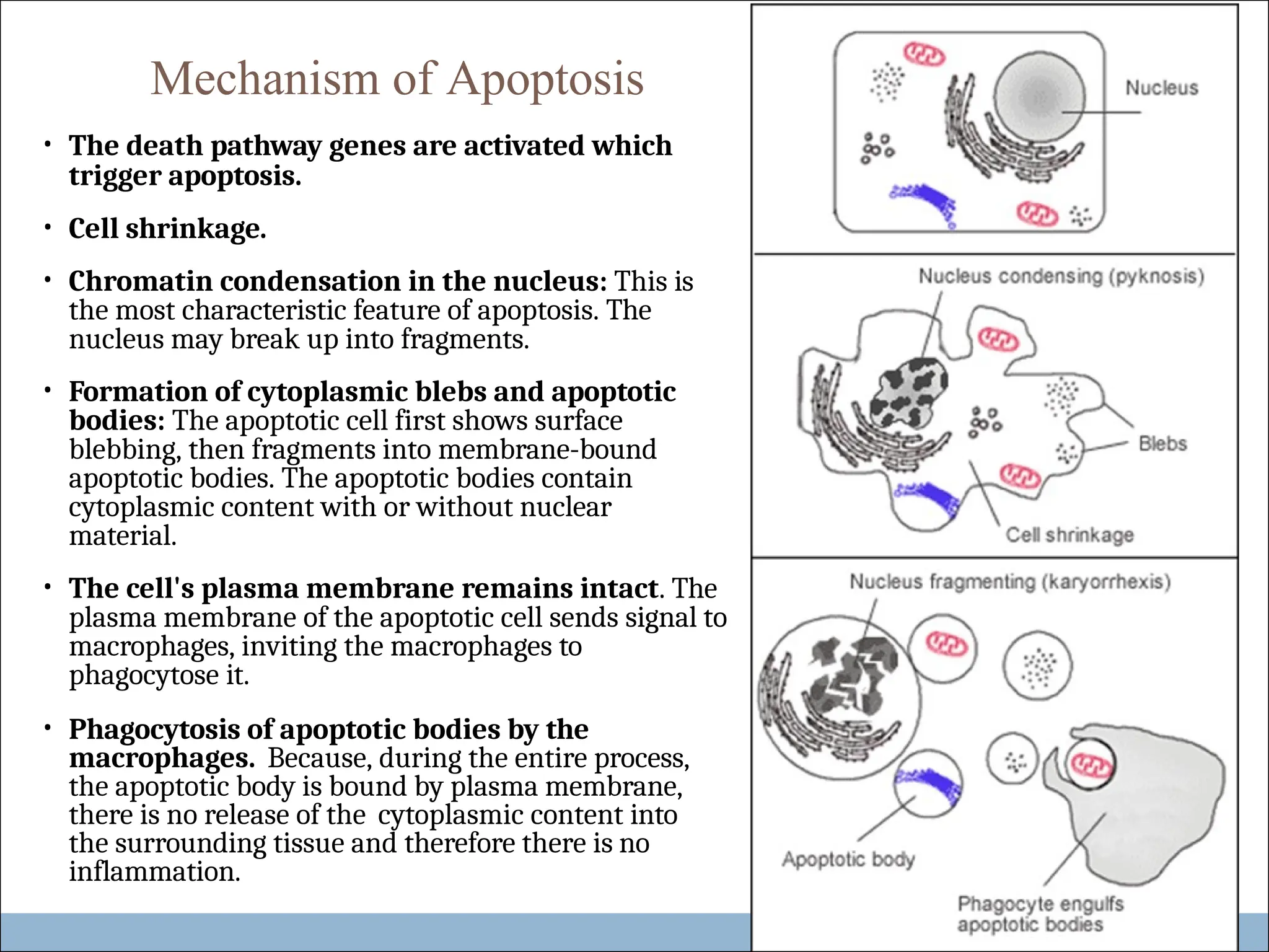
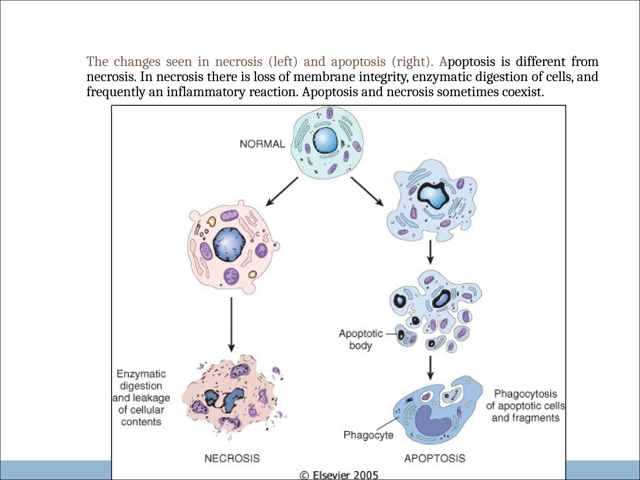
Chapter :
CELL INJURY
Apoptosis
APOPTOSIS
⚫ Apoptosis is programmed cell death. Apoptosis means “falling off”. It is a
type of cell suicide.
⚫ Is results from activation of ‘death pathway genes’.
⚫ It is a pathway of cell death in which cells destined to die activate their
own enzymes to degrade their own nuclear DNA and proteins.
It can be
⚫ Physiological/ adapative.
⚫ Pathologic.
NOTE: Apoptosis and necrosis can sometimes coexist.
Apoptosis in Physiologic Situations
 The programmed destruction of cells during
embryogenesis.
 Hormone-dependent: e.g. endometrial cell breakdown
during the menstrual cycle, the regression of the lactating
breast after weaning, and prostatic atrophy after castration
(adaptive atrophy).
 Apoptosis in proliferating cells e.g. intestinal epithelial
lining is always being replaced.
 Cells that after performing their function undergo
apoptosis e.g. neutrophils and lymphocytes in
inflammation.
 Sometimes body produces harmful lymphocytes and they
are also destroyed by apoptosis.
Apoptosis in Pathologic Conditions
• Cell death produced by injury e.g. radiation.
• In certain diseases e.g. in viral hepatitis the infected hepatocytes
undergo apoptosis (acidophilic bodies) or injury of skin epidermal
cells (keratinocytes) leads to apoptosis of keratinocytes (Civatte
bodies).
• Pathologic atrophy in organs e.g. pancreas, parotid gland, and kidney
• Corticosteroid induced atrophy of the neonatal thymus
• Cell death in tumors (usually accompanied by necrosis).
Mechanism of Apoptosis
• The death pathway genes are activated which
trigger apoptosis.
• Cell shrinkage.
• Chromatin condensation in the nucleus: This is
the most characteristic feature of apoptosis. The
nucleus may break up into fragments.
• Formation of cytoplasmic blebs and apoptotic
bodies: The apoptotic cell first shows surface
blebbing, then fragments into membrane-bound
apoptotic bodies. The apoptotic bodies contain
cytoplasmic content with or without nuclear
material.
• The cell's plasma membrane remains intact. The
plasma membrane of the apoptotic cell sends signal to
macrophages, inviting the macrophages to
phagocytose it.
• Phagocytosis of apoptotic bodies by the
macrophages. Because, during the entire process,
the apoptotic body is bound by plasma membrane,
there is no release of the cytoplasmic content into
the surrounding tissue and therefore there is no
inflammation.
Morphology of
Apoptosis
• On histology apoptosis
involves single cells or small
clusters of cells. The apoptotic
cell appears as a round or oval
mass of intensely eosinophilic
cytoplasm with dense nucleus.
There is no inflammation.
apoptosis in liver
cell
apoptosis in
epidermis.
Important enzymes of apoptosis
1. Cysteine proteases named caspases
2. Ca2+ dependent endonucleases
3. Mg2+ dependent endonucleases
Regulation of apoptosis
It is mediated by a number of genes and their products e.g:
⚫ bcl-2 gene inhibits apoptosis
⚫ bax genes facilitates apoptosis
⚫ p53 facilitates apoptosis by inhibiting bcl2 and promoting
bax genes.
The changes seen in necrosis (left) and apoptosis (right). Apoptosis is different from
necrosis. In necrosis there is loss of membrane integrity, enzymatic digestion of cells, and
frequently an inflammatory reaction. Apoptosis and necrosis sometimes coexist.
Feature Necrosis Apoptosis
Cell size Enlarged (swelling) Reduced (shrinkage)
Nucleus Pyknosis
Karyorrhexis
Fragmentation into nucleosome size
fragments
karyolysis
Plasma
membrane
Disrupted Intact; altered structure, especially
orientation of lipids
Cellular
contents
Enzymatic digestion; may
leak out of cell
Intact; may be released in apoptotic bodies
Adjacent
inflammation
Frequent No
Physiologic Invariably pathologic Often physiologic, means of eliminating
or pathologic (culmination of irreversible unwanted cells; may be pathologic after some
role cell injury) forms of cell injury, especially DNA damage
THANK YOU

5. cell injury...APOPTOSIS ppt.programmed cell death

  • 1.
  • 2.
    APOPTOSIS ⚫ Apoptosis isprogrammed cell death. Apoptosis means “falling off”. It is a type of cell suicide. ⚫ Is results from activation of ‘death pathway genes’. ⚫ It is a pathway of cell death in which cells destined to die activate their own enzymes to degrade their own nuclear DNA and proteins. It can be ⚫ Physiological/ adapative. ⚫ Pathologic. NOTE: Apoptosis and necrosis can sometimes coexist.
  • 3.
    Apoptosis in PhysiologicSituations  The programmed destruction of cells during embryogenesis.  Hormone-dependent: e.g. endometrial cell breakdown during the menstrual cycle, the regression of the lactating breast after weaning, and prostatic atrophy after castration (adaptive atrophy).  Apoptosis in proliferating cells e.g. intestinal epithelial lining is always being replaced.  Cells that after performing their function undergo apoptosis e.g. neutrophils and lymphocytes in inflammation.  Sometimes body produces harmful lymphocytes and they are also destroyed by apoptosis.
  • 4.
    Apoptosis in PathologicConditions • Cell death produced by injury e.g. radiation. • In certain diseases e.g. in viral hepatitis the infected hepatocytes undergo apoptosis (acidophilic bodies) or injury of skin epidermal cells (keratinocytes) leads to apoptosis of keratinocytes (Civatte bodies). • Pathologic atrophy in organs e.g. pancreas, parotid gland, and kidney • Corticosteroid induced atrophy of the neonatal thymus • Cell death in tumors (usually accompanied by necrosis).
  • 5.
    Mechanism of Apoptosis •The death pathway genes are activated which trigger apoptosis. • Cell shrinkage. • Chromatin condensation in the nucleus: This is the most characteristic feature of apoptosis. The nucleus may break up into fragments. • Formation of cytoplasmic blebs and apoptotic bodies: The apoptotic cell first shows surface blebbing, then fragments into membrane-bound apoptotic bodies. The apoptotic bodies contain cytoplasmic content with or without nuclear material. • The cell's plasma membrane remains intact. The plasma membrane of the apoptotic cell sends signal to macrophages, inviting the macrophages to phagocytose it. • Phagocytosis of apoptotic bodies by the macrophages. Because, during the entire process, the apoptotic body is bound by plasma membrane, there is no release of the cytoplasmic content into the surrounding tissue and therefore there is no inflammation.
  • 7.
    Morphology of Apoptosis • Onhistology apoptosis involves single cells or small clusters of cells. The apoptotic cell appears as a round or oval mass of intensely eosinophilic cytoplasm with dense nucleus. There is no inflammation. apoptosis in liver cell apoptosis in epidermis.
  • 8.
    Important enzymes ofapoptosis 1. Cysteine proteases named caspases 2. Ca2+ dependent endonucleases 3. Mg2+ dependent endonucleases Regulation of apoptosis It is mediated by a number of genes and their products e.g: ⚫ bcl-2 gene inhibits apoptosis ⚫ bax genes facilitates apoptosis ⚫ p53 facilitates apoptosis by inhibiting bcl2 and promoting bax genes.
  • 9.
    The changes seenin necrosis (left) and apoptosis (right). Apoptosis is different from necrosis. In necrosis there is loss of membrane integrity, enzymatic digestion of cells, and frequently an inflammatory reaction. Apoptosis and necrosis sometimes coexist.
  • 10.
    Feature Necrosis Apoptosis Cellsize Enlarged (swelling) Reduced (shrinkage) Nucleus Pyknosis Karyorrhexis Fragmentation into nucleosome size fragments karyolysis Plasma membrane Disrupted Intact; altered structure, especially orientation of lipids Cellular contents Enzymatic digestion; may leak out of cell Intact; may be released in apoptotic bodies Adjacent inflammation Frequent No Physiologic Invariably pathologic Often physiologic, means of eliminating or pathologic (culmination of irreversible unwanted cells; may be pathologic after some role cell injury) forms of cell injury, especially DNA damage
  • 11.