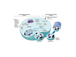
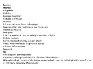
APOPTOSIS
DR PREETI AGRAWAL
Definition – It is a form of ‘coordinated and
internally programmed cell death’
Apoptosis is a pathway of cell death that is
induced by a tightly regulated suicide program
in which cells destined to die activate enzymes
that degrade the cells' own nuclear DNA and
nuclear and cytoplasmic proteins.
Apoptosis is responsible for mediating cell death in wide
variety of physiologic and pathologic processes as under
1. Apoptosis in Physiologic Situations -Death by apoptosis is
a normal phenomenon that serves to eliminate cells that
are no longer needed, and to maintain a steady number of
various cell populations in tissues.eg.
• The programmed destruction of cells during
embryogenesis, including implantation, organogenesis,
developmental involution, and metamorphosis
• Involution of hormone-dependent tissues upon hormone
withdrawal, the regression of the lactating breast after
weaning, and prostatic atrophy after castration.
• Cell loss in proliferating cell populations, such as immature
lymphocytes in the bone marrow
2. Apoptosis in Pathologic Conditions -Apoptosis eliminates
cells that are injured beyond repair without eliciting a host
reaction, thus limiting collateral tissue damage.
• DNA damage. Radiation, cytotoxic anticancer drugs, and
hypoxia can damage DNA, either directly or via production
of free radicals. These injurious stimuli can cause
apoptosis if the insult is mild, but larger doses of the same
stimuli may result in necrotic cell death.
• Accumulation of misfolded proteins. Improperly folded
proteins may arise because of mutations in the genes
encoding these proteins or because of extrinsic factors,
such as damage caused by free radicals. Apoptosis caused
by the accumulation of misfolded proteins has been
invoked as the basis of several degenerative diseases of
the central nervous system and other organs.
• Cell death in certain viral infections
• Pathologic atrophy in parenchymal organs after duct
obstruction, such as occurs in the pancreas, parotid gland,
and kidney.
MORPHOLOGIC AND BIOCHEMICAL CHANGES IN
APOPTOSIS
Morphology. The following morphologic features, some
best seen with the electron microscope, characterize cells
undergoing apoptosis
Cell shrinkage. The cell is smaller in size; the cytoplasm is
dense and the organelles, though relatively normal, are
more tightly packed.
Chromatin condensation. This is the most characteristic
feature of apoptosis. The chromatin aggregates peripherally,
under the nuclear membrane, into dense masses of various
shapes and sizes. The nucleus itself may break up, producing
two or more fragments.
Formation of cytoplasmic blebs and apoptotic bodies. The
apoptotic cell first shows extensive surface blebbing, then
undergoes fragmentation into membrane-bound apoptotic
bodies composed of cytoplasm and tightly packed
organelles, with or without nuclear fragments.
Phagocytosis of apoptotic cells or cell bodies, usually by
macrophages. The apoptotic bodies are rapidly ingested by
phagocytes and degraded by the phagocyte's lysosomal
enzymes.
On histologic examination, in tissues stained with hematoxylin
and eosin, the apoptotic cell appears as a round or oval mass
of intensely eosinophilic cytoplasm with fragments of dense
nuclear chromatin
Biochemical Features of Apoptosis-
• Activation of Caspases – caspases are family of
cysteine proteases. The term caspase is based on two
properties of this family of enzymes:
the “c” refers to a cysteine protease and
“aspase” refers to the unique ability of these
enzymes to cleave after aspartic acid residues.
The caspase family. can be divided functionally into
two groups—initiator and executioner—depending on
the order in which they are activated during
apoptosis. Initiator caspases include caspase-8 and
caspase-9. Several other caspases, including caspase-
3 and caspase-6,
• DNA and Protein Breakdown. Apoptotic cells exhibit a
characteristic breakdown of DNA into large 50- to 300-
kilobase pieces. Subsequently, there is cleavage of DNA by
Ca2+- and Mg2+-dependent endonucleases into fragments
whose sizes are multiples of 180 to 200 base pairs, reflecting
cleavage between nucleosomal subunits. The fragments may
be visualized by electrophoresis as DNA “ladders”
• Membrane Alterations and Recognition by Phagocytes.
movement of some phospholipids (notably
phosphatidylserine) from the inner leaflet to the outer leaflet
of the membrane, where they are recognized by a number of
receptors on phagocytes. annexin V staining is commonly
used to identify apoptotic cells
MECHANISMS OF APOPTOSIS
All cells contain intrinsic mechanisms that signal death or
survival, and apoptosis results from an imbalance in these
signals.
The process of apoptosis may be divided into
an initiation phase, during which some caspases become
catalytically active, and
an execution phase, during which other caspases trigger the
degradation of critical cellular components.
Initiation of apoptosis occurs principally by signals from two
distinct pathways: the intrinsic, or mitochondrial, pathway,
and
the extrinsic, or death receptor–initiated, pathway
Feature
Necrosis
Apoptosis
Cell size
Enlarged (swelling)
Reduced (shrinkage)
Nucleus
Pyknosis → karyorrhexis → karyolysis
Fragmentation into nucleosome size fragments
Plasma membrane
Disrupted
Intact; altered structure, especially orientation of lipids
Cellular contents
Enzymatic digestion; may leak out of cell
Intact; may be released in apoptotic bodies
Adjacent inflammation
Frequent
No
Physiologic or pathologic role
Invariably pathologic (culmination of irreversible cell injury)
Often physiologic, means of eliminating unwanted cells; may be pathologic after some forms
of cell injury, especially DNA damage

Apoptosis

  • 1.
  • 2.
    Definition – Itis a form of ‘coordinated and internally programmed cell death’ Apoptosis is a pathway of cell death that is induced by a tightly regulated suicide program in which cells destined to die activate enzymes that degrade the cells' own nuclear DNA and nuclear and cytoplasmic proteins.
  • 3.
    Apoptosis is responsiblefor mediating cell death in wide variety of physiologic and pathologic processes as under 1. Apoptosis in Physiologic Situations -Death by apoptosis is a normal phenomenon that serves to eliminate cells that are no longer needed, and to maintain a steady number of various cell populations in tissues.eg. • The programmed destruction of cells during embryogenesis, including implantation, organogenesis, developmental involution, and metamorphosis • Involution of hormone-dependent tissues upon hormone withdrawal, the regression of the lactating breast after weaning, and prostatic atrophy after castration. • Cell loss in proliferating cell populations, such as immature lymphocytes in the bone marrow
  • 4.
    2. Apoptosis inPathologic Conditions -Apoptosis eliminates cells that are injured beyond repair without eliciting a host reaction, thus limiting collateral tissue damage. • DNA damage. Radiation, cytotoxic anticancer drugs, and hypoxia can damage DNA, either directly or via production of free radicals. These injurious stimuli can cause apoptosis if the insult is mild, but larger doses of the same stimuli may result in necrotic cell death. • Accumulation of misfolded proteins. Improperly folded proteins may arise because of mutations in the genes encoding these proteins or because of extrinsic factors, such as damage caused by free radicals. Apoptosis caused by the accumulation of misfolded proteins has been invoked as the basis of several degenerative diseases of the central nervous system and other organs.
  • 5.
    • Cell deathin certain viral infections • Pathologic atrophy in parenchymal organs after duct obstruction, such as occurs in the pancreas, parotid gland, and kidney.
  • 6.
    MORPHOLOGIC AND BIOCHEMICALCHANGES IN APOPTOSIS Morphology. The following morphologic features, some best seen with the electron microscope, characterize cells undergoing apoptosis Cell shrinkage. The cell is smaller in size; the cytoplasm is dense and the organelles, though relatively normal, are more tightly packed. Chromatin condensation. This is the most characteristic feature of apoptosis. The chromatin aggregates peripherally, under the nuclear membrane, into dense masses of various shapes and sizes. The nucleus itself may break up, producing two or more fragments.
  • 7.
    Formation of cytoplasmicblebs and apoptotic bodies. The apoptotic cell first shows extensive surface blebbing, then undergoes fragmentation into membrane-bound apoptotic bodies composed of cytoplasm and tightly packed organelles, with or without nuclear fragments. Phagocytosis of apoptotic cells or cell bodies, usually by macrophages. The apoptotic bodies are rapidly ingested by phagocytes and degraded by the phagocyte's lysosomal enzymes.
  • 8.
    On histologic examination,in tissues stained with hematoxylin and eosin, the apoptotic cell appears as a round or oval mass of intensely eosinophilic cytoplasm with fragments of dense nuclear chromatin
  • 9.
    Biochemical Features ofApoptosis- • Activation of Caspases – caspases are family of cysteine proteases. The term caspase is based on two properties of this family of enzymes: the “c” refers to a cysteine protease and “aspase” refers to the unique ability of these enzymes to cleave after aspartic acid residues. The caspase family. can be divided functionally into two groups—initiator and executioner—depending on the order in which they are activated during apoptosis. Initiator caspases include caspase-8 and caspase-9. Several other caspases, including caspase- 3 and caspase-6,
  • 10.
    • DNA andProtein Breakdown. Apoptotic cells exhibit a characteristic breakdown of DNA into large 50- to 300- kilobase pieces. Subsequently, there is cleavage of DNA by Ca2+- and Mg2+-dependent endonucleases into fragments whose sizes are multiples of 180 to 200 base pairs, reflecting cleavage between nucleosomal subunits. The fragments may be visualized by electrophoresis as DNA “ladders” • Membrane Alterations and Recognition by Phagocytes. movement of some phospholipids (notably phosphatidylserine) from the inner leaflet to the outer leaflet of the membrane, where they are recognized by a number of receptors on phagocytes. annexin V staining is commonly used to identify apoptotic cells
  • 11.
    MECHANISMS OF APOPTOSIS Allcells contain intrinsic mechanisms that signal death or survival, and apoptosis results from an imbalance in these signals. The process of apoptosis may be divided into an initiation phase, during which some caspases become catalytically active, and an execution phase, during which other caspases trigger the degradation of critical cellular components. Initiation of apoptosis occurs principally by signals from two distinct pathways: the intrinsic, or mitochondrial, pathway, and the extrinsic, or death receptor–initiated, pathway
  • 14.
    Feature Necrosis Apoptosis Cell size Enlarged (swelling) Reduced(shrinkage) Nucleus Pyknosis → karyorrhexis → karyolysis Fragmentation into nucleosome size fragments Plasma membrane Disrupted Intact; altered structure, especially orientation of lipids Cellular contents Enzymatic digestion; may leak out of cell Intact; may be released in apoptotic bodies Adjacent inflammation Frequent No Physiologic or pathologic role Invariably pathologic (culmination of irreversible cell injury) Often physiologic, means of eliminating unwanted cells; may be pathologic after some forms of cell injury, especially DNA damage