Downloaded 284 times


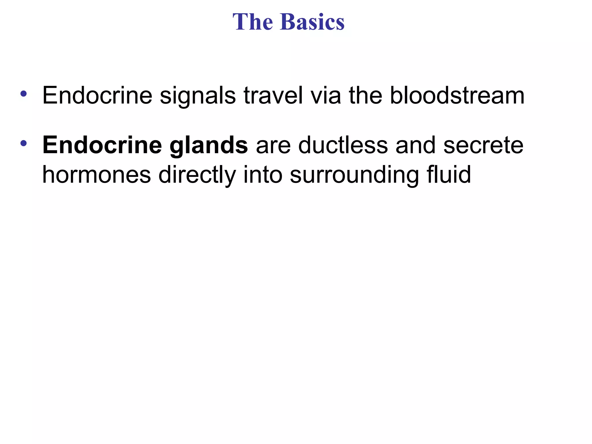
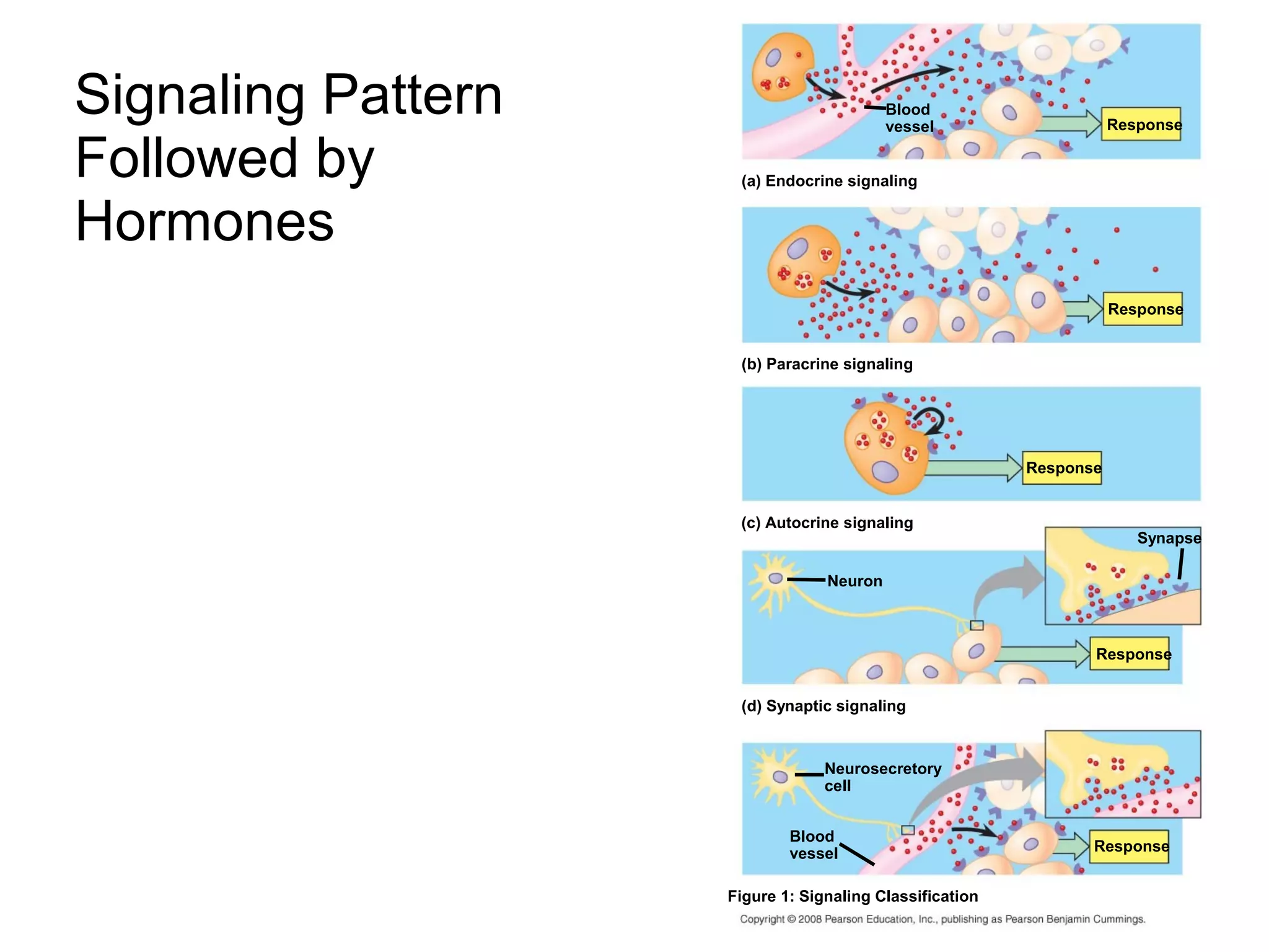


The document summarizes key aspects of the endocrine system and hormone signaling. It describes how hormones are chemical messengers that circulate through the bloodstream and activate target cells. The endocrine system works alongside the nervous system to regulate slower but longer-acting responses, such as reproduction and metabolism. Examples are provided of hormone types, the mechanisms of hormone signaling, feedback loops that regulate hormone levels, and specific endocrine glands like the pituitary, thyroid, and parathyroid glands. Diseases associated with hormonal imbalances like diabetes and hyperthyroidism are also mentioned.