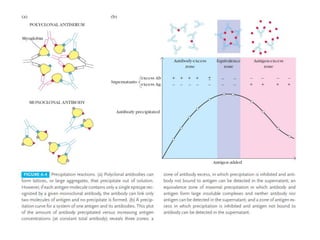
ANTIBODY CLASSES AND
FUNCTION
BY
VASANTHA KUMAR G
DEPARTMENT OF INDUSTRIAL BIOTECHNOLOGY
GOVERNMENT COLLEGE OF TECHNOLOGY
INTRODUCTION
• Antibodies are the antigen binding proteins present on
the B-cell membrane and secreted by plasma cells.
• These are of two types
1. membrane bound antibodies – confers antigenic
specificity on B cells.
2. secreted antibodies – circulate in blood and serve as
the effectors of humoral immunity.
• Antibodies reside in the serum.
STRUCTURE OF ANTIBODIES
• Antibody molecules have a common structure
of four peptide chains. This structure consists
of two
1. Two identical light (L) chains, polypeptides of
about 25,000 molecular weight
2. Two identical heavy (H) chains, larger
polypeptides of molecular weight 50,000 or
more.
• Each light chain is bound to a heavy chain by a
disulfide bond and by non-covalent
interactions.
• The two heavy chain were also joined by this
forces.
• Exact number and precise positions of these
interchain disulfide bonds differs among
antibody classes and subclasses.
• Results in the formation of basic four-chain
(H-L)2 antibody structure, a dimer of dimers.
• first 110 or so amino acids of the amino-
terminal region of a light or heavy chain varies
greatly among antibodies of different
specificity called V regions.
1. VL in light chain
2. VH in heavy chain
• Most of the differences among antibodies fall
within areas of the V regions called
complementarity-determining regions
(CDRs), that constitute the antigen binding
site of the antibody molecule.
• The regions of relatively constant sequence
beyond the variable regions have been
dubbed C regions, CL on the light chain and CH
on the heavy chain.
• there were two light chain types, kappa (κ)
and lambda (λ).
• The amino acid sequences of light chains
show minor differences that are used to
classify light chains into subtypes.
• In mice, there are three subtypes (1, 2, and
3); in humans, there are four subtypes.
• five different heavy-chain constant (C) regions
(μ,δ,γ,ε and α ).
• Each of these five different heavy chains is called
an isotype.
• The length of the constant regions is
approximately 330 amino acids for δ,γ and α and
440 amino acids for μ and ε.
• The heavy chains of a given antibody molecule
determine the class of that antibody: IgM(μ),
IgG(γ), IgA(α ), IgD(δ), or IgE(ε).
• A single antibody molecule has two identical
heavy chains and two identical light
chains,H2L2, or a multiple (H2L2)n of this basic
four-chain structure.
FUNCTIONS
• antibodies generally do not kill or remove pathogens
solely by binding to them.
• But invoke responses—effector functions—that will
result in removal of the antigen and death of the
pathogen.
• variable regions of antibody are the sole agents of
binding to antigen.
• the heavy-chain constant region (CH) is responsible for
a variety of collaborative interactions with other
proteins, cells, and tissues that result in the effector
functions of the humoral response.
• Not all classes of immunoglobulin have the
same functional properties as there are five
different types of heavy chain.
1. OPSONIZATION
• The process of promotion of phagocytosis of
antigens by macrophages and neutrophils.
• Fc receptors (FcR), which can bind the constant
region of Ig molecules, are present on the
surfaces of macrophages and neutrophils.
• The binding of phagocyte Fc receptors with
several antibody molecules complexed with the
same target, such as a bacterial cell, produces an
interaction that results in the binding of the
pathogen to the phagocyte membrane.
• This initiates a signal-transduction pathway
that results in the phagocytosis of the antigen-
antibody complex.
• Processes that take place in phagocytic cells
include
1. enzymatic digestion
2. oxidative damage
3. membrane-disrupting effects of antibacterial
peptides
2. ACTIVATION OF COMPLEMENT
SYSTEM
• IgM and, in humans, most IgG subclasses can
activate a collection of serum glycoproteins called
the complement system.
• An important byproduct of the complement
activation pathway is a protein fragment called
C3b, which binds nonspecifically to cell- and
antigen-antibody complexes.
• Many cell types - for example, red blood cells and
macrophages - have receptors for C3b and so
bind cells or complexes to which C3b has
adhered.
• Binding of adherent C3b by macrophages
leads to phagocytosis of the cells or molecular
complexes attached to C3b.
• The collaboration between antibody and the
complement system is important for the
inactivation and removal of antigens and the
killing of pathogens.
3. ANTIBODY DEPENDENT CELL
MEDIATED CYTOTOXICITY[ADCC]
• The linking of antibody bound to target cells
(virus infected cells of the host) with the Fc
receptors of a number of cell types,
particularly natural killer (NK) cells, can direct
the cytotoxic activities of effector cell on the
target cell.
• The antibody acts as a newly acquired
receptor enabling the attacking cell to
recognize and kill the target cell.
4. CROSSING OF EPITHELIAL LAYERS BY
TRANSCYTOSIS
• Transcytosis – transfer across the interior of
the cells.
• The delivery of antibody to the mucosal
surfaces of the respiratory, gastrointestinal,
and urogenital tracts, as well as its export to
breast milk, requires the movement of
immunoglobulin across epithelial layers, by
transcytosis.
• IgA is the major antibody species that undergoes
such transcytosis, although IgM can also be
transported to mucosal surfaces.
• The capacity to be transported depends on
properties of the constant region.
• In humans, the transfer takes place from
maternal to fetus during the third trimester of
gestation.
• The transfer of IgG from mother to fetus is a form
of passive immunization.
CLASSES OF IMMUNOGLOBINS
Based on the amino acid sequences in the
polypeptide chain are the immunoglobulins are
classified into five different types ,
1. Immunoglobulin G (IgG)
2. Immunoglobulin M (IgM)
3. Immunoglobulin A (IgA)
4. Immunoglobulin E (IgE)
5. Immunoglobulin D (IgD)
Immunoglobulin G (IgG)
• the most abundant class in serum, constitutes
about 80% of the total serum
immunoglobulin.
• consists of two γ heavy chains and two λ or
two κ light chains.
• four human IgG subclasses, distinguished by
differences in γ - chain sequence.
• IgG1, IgG2, IgG3, and IgG4 (acording to their
decreasing average serum concentrations)
• IgG1, IgG3, and IgG4 readily cross the placenta
and protects the developing fetus.
• IgG3 is the most effective complement activator,
followed by IgG1 and IgG2 ; IgG4 is not able to
activate complement at all.
• IgG1 and IgG3 bind with high affinity to Fc
receptor on phagocytic cells and thus mediate
opsonization. While, IgG4 has an intermediate
affinity, and IgG2 has an extremely low affinity.
Immunoglobulin M (IgM)
• 5%–10% of the total serum immunoglobulin.
• average serum concentration of 1.5 mg/ml.
• Monomeric IgM, 180 kDA is expressed as
membrane-bound antibody on B cells.
• IgM is secreted by plasma cells as a pentamer.
• arranged with their Fc regions in the center of
the pentamer and the ten antigen-binding
sites on the periphery of the molecule.
• Fc-linked polypeptide called the J (joining)
chain, is disulfide-bonded to the carboxyl-
terminal cysteine residue of two of the ten
chains
• Required for polymerization of the monomers
to form pentameric IgM.
• IgM is the first immunoglobulin class
produced in a primary response to an antigen,
and to be synthesized by the neonate.
• Has a higher valency than the other isotypes.
• IgM is more efficient than other isotypes in
binding antigens with many repeating
epitopes.
• more efficient than IgG at activating
complement.
• found in very low concentrations in the
intercellular tissue fluids.
• presence of the J chain allows IgM to
transport across epithelial linings to enter the
external secretions that bathe mucosal
surfaces.
Immunoglobulin A (IgA)
• IgA constitutes only 10%–15% of the total
immunoglobulin in serum
• it is the predominant immunoglobulin in
external secretions.
• exists primarily as a monomer.
• IgA-secreting plasma cells are concentrated
along mucous membrane surfaces.
• IgA of external secretions called secretory IgA,
consists of
1. a dimer or tetramer
2. a J-chain polypeptide
• a 70,000-MW polypeptide chain called secretory
component , derived from the receptor that is
responsible for transporting polymeric IgA across
cell membranes, it masks sites susceptible to
protease cleavage in the hinge region of
secretory IgA.
• Binding of secretory IgA to bacterial and viral
surface antigens prevents attachment of the
pathogens to the mucosal cells.
• Complexes of secretory IgA and antigen are easily
entrapped in mucus and then eliminated by the
ciliated epithelial cells.
• Important line of defense against bacteria such as
Salmonella, Vibrio cholerae, and Neisseria
gonorrhoeae and viruses such as polio,
influenza, and reovirus.
Immunoglobulin E (IgE)
• serum concentration 0.3 μg/ml.
• mediate the immediate hypersensitivity
reactions.
• P-K reaction (named for its originators,
Prausnitz and Kustner), was the basis for the
first biological assay for IgE activity.
• identification of IgE was accomplished by K.
and T. Ishizaka in 1966,
• IgE binds to Fc receptors on the membranes of
blood basophils and tissue mast cells.
• Cross-linkage of receptor bound IgE molecules
by antigen (allergen) induces degranulation
which gives rise to allergic manifestations.
Immunoglobulin D (IgD)
• first discovered when a patient developed a
multiple myeloma.
• serum concentration 30 μg/ml.
• constitutes about 0.2% of the total
immunoglobulin in serum.
• IgD together with IgM, is the major
membrane bound immunoglobulin expressed
by mature B cells.
Antigenic Determinants
on Immunoglobulins(Antibodies)
• antigenic determinants or epitopes on
immunoglobulin molecules fall into three
major categories:
1. Isotypic determinants.
2. Allotypic determinants.
3. Idiotypic determinants.
Isotype
• These are constant-region determinants.
• Each isotype is encoded by a separate constant
region gene, and all members of a species carry
the same gene with multiple alleles.
• Different species express different isotypes.
• antibody from other species will be recognized
as foreign, inducing an antibody response to the
isotypic determinants on the foreign antibody.
• Anti-isotype antibodies are produced by this
method for research purposes.
Allotype
• although same type of gene is inherited within
individuals of same species multiple alleles exist
for some of the genes.
• That encode subtle amino acid differences, called
allotypic determinants.
• occur in some, but not all, members of a species.
• The sum of the individual allotypic determinants
displayed by an antibody determines its allotype.
• allotypes have been characterized for all four
IgG subclasses, for one IgA subclass, and for
the κ light chain.
• The γ-chain allotypes are referred to as Gm
markers. At least 25 different Gm allotypes
have been identified.
• Each of these allotypic determinants
represents differences in one to four amino
acids.
• Antibody to allotypic determinants can be
produced by injecting antibodies from one
member of a species into another member of
the same species who carries different
allotypic determinants.
Idiotype
• unique amino acid sequence of the VH and VL
domains can function as a set of antigenic
determinants.
• Each individual antigenic determinant of the
variable region is referred to as an idiotope.
• arise from the sequence of the heavy- and
light-chain variable regions.
• sum of the individual idiotopes is called the
idiotype of the antibody.
• Injection of monoclonal antibody into a
recipient who is genetically identical to the
donor will result in the formation of anti-
idiotype antibody to the idiotypic
determinants.
Monoclonal Antibodies
• Antibodies derived from a single B cell clone and
is a homogeneous collection of binding sites.
• In 1975, Georges Köhler and Cesar Milstein
devised a method for preparing monoclonal
antibody.
• By fusing a normal activated, antibody-producing
B cell with a myeloma cell (a cancerous plasma
cell), a hybrid cell were generated, called a
hybridoma.
• Hybridomas possessed the immortal growth
properties of the myeloma cell and secretes
the antibody produced by the B cell.
• Then it is cultured indefinitely.
Applications
• Monoclonal antibodies were used primarily
as in vitro diagnostic reagents.
1. detecting pregnancy
2. diagnosing numerous pathogenic
microorganisms
3. measuring the blood levels of various drugs.
4. matching histocompatibility antigens
5. detecting antigens shed by certain tumors.
• Radiolabeled monoclonal antibodies used in
vivo for detecting or locating tumor antigens.
• Example : antibody to breast-cancer cells
labelled with iodine – 131.
• Immunotoxins composed of tumor-specific
monoclonal antibodies coupled to lethal
toxins are potentially valuable therapeutic
reagents.
• Toxins used are
1. ricin
2. Shigella toxin
3. diphtheria toxin.
• toxins are so potent that a single molecule has
been shown to kill a cell.
Abzymes
• Antibodies which catalyzes a chemical reaction
by lowering the activation energy similar to
enzymes are called Abzymes(catalytic antibody).
• A hapten-carrier complex was synthesized in
which the hapten structurally resembled of an
ester undergoing hydrolysis.
• Spleen cells from mice immunized with this were
fused with myeloma cells to generate
monoclonal antihapten monoclonal antibodies.
• these monoclonal antibodies when incubated
with an ester substrate, some of them
accelerated hydrolysis by about 1000-fold.
• A central goal of catalytic antibody research is
the derivation of a abzymes that acts as
restriction enzymes which cut DNA at specific
sites.
Antigen – Antibody Interaction
• The noncovalent interactions that form the basis
of antigen-antibody (Ag-Ab) binding include
1. hydrogen bonds
2. Ionic bonds
3. hydrophobic interactions
4. van der Waals interactions
• operates over a very short distance, generally
about 1 angstrom( Å).
• Antibody Affinity
1. Quantitative Measure of Binding Strength.
2. The combined strength of the noncovalent
interactions between a single antigen-
binding site on an antibody and a single
epitope is the affinity of the antibody for
that epitope.
• Antibody Avidity
1. The strength of multiple interactions
between a multivalent antibody and a
antigen containing repeating antigenic
determinants is called the avidity.
2. interaction of an antibody molecule with an
antigen molecule at one site will increase the
probability of reaction between those two
molecules at a second site.
• better measure of antibody’s binding capacity
within biological systems than the affinity.
• High avidity can compensate for low affinity.
Cross Reactivity
• antibody elicited by one antigen can cross-
react with an unrelated antigen if two
different antigens share an identical or very
similar epitope.
• However, antibody’s affinity for the cross-
reacting epitope is usually less.
• often observed among polysaccharide
antigens that contain similar oligosaccharide
residues.
• Basis for ABO blood typing tests in which
antibodies elicited against microbial antigens
cross – react with the with the blood cell
antigens, forming agglutination.
• Antibodies raised against cell-wall proteins
called M antigens in Streptococcus pyogenes,
cross react with several myocardial and
skeletal muscle proteins.
• vaccinia virus,which causes cowpox, expresses
cross-reacting epitopes with variola virus, the
causative agent of smallpox.
Precipitation Reaction
• Antibody and soluble antigen interacting in
aqueous solution form a lattice that
eventually develops into a visible precipitate.
• Antibodies that aggregate soluble antigens are
called precipitins.
• occurs more slowly and often takes a day or
two to reach completion.
• Conditions for formation of precipitation are
1. The antibody must be bivalent.
2. the antigen must be either bivalent or
polyvalent; that is, it must have at least two
copies of the same epitope, or have different
epitopes that react with different antibodies
present in polyclonal antisera.
• Precipitate doesn’t form with a monoclonal
antibody.
Agglutination Reactions
• The interaction between antibody and a
particulate antigen results in visible clumping
called agglutination.
• Antibodies that produce such reactions are
called agglutinins.
• similar in principle to precipitation reactions.
• Inhibition of agglutination reactions due to
excess antibodies is called prozone effect.
• antibodies that bind to the antigen but do not
induce agglutination due to restricted
flexibility in the hinge region, are called
incomplete antibodies, which are often of the
IgG class.
REFERENCE
• Kuby ; Immunology ; 5th edition

Antibodies classes and function

  • 1.
    ANTIBODY CLASSES AND FUNCTION BY VASANTHAKUMAR G DEPARTMENT OF INDUSTRIAL BIOTECHNOLOGY GOVERNMENT COLLEGE OF TECHNOLOGY
  • 2.
    INTRODUCTION • Antibodies arethe antigen binding proteins present on the B-cell membrane and secreted by plasma cells. • These are of two types 1. membrane bound antibodies – confers antigenic specificity on B cells. 2. secreted antibodies – circulate in blood and serve as the effectors of humoral immunity. • Antibodies reside in the serum.
  • 3.
    STRUCTURE OF ANTIBODIES •Antibody molecules have a common structure of four peptide chains. This structure consists of two 1. Two identical light (L) chains, polypeptides of about 25,000 molecular weight 2. Two identical heavy (H) chains, larger polypeptides of molecular weight 50,000 or more.
  • 4.
    • Each lightchain is bound to a heavy chain by a disulfide bond and by non-covalent interactions. • The two heavy chain were also joined by this forces. • Exact number and precise positions of these interchain disulfide bonds differs among antibody classes and subclasses.
  • 5.
    • Results inthe formation of basic four-chain (H-L)2 antibody structure, a dimer of dimers. • first 110 or so amino acids of the amino- terminal region of a light or heavy chain varies greatly among antibodies of different specificity called V regions. 1. VL in light chain 2. VH in heavy chain
  • 7.
    • Most ofthe differences among antibodies fall within areas of the V regions called complementarity-determining regions (CDRs), that constitute the antigen binding site of the antibody molecule. • The regions of relatively constant sequence beyond the variable regions have been dubbed C regions, CL on the light chain and CH on the heavy chain.
  • 9.
    • there weretwo light chain types, kappa (κ) and lambda (λ). • The amino acid sequences of light chains show minor differences that are used to classify light chains into subtypes. • In mice, there are three subtypes (1, 2, and 3); in humans, there are four subtypes.
  • 10.
    • five differentheavy-chain constant (C) regions (μ,δ,γ,ε and α ). • Each of these five different heavy chains is called an isotype. • The length of the constant regions is approximately 330 amino acids for δ,γ and α and 440 amino acids for μ and ε. • The heavy chains of a given antibody molecule determine the class of that antibody: IgM(μ), IgG(γ), IgA(α ), IgD(δ), or IgE(ε).
  • 11.
    • A singleantibody molecule has two identical heavy chains and two identical light chains,H2L2, or a multiple (H2L2)n of this basic four-chain structure.
  • 13.
    FUNCTIONS • antibodies generallydo not kill or remove pathogens solely by binding to them. • But invoke responses—effector functions—that will result in removal of the antigen and death of the pathogen. • variable regions of antibody are the sole agents of binding to antigen. • the heavy-chain constant region (CH) is responsible for a variety of collaborative interactions with other proteins, cells, and tissues that result in the effector functions of the humoral response.
  • 14.
    • Not allclasses of immunoglobulin have the same functional properties as there are five different types of heavy chain.
  • 15.
    1. OPSONIZATION • Theprocess of promotion of phagocytosis of antigens by macrophages and neutrophils. • Fc receptors (FcR), which can bind the constant region of Ig molecules, are present on the surfaces of macrophages and neutrophils. • The binding of phagocyte Fc receptors with several antibody molecules complexed with the same target, such as a bacterial cell, produces an interaction that results in the binding of the pathogen to the phagocyte membrane.
  • 16.
    • This initiatesa signal-transduction pathway that results in the phagocytosis of the antigen- antibody complex. • Processes that take place in phagocytic cells include 1. enzymatic digestion 2. oxidative damage 3. membrane-disrupting effects of antibacterial peptides
  • 17.
    2. ACTIVATION OFCOMPLEMENT SYSTEM • IgM and, in humans, most IgG subclasses can activate a collection of serum glycoproteins called the complement system. • An important byproduct of the complement activation pathway is a protein fragment called C3b, which binds nonspecifically to cell- and antigen-antibody complexes. • Many cell types - for example, red blood cells and macrophages - have receptors for C3b and so bind cells or complexes to which C3b has adhered.
  • 18.
    • Binding ofadherent C3b by macrophages leads to phagocytosis of the cells or molecular complexes attached to C3b. • The collaboration between antibody and the complement system is important for the inactivation and removal of antigens and the killing of pathogens.
  • 19.
    3. ANTIBODY DEPENDENTCELL MEDIATED CYTOTOXICITY[ADCC] • The linking of antibody bound to target cells (virus infected cells of the host) with the Fc receptors of a number of cell types, particularly natural killer (NK) cells, can direct the cytotoxic activities of effector cell on the target cell. • The antibody acts as a newly acquired receptor enabling the attacking cell to recognize and kill the target cell.
  • 20.
    4. CROSSING OFEPITHELIAL LAYERS BY TRANSCYTOSIS • Transcytosis – transfer across the interior of the cells. • The delivery of antibody to the mucosal surfaces of the respiratory, gastrointestinal, and urogenital tracts, as well as its export to breast milk, requires the movement of immunoglobulin across epithelial layers, by transcytosis.
  • 21.
    • IgA isthe major antibody species that undergoes such transcytosis, although IgM can also be transported to mucosal surfaces. • The capacity to be transported depends on properties of the constant region. • In humans, the transfer takes place from maternal to fetus during the third trimester of gestation. • The transfer of IgG from mother to fetus is a form of passive immunization.
  • 22.
    CLASSES OF IMMUNOGLOBINS Basedon the amino acid sequences in the polypeptide chain are the immunoglobulins are classified into five different types , 1. Immunoglobulin G (IgG) 2. Immunoglobulin M (IgM) 3. Immunoglobulin A (IgA) 4. Immunoglobulin E (IgE) 5. Immunoglobulin D (IgD)
  • 23.
    Immunoglobulin G (IgG) •the most abundant class in serum, constitutes about 80% of the total serum immunoglobulin. • consists of two γ heavy chains and two λ or two κ light chains. • four human IgG subclasses, distinguished by differences in γ - chain sequence. • IgG1, IgG2, IgG3, and IgG4 (acording to their decreasing average serum concentrations)
  • 25.
    • IgG1, IgG3,and IgG4 readily cross the placenta and protects the developing fetus. • IgG3 is the most effective complement activator, followed by IgG1 and IgG2 ; IgG4 is not able to activate complement at all. • IgG1 and IgG3 bind with high affinity to Fc receptor on phagocytic cells and thus mediate opsonization. While, IgG4 has an intermediate affinity, and IgG2 has an extremely low affinity.
  • 27.
    Immunoglobulin M (IgM) •5%–10% of the total serum immunoglobulin. • average serum concentration of 1.5 mg/ml. • Monomeric IgM, 180 kDA is expressed as membrane-bound antibody on B cells. • IgM is secreted by plasma cells as a pentamer. • arranged with their Fc regions in the center of the pentamer and the ten antigen-binding sites on the periphery of the molecule.
  • 29.
    • Fc-linked polypeptidecalled the J (joining) chain, is disulfide-bonded to the carboxyl- terminal cysteine residue of two of the ten chains • Required for polymerization of the monomers to form pentameric IgM. • IgM is the first immunoglobulin class produced in a primary response to an antigen, and to be synthesized by the neonate.
  • 30.
    • Has ahigher valency than the other isotypes. • IgM is more efficient than other isotypes in binding antigens with many repeating epitopes. • more efficient than IgG at activating complement. • found in very low concentrations in the intercellular tissue fluids.
  • 31.
    • presence ofthe J chain allows IgM to transport across epithelial linings to enter the external secretions that bathe mucosal surfaces.
  • 32.
    Immunoglobulin A (IgA) •IgA constitutes only 10%–15% of the total immunoglobulin in serum • it is the predominant immunoglobulin in external secretions. • exists primarily as a monomer. • IgA-secreting plasma cells are concentrated along mucous membrane surfaces.
  • 34.
    • IgA ofexternal secretions called secretory IgA, consists of 1. a dimer or tetramer 2. a J-chain polypeptide • a 70,000-MW polypeptide chain called secretory component , derived from the receptor that is responsible for transporting polymeric IgA across cell membranes, it masks sites susceptible to protease cleavage in the hinge region of secretory IgA.
  • 37.
    • Binding ofsecretory IgA to bacterial and viral surface antigens prevents attachment of the pathogens to the mucosal cells. • Complexes of secretory IgA and antigen are easily entrapped in mucus and then eliminated by the ciliated epithelial cells. • Important line of defense against bacteria such as Salmonella, Vibrio cholerae, and Neisseria gonorrhoeae and viruses such as polio, influenza, and reovirus.
  • 38.
    Immunoglobulin E (IgE) •serum concentration 0.3 μg/ml. • mediate the immediate hypersensitivity reactions. • P-K reaction (named for its originators, Prausnitz and Kustner), was the basis for the first biological assay for IgE activity. • identification of IgE was accomplished by K. and T. Ishizaka in 1966,
  • 40.
    • IgE bindsto Fc receptors on the membranes of blood basophils and tissue mast cells. • Cross-linkage of receptor bound IgE molecules by antigen (allergen) induces degranulation which gives rise to allergic manifestations.
  • 42.
    Immunoglobulin D (IgD) •first discovered when a patient developed a multiple myeloma. • serum concentration 30 μg/ml. • constitutes about 0.2% of the total immunoglobulin in serum. • IgD together with IgM, is the major membrane bound immunoglobulin expressed by mature B cells.
  • 44.
    Antigenic Determinants on Immunoglobulins(Antibodies) •antigenic determinants or epitopes on immunoglobulin molecules fall into three major categories: 1. Isotypic determinants. 2. Allotypic determinants. 3. Idiotypic determinants.
  • 45.
    Isotype • These areconstant-region determinants. • Each isotype is encoded by a separate constant region gene, and all members of a species carry the same gene with multiple alleles. • Different species express different isotypes. • antibody from other species will be recognized as foreign, inducing an antibody response to the isotypic determinants on the foreign antibody.
  • 46.
    • Anti-isotype antibodiesare produced by this method for research purposes.
  • 47.
    Allotype • although sametype of gene is inherited within individuals of same species multiple alleles exist for some of the genes. • That encode subtle amino acid differences, called allotypic determinants. • occur in some, but not all, members of a species. • The sum of the individual allotypic determinants displayed by an antibody determines its allotype.
  • 48.
    • allotypes havebeen characterized for all four IgG subclasses, for one IgA subclass, and for the κ light chain. • The γ-chain allotypes are referred to as Gm markers. At least 25 different Gm allotypes have been identified. • Each of these allotypic determinants represents differences in one to four amino acids.
  • 49.
    • Antibody toallotypic determinants can be produced by injecting antibodies from one member of a species into another member of the same species who carries different allotypic determinants.
  • 50.
    Idiotype • unique aminoacid sequence of the VH and VL domains can function as a set of antigenic determinants. • Each individual antigenic determinant of the variable region is referred to as an idiotope. • arise from the sequence of the heavy- and light-chain variable regions. • sum of the individual idiotopes is called the idiotype of the antibody.
  • 51.
    • Injection ofmonoclonal antibody into a recipient who is genetically identical to the donor will result in the formation of anti- idiotype antibody to the idiotypic determinants.
  • 53.
    Monoclonal Antibodies • Antibodiesderived from a single B cell clone and is a homogeneous collection of binding sites. • In 1975, Georges Köhler and Cesar Milstein devised a method for preparing monoclonal antibody. • By fusing a normal activated, antibody-producing B cell with a myeloma cell (a cancerous plasma cell), a hybrid cell were generated, called a hybridoma.
  • 54.
    • Hybridomas possessedthe immortal growth properties of the myeloma cell and secretes the antibody produced by the B cell. • Then it is cultured indefinitely.
  • 56.
    Applications • Monoclonal antibodieswere used primarily as in vitro diagnostic reagents. 1. detecting pregnancy 2. diagnosing numerous pathogenic microorganisms 3. measuring the blood levels of various drugs. 4. matching histocompatibility antigens 5. detecting antigens shed by certain tumors.
  • 57.
    • Radiolabeled monoclonalantibodies used in vivo for detecting or locating tumor antigens. • Example : antibody to breast-cancer cells labelled with iodine – 131. • Immunotoxins composed of tumor-specific monoclonal antibodies coupled to lethal toxins are potentially valuable therapeutic reagents.
  • 58.
    • Toxins usedare 1. ricin 2. Shigella toxin 3. diphtheria toxin. • toxins are so potent that a single molecule has been shown to kill a cell.
  • 59.
    Abzymes • Antibodies whichcatalyzes a chemical reaction by lowering the activation energy similar to enzymes are called Abzymes(catalytic antibody). • A hapten-carrier complex was synthesized in which the hapten structurally resembled of an ester undergoing hydrolysis. • Spleen cells from mice immunized with this were fused with myeloma cells to generate monoclonal antihapten monoclonal antibodies.
  • 60.
    • these monoclonalantibodies when incubated with an ester substrate, some of them accelerated hydrolysis by about 1000-fold. • A central goal of catalytic antibody research is the derivation of a abzymes that acts as restriction enzymes which cut DNA at specific sites.
  • 61.
    Antigen – AntibodyInteraction • The noncovalent interactions that form the basis of antigen-antibody (Ag-Ab) binding include 1. hydrogen bonds 2. Ionic bonds 3. hydrophobic interactions 4. van der Waals interactions • operates over a very short distance, generally about 1 angstrom( Å).
  • 62.
    • Antibody Affinity 1.Quantitative Measure of Binding Strength. 2. The combined strength of the noncovalent interactions between a single antigen- binding site on an antibody and a single epitope is the affinity of the antibody for that epitope.
  • 63.
    • Antibody Avidity 1.The strength of multiple interactions between a multivalent antibody and a antigen containing repeating antigenic determinants is called the avidity. 2. interaction of an antibody molecule with an antigen molecule at one site will increase the probability of reaction between those two molecules at a second site.
  • 64.
    • better measureof antibody’s binding capacity within biological systems than the affinity. • High avidity can compensate for low affinity.
  • 65.
    Cross Reactivity • antibodyelicited by one antigen can cross- react with an unrelated antigen if two different antigens share an identical or very similar epitope. • However, antibody’s affinity for the cross- reacting epitope is usually less. • often observed among polysaccharide antigens that contain similar oligosaccharide residues.
  • 66.
    • Basis forABO blood typing tests in which antibodies elicited against microbial antigens cross – react with the with the blood cell antigens, forming agglutination. • Antibodies raised against cell-wall proteins called M antigens in Streptococcus pyogenes, cross react with several myocardial and skeletal muscle proteins.
  • 67.
    • vaccinia virus,whichcauses cowpox, expresses cross-reacting epitopes with variola virus, the causative agent of smallpox.
  • 68.
    Precipitation Reaction • Antibodyand soluble antigen interacting in aqueous solution form a lattice that eventually develops into a visible precipitate. • Antibodies that aggregate soluble antigens are called precipitins. • occurs more slowly and often takes a day or two to reach completion.
  • 69.
    • Conditions forformation of precipitation are 1. The antibody must be bivalent. 2. the antigen must be either bivalent or polyvalent; that is, it must have at least two copies of the same epitope, or have different epitopes that react with different antibodies present in polyclonal antisera. • Precipitate doesn’t form with a monoclonal antibody.
  • 71.
    Agglutination Reactions • Theinteraction between antibody and a particulate antigen results in visible clumping called agglutination. • Antibodies that produce such reactions are called agglutinins. • similar in principle to precipitation reactions. • Inhibition of agglutination reactions due to excess antibodies is called prozone effect.
  • 72.
    • antibodies thatbind to the antigen but do not induce agglutination due to restricted flexibility in the hinge region, are called incomplete antibodies, which are often of the IgG class.
  • 73.
    REFERENCE • Kuby ;Immunology ; 5th edition