The document discusses antepartum fetal monitoring techniques used to assess fetal well-being, including fetal movement counting, assessment of uterine growth, antepartum fetal heart rate testing (nonstress test), biophysical profile, and Doppler velocimetry. It describes how uteroplacental insufficiency can lead to a theoretical scheme of fetal deterioration and outlines conditions that place the fetus at risk. Details are provided on performing and interpreting the nonstress test used to detect fetal distress.
The document discusses various factors that can impact the fetus during labor, including uterine contractions, cord accidents, and head compression. It also reviews different methods for intrapartum fetal monitoring, such as cardiotocography (CTG), to detect hypoxia and acidosis in the fetus so timely interventions can be provided. The goal of fetal monitoring is to improve perinatal outcomes by identifying any non-reassuring signs on the CTG tracing and addressing potentially correctable causes to prevent fetal asphyxia and injury.
This document provides an overview of cardiotocography (CTG) monitoring during labor. It discusses the fetal response to hypoxia, indications for CTG monitoring such as high-risk pregnancies or when oxytocin is used, and the types of CTG monitoring including external and internal methods. Important considerations for CTG monitoring are highlighted, such as it being a screening rather than diagnostic tool with both high negative but low positive predictive value. The steps of external and internal CTG monitoring are outlined, as are the components of a CTG paper tracing. The document concludes with notes on CTG monitoring for litigation and education/training purposes.
This document discusses various methods for assessing fetal wellbeing, including fetal movement counting, fetal heart rate monitoring, biophysical profile testing, and Doppler ultrasound. It provides details on different fetal heart rate patterns seen on cardiotocography and how to interpret them. Additionally, it explains the procedures and interpretations for non-stress tests, contraction stress tests, vibroacoustic stimulation, and biophysical profiles. The ideal goals for fetal testing are described as quick, easy to perform, reproducible results that can reliably identify compromised fetuses when intervention could improve outcomes, without false alarms for healthy fetuses.
Intrauterine growth restriction (IUGR) refers to fetal growth that fails to reach the fetus's growth potential. There are two main types - symmetrical and asymmetrical IUGR. Symmetrical IUGR affects all body parts equally while asymmetrical IUGR spares the brain by preferentially shunting nutrients to the head. IUGR can be diagnosed through ultrasound measurements, history and physical exam. Management may include testing to find the cause, ongoing monitoring, and delivery depending on fetal status. Complications of IUGR include increased risk of stillbirth, asphyxia, and problems for the newborn like hypoglycemia.
This document discusses various methods for assessing fetal well-being, including the biophysical profile (BPP), modified BPP, Doppler ultrasound of the umbilical artery (UA) and middle cerebral artery (MCA), and fetal breathing movements. The BPP assigns scores based on 5 components to evaluate fetal condition. Doppler ultrasound of the UA and MCA can identify fetuses at risk, with abnormal UA Doppler indicating uteroplacental insufficiency and intrauterine growth restriction. MCA Doppler detects the brain-sparing effect in which blood is redistributed from the UA to the brain in compromised fetuses.
This document discusses color Doppler ultrasound techniques for fetal surveillance. It describes the anatomy of fetal and placental circulation and examines the uterine, umbilical and middle cerebral arteries. Waveform analysis using indices like S/D ratio, resistance index and pulsatility index is discussed. Normal and abnormal Doppler findings are presented along with their clinical significance and management. Precise techniques for imaging and interpreting various fetal blood vessels are provided.
This document presents a case of a 23-year-old pregnant woman in her third trimester who is presenting with cessation of menses for 8.5 months and ghabrahat (anxiety) for 3 days. Her obstetric history and examination are presented. Based on her last menstrual period and ultrasound dating, her gestational age does not match. Doppler ultrasound of the umbilical and middle cerebral arteries will be performed next to investigate possible fetal growth restriction, oligohydramnios, or intrauterine fetal demise. Abnormal Doppler findings would indicate increased risk of adverse perinatal outcomes.
This document discusses the role of color Doppler ultrasound in antepartum fetal surveillance. It begins by outlining the purposes of fetal surveillance, which include reducing fetal death and optimizing delivery timing. It then discusses various maternal and fetal conditions that require increased surveillance due to risks of chronic hypoxia. The document covers different methods of antepartum surveillance and provides detailed explanations of Doppler ultrasound principles, techniques like uterine and umbilical artery Doppler, and how abnormal Doppler readings can predict complications like fetal growth restriction.
A biophysical profile is a prenatal test which is used to check on a baby's well-being. The test combines the fetal heart rate monitoring (NST- Non Stress Test) and fetal ultrasound to evaluate a Fetal heart rate, movements, breathing, muscle tone and amniotic fluid level.
This document provides information on antepartum and intrapartum fetal surveillance. It discusses various testing modalities used in antepartum surveillance such as fetal movement counting, non-stress testing, biophysical profile, and Doppler velocimetry. It also describes parameters assessed in intrapartum surveillance including fetal heart rate monitoring patterns such as baseline rate, variability, accelerations, and decelerations. The goal of both antepartum and intrapartum surveillance is to detect fetal hypoxia and intervene early to prevent injury or death.
This document discusses various methods of antepartum fetal surveillance including fetal movement counting, non-stress tests, contraction stress tests, biophysical profiles, and Doppler ultrasounds. It provides details on how each test is performed and interpreted, and what outcomes they can predict regarding fetal wellbeing and risk of complications. The goal of antepartum fetal surveillance is to monitor the fetus, identify any risks, and prevent fetal death or neonatal complications through timely medical intervention when needed.
This document discusses the diagnosis and management of morbidly adherent placenta (MAP). It notes that the incidence of MAP has increased substantially in recent decades. Ultrasound is the primary tool for antenatal diagnosis, with findings like myometrial thinning and placental lacunae. MRI can be used as an adjunct. Treatment options include preterm cesarean hysterectomy or conservative approaches like leaving the placenta in situ or attempting placental resection. Conservative approaches aim to reduce morbidity while preserving fertility but carry risks of hemorrhage.
This document summarizes antepartum fetal assessment techniques. It describes the aims of fetal monitoring as ensuring fetal growth and well-being. Various clinical evaluation methods are outlined, including fetal movements, breathing, biophysical profile, amniotic fluid volume, and Doppler velocimetry. Specific tests like non-stress tests and contraction stress tests are also defined. The document provides details on interpreting test results and guidelines for testing frequency from organizations like ACOG. The overall purpose is to screen for high-risk factors affecting the fetus and guide management to improve perinatal outcomes.
This document discusses antepartum fetal assessment. It begins by defining antepartum fetal surveillance as the assessment of fetal well-being before labor onset. The goals are early detection of at-risk fetuses to allow timely management and avoiding unnecessary interventions. It then lists various maternal, fetal, and pregnancy conditions that warrant fetal surveillance. The document goes on to describe multiple methods of antepartum assessment including ultrasound, non-stress tests, biophysical profiles, and Doppler studies. It provides details on interpreting and acting on the results of these tests to monitor fetal health and determine need for delivery.
The document discusses various methods for assessing fetal well-being during pregnancy, including clinical evaluation, biochemical tests, and biophysical tests. Clinical evaluation involves monitoring the mother's weight, blood pressure, fetal growth and movement. Biophysical tests examine the fetus's heart rate and movement patterns in response to stimuli. These include the non-stress test (NST), biophysical profile (BPP), and contraction stress test. Ultrasound evaluates fetal anatomy and growth, while Doppler ultrasound assesses blood flow in vessels. Together these tools provide information on the fetus's health and help guide management to improve birth outcomes.
Preterm labor is defined as labor beginning before 37 weeks of gestation. It occurs in 7-12% of pregnancies worldwide and is a major cause of neonatal mortality and morbidity. Risk factors include infections, uterine distention from multiples, short cervical length on ultrasound, prior preterm births, and short inter-pregnancy intervals. Diagnosis involves assessing cervical dilation and effacement on exam along with fetal fibronectin testing and ultrasound evaluation of the cervix. Management aims to delay delivery as long as possible to improve neonatal outcomes.
This document discusses umbilical and uterine artery Doppler ultrasound. It notes that umbilical artery Doppler is useful for predicting abnormal fetal outcomes, with a resistance index above 0.72 outside normal limits after 26 weeks. Absent or reversed end diastolic flow in the umbilical artery indicates fetal distress and need for monitoring or delivery. Uterine artery Doppler has limited use in predicting fetal growth restriction but can suggest maternal versus fetal causes. An abnormal uterine Doppler with decreased diastolic flow or persistence of a diastolic notch after 24 weeks can help predict preeclampsia. Fortnightly umbilical artery Doppler scans are recommended when growth is not maintained or abdominal circumference is below the third percentile.
This document discusses placenta accreta syndrome, including risk factors, diagnostic methods, and management strategies. It begins with an overview of placenta accreta classifications. Ultrasound and MRI are important diagnostic tools, with ultrasound being the primary method. Risk factors include prior c-sections, placenta previa, and uterine surgeries. Early diagnosis allows for elective c-section and interventions like arterial embolization to reduce bleeding. Hysterectomy is often needed to control hemorrhage but conservative approaches aim to preserve the uterus. Proper multidisciplinary care and prevention of delays in management can improve outcomes for this serious condition.
CTG in simple methods in fetal assessment according to RCOG guidelines.
easy and concise
feel free to download
by OSAMA AKL
MRCOG instructor
contact me on WhatsApp 00201008067383
Clinical application of doppler in obstetrics newayesha iffat
This document discusses the clinical application of Doppler ultrasound in obstetrics. It describes how Doppler of the umbilical artery is used to monitor fetal well-being and detect conditions like intrauterine growth restriction. It outlines the normal parameters assessed and how they change with gestation. Abnormal Doppler findings like absent or reversed end diastolic flow indicate placental insufficiency. Middle cerebral artery Doppler and other assessments are also described.
This document discusses various methods for assessing fetal well-being, including fetal movement counting, fetal heart rate monitoring, biophysical profiling, and Doppler ultrasound. It provides details on techniques such as the non-stress test (NST), contraction stress test (CST), and biophysical profile (BPP). Each method is described, including how it is performed, interpreted, advantages, and disadvantages. The document emphasizes that no single test exists that can perfectly identify a compromised fetus at a stage when intervention improves outcomes, without also identifying healthy fetuses as abnormal.
1. Intrauterine growth restriction (IUGR) is a complication of pregnancy where the fetus does not attain its full growth potential, affecting up to 10% of pregnancies.
2. Risk factors for IUGR include maternal conditions, fetal anomalies, infections, and placental insufficiency. Abnormal umbilical artery Doppler is associated with increased risk of adverse outcomes.
3. Serial ultrasounds and Doppler studies are used to monitor fetal growth and well-being. Timing of delivery depends on gestational age and severity of IUGR.
Ultrasound is useful in the first trimester for evaluating bleeding, pain, gestational sac location and development. A gestational sac is normally visible by 4 weeks ultrasound. The yolk sac appears by 5 weeks and the embryo with cardiac activity by 6 weeks. Abnormal findings include lack of growth, irregular sac shape, large yolk sac size. Doppler can assess blood flow. Ectopic pregnancies can be detected by visualizing an embryo outside the uterus combined with serum hCG levels. Multiple pregnancies are determined by membrane thickness and number of yolk sacs.
This document discusses intrapartum fetal monitoring during labor. It covers the three main risk factors for the fetus during labor: uterine contractions, cord accidents, and head compression. It describes how uterine contractions can affect fetal oxygenation and blood flow. It also discusses the different methods for fetal monitoring, including intermittent auscultation, cardiotocography (CTG), scalp stimulation, and fetal scalp sampling. Key aspects of interpreting a CTG trace such as baseline fetal heart rate, variability, accelerations, and decelerations are explained. The document provides guidance on evaluating suspicious or non-reassuring CTG traces and implementing corrective measures to improve the fetal condition.
This document provides information on intrapartum fetal monitoring techniques including fetal heart rate monitoring, indications for continuous electronic fetal monitoring, interpretation of fetal heart rate patterns, and management of non-reassuring fetal status. It discusses techniques like intermittent auscultation, electronic fetal monitoring, fetal scalp pH testing, pulse oximetry, and lactate testing. The goal of intrapartum monitoring is timely identification and rescue of fetuses at risk for neonatal morbidity from hypoxic insult during labor and delivery.
Undergraduate course lectuers in Obstetrics&Gynecology
Prepared by DR Manal Behery
Assistant Professor in OB&GYNE ,Faculty of medicine,Zagazig University
This document provides an overview of various biophysical tests used to assess fetal wellbeing, including fetal movement count, FHR monitoring, cardiotocography, non-stress test, contraction stress test, amniotic fluid volume assessment, ultrasound, biophysical profile, modified biophysical profile, Doppler velocimetry, CT scan, and MRI scan. It describes the procedures, interpretations, and indications for each test. Overall, the document serves as a comprehensive reference for common biophysical assessments used in antenatal care.
This document discusses intrapartum fetal heart rate (FHR) monitoring. It finds that while continuous electronic FHR monitoring increases cesarean rates compared to intermittent auscultation, neither approach improves neonatal outcomes. For low-risk women, routine electronic monitoring is not recommended. High-risk pregnancies should be continuously monitored. FHR patterns are categorized as reassuring, nonreassuring, or indeterminate. Nonreassuring patterns may indicate fetal acidosis and require interventions like oxygen, fluids, or discontinuing uterotonic drugs.
This document outlines maternal and fetal medical complications that may occur during pregnancy in two parts. Part I discusses maternal medical complications affecting various body systems including neurology, psychiatry, endocrinology, pulmonology, cardiology, hematology, gastroenterology, nephrology, dermatology, rheumatology, infectious diseases, oncology, and trauma/critical care. Part II covers fetal complications such as multiple gestations, growth disorders, infections, immune disorders, fetal death, and antenatal testing.
Mini-mental state examination in patients with hepatic encephalopathy and liv...Enrique Moreno Gonzalez
Mini-Mental State Examination (MMSE) is one of the most commonly used methods in the assessment of cognitive mental status. MMSE has been used in hepatology but its usefulness in the evaluation of hepatic encephalopathy (HE) has never been properly assessed. The aim of the study was to investigate the value of MMSE in detection of HE in atients with cirrhosis.
The document discusses various physiological changes that occur in pregnancy across multiple body systems. The uterus increases dramatically in size from 70g and 10mL non-pregnant to approximately 1100g and 5L by the end of pregnancy. Hormonal changes include increased estrogen, progesterone, hCG, hPL, prolactin, IGF, and decreased hGH levels. This leads to adaptations in various organ systems like increased blood volume by 45%, enlarged heart and increased cardiac output, mild anemia and thrombocytopenia, immunosuppression to tolerate the fetus, and metabolic changes in carbohydrate and fat metabolism. Respiration is also altered to support higher oxygen demands.
1) The document discusses various methods for assessing fetal well-being during labor, including assessment of uterine growth, fetal movement counting, antepartum fetal heart rate testing (NST and CST), biophysical profile, modified biophysical profile, and umbilical artery Doppler velocimetry.
2) It describes meconium aspiration syndrome, noting that it occurs when meconium is aspirated before or during birth, usually due to fetal hypoxic stress. Diagnosis involves history of meconium in amniotic fluid and respiratory distress in the newborn. Treatment involves intubation, suctioning, and NICU care.
3) Risk factors for meconium aspiration
Assessment of fetal wellbeing in pregnancy and labour 716
This document provides recommendations for antepartum and intrapartum fetal monitoring techniques based on evidence from studies and clinical experience. It summarizes various techniques for antepartum surveillance including fetal movement counting, non-stress tests, biophysical profiles, and Doppler assessments. For intrapartum monitoring, it recommends that intermittent auscultation be used for most low-risk pregnancies, while electronic fetal monitoring is appropriate for high-risk cases. It also provides guidance on interpreting fetal heart rate tracings and responding to atypical patterns through scalp stimulation or blood sampling. The overall goal is to prevent fetal injury through optimal timing of delivery while also avoiding unnecessary interventions.
This document discusses various antepartum fetal assessment tests including fetal movement counting, nonstress tests (NST), biophysical profiles (BPP), contraction stress tests (CST), and Doppler flow studies. The NST evaluates fetal heart rate patterns in response to movement or stimulation to assess well-being. The BPP comprehensively evaluates fetal tone, movement, breathing and amniotic fluid volume. The CST assesses fetal heart rate patterns during induced contractions to identify signs of distress. Doppler flow studies evaluate umbilical artery blood flow waveforms to identify signs of placental insufficiency. Together these tests aim to monitor fetal well-being during pregnancy and identify those in need of delivery.
This document provides an overview of antepartum fetal surveillance methods. It discusses that the majority of fetal deaths occur in the antepartum period due to causes like fetal hypoxia, maternal complications, congenital malformations, and unexplained causes. The primary objective of antenatal fetal assessment is to avoid fetal death. It then describes various monitoring methods including clinical monitoring, special investigations like biochemical tests, cytogenetic tests, biophysical profiling, and Doppler ultrasound assessments of the fetus and amniotic fluid volume.
This document provides an overview of obstetrics exam questions, cases, and notes on topics like fetal monitoring, biophysical profile (BPP) scoring, Doppler ultrasound, fetal heart rate patterns, and fetal assessment tests. It includes 26 multiple choice questions on these topics, along with brief explanations of answers. The key points covered are the criteria for normal vs abnormal test results on non-stress tests (NST), BPP, oxytocin challenge test (OCT), and definitions of different types of fetal heart rate decelerations and their clinical significance.
1. This document provides information on psychiatric nursing for the July 2012 PNLE exam, including topics on neurotransmitters, therapeutic and non-therapeutic communication techniques, defense mechanisms, the nurse-patient relationship, anxiety, depression, bipolar disorder, mood disorders, schizophrenia, and antidepressant medications.
2. Key aspects of neurotransmitters like dopamine, norepinephrine, serotonin, acetylcholine, and GABA are discussed. Therapeutic communication techniques include silence, reflection, and clarification, while non-therapeutic techniques include reassurance and advising.
3. Common defense mechanisms, the phases of the nurse-patient relationship, levels of anxiety and interventions, and assessments and treatments for various mood and
The document provides an overview of electronic fetal monitoring, including:
1. It defines the objectives of explaining fetal heart rate patterns using standard terminology and identifying normal and abnormal patterns on fetal heart rate tracings.
2. It reviews elements of the fetal heart rate tracing like baseline rate, variability, accelerations, and decelerations using standard definitions from NICHD.
3. It provides examples of fetal heart rate tracings and asks the reader to interpret them using the described terminology.
Instruments used in gynecology and obstetrics ~ young doctors research forumJonathan Bwalya
This document provides descriptions of various instruments used in gynecology and obstetrics, including their uses, indications, and brief descriptions of procedures. It summarizes over 20 different instruments such as forceps, dilators, speculums, retractors, and needles. For each instrument, it provides a brief description of what it is used for and how it is used in relevant procedures.
This document provides guidance on interpreting cardiotocography (CTG) readings during labor and delivery. It discusses how to prepare for and perform CTG monitoring, including setting up the machine, positioning the patient, and differentiating the maternal and fetal heart rates. It then describes how to interpret various features of the CTG tracing such as the baseline fetal heart rate, variability, accelerations, decelerations, and overall patterns. Recommendations are provided on the actions to take based on whether the CTG reading is normal, suspicious, or pathological.
During pregnancy, the female body undergoes many physiological changes to support the growing fetus. The genital organs like the uterus, cervix, and breasts enlarge and the vascularity increases. The cardiovascular system works harder - blood volume, heart rate, and cardiac output increase. Respiration also increases to supply more oxygen to the mother and fetus. Hormonal changes driven by the placenta result in further physical adaptations like skin pigmentation and breast development. These changes help create a healthy environment for the baby to develop over the 9 months of pregnancy.
This document provides information on physiological changes, nursing interventions, complications, and psychological adaptation during the postpartum period. [1] Key physiological changes include lochia, uterine involution, breast changes, and common discomforts like perineal pain and breast engorgement. [2] Nursing interventions focus on monitoring vital signs, fundal checks, assessing lochia, encouraging feeding and ambulation. [3] Potential postpartum complications discussed are hemorrhage, thrombophlebitis, infection, and mood disorders.
This document discusses various methods of antenatal fetal surveillance to monitor fetal well-being and detect any issues. It describes clinical tests like fundal height measurements, biochemical tests like estriol levels, and biophysical tests like fetal movement counts and non-stress tests. The aim is to determine gestational age, check for fetal anomalies, detect growth abnormalities, and identify acute or chronic fetal hypoxia through regular surveillance. This monitoring is especially important for high-risk pregnancies.
Pregnant women have a 2-4x increased risk of venous thromboembolism (VTE) compared to non-pregnant women. While the left leg is most commonly affected, pelvic and ovarian vein thromboses can also occur. Diagnosis of VTE in pregnancy poses challenges due to atypical symptoms and limitations of diagnostic tests due to radiation exposure risk to the fetus. Low molecular weight heparin is the preferred treatment for VTE in pregnancy due to its safety profile and lack of placental crossing. Untreated VTE can have severe consequences, so diagnosis and treatment are important despite diagnostic limitations.
This document discusses abortion and spontaneous abortion. It defines abortion as pregnancy termination prior to 28 weeks or less than 500g birth weight, which can occur spontaneously or intentionally. Spontaneous abortion, also called miscarriage, is defined as abortion occurring without medical intervention. The document then discusses the pathology, etiology, and maternal and fetal factors involved in spontaneous abortion. It also discusses incompetent cervix as a cause of recurrent early pregnancy loss and describes treatments like cerclage procedures.
Prolonged pregnancy and induction of labour were discussed. Prolonged pregnancy is defined as 42 weeks or more of gestation and can increase risks to the fetus including post-maturity syndrome. Management options for prolonged pregnancy include induction of labour or continued monitoring depending on cervical status and dates certainty. Induction of labour is the artificial initiation of uterine contractions prior to spontaneous onset to achieve delivery.
Premature & Growth retarded infants - Part 1Eneutron
The document discusses preterm and growth-retarded infants. It defines preterm infants as those delivered before 37 weeks gestation or weighing less than 2500g. Growth-retarded infants may also weigh less than 2500g. The document classifies newborns based on birth weight and discusses survival rates, which depend on factors like birth weight, hospital care available, and whether delivery occurs in a hospital equipped for neonatal care. Causes of preterm birth and growth retardation include fetal, placental, maternal, and environmental factors. Management of growth-retarded infants includes examination for abnormalities and testing for infections.
POST DATED PREGNANCY AND INTRA-UTERINE FETAL DEATH, IUFD, Mob: 7289915430, w...Pradeep Garg
This document discusses post-dated pregnancy and intra-uterine fetal death (IUFD). It defines IUFD as the death of a fetus in the uterus and lists various potential causes including pregnancy complications, fetal issues, and idiopathic causes. The document outlines methods for diagnosing IUFD such as symptoms, signs, investigations including ultrasound and biophysical profile, and management approaches including expectant management, induction of labor, and fetal surveillance. It also discusses post-dated pregnancy risks and recommendations for induction of labor at or beyond 41 weeks gestation.
This document discusses early pregnancy complications, specifically ectopic pregnancy. It defines ectopic pregnancy as any pregnancy implanted outside the uterine cavity, most commonly in the fallopian tubes. Risk factors for ectopic pregnancy include infections, prior surgery, infertility treatments and IUD use. Diagnosis involves transvaginal ultrasound, beta-HCG levels, and sometimes laparoscopy. Treatment options include expectant management, surgery (laparotomy or laparoscopy) or medical management with methotrexate. The success of methotrexate treatment depends on factors like beta-HCG levels and mass size. Close surveillance of beta-HCG levels is required after any treatment.
The document discusses ectopic pregnancy, which occurs when a fertilized egg implants and grows outside the uterus, usually in the fallopian tubes. It notes that ectopic pregnancies have increased in incidence due to factors like PID, IUD use, and ART. The document covers the signs, symptoms, risk factors, diagnosis, and management of ectopic pregnancies. It emphasizes the importance of early diagnosis and treatment to prevent life-threatening tubal rupture.
The document discusses various methods for fetal monitoring during pregnancy. It describes the aims of fetal monitoring including assessing fetal well-being, growth, abnormalities, and gestational age. Common monitoring methods discussed include weight gain monitoring, fundal height measurements, fetal kick counts, diagnostic ultrasound scans, and cardiotocography. The document provides details on various ultrasound measurements and assessments that can be made including fetal anatomy, growth, amniotic fluid, umbilical cord doppler, and biophysical screening tests.
This document discusses various methods of assessing fetal well-being during pregnancy, known as antepartum fetal monitoring. It describes tests such as fetal movement counting, non-stress tests, biophysical profiles, and Doppler velocimetry that evaluate factors like fetal heart rate, movement, tone and amniotic fluid to detect any complications. The goal is to allow intervention before fetal death or damage from hypoxia while avoiding unnecessary early delivery. Each test has benefits and limitations in accurately detecting issues with the placenta or fetus.
An obstetrical emergency is a suddenly developing life-threatening condition related to pregnancy or delivery that requires urgent medical intervention to prevent maternal death. Common emergencies include hemorrhage, hypertensive disorders, abdominal issues, umbilical cord prolapse, shoulder dystocia, amniotic fluid embolism, and postpartum psychosis. Prompt recognition and treatment are essential to prevent maternal mortality from conditions like ruptured uterus, preeclampsia, eclampsia, and excessive bleeding.
ASSESSMENT OF FETAL WELL BEING in obstetric bms.pptxByamugishaJames
This document discusses various methods for assessing fetal well-being during pregnancy. It describes biochemical techniques like maternal serum alpha-fetoprotein levels, clinical techniques like fetal movement counting and abdominal exams, and biophysical techniques like non-stress tests, biophysical profiles, and fetal heart rate monitoring. The goals of fetal surveillance are to identify fetuses at risk for preventable problems and intervene early. High-risk pregnancies require more frequent monitoring to detect signs of fetal compromise.
1) Antepartum haemorrhage is defined as bleeding from or into the genital tract after 28 weeks of pregnancy but before birth. Placenta praevia and abruption placentae are two common causes.
2) Placenta praevia occurs when the placenta implants partially or fully in the lower uterine segment. It can cause painless vaginal bleeding and is diagnosed using ultrasound.
3) Abruption placentae is the premature separation of a normally implanted placenta and can cause abdominal pain with concealed or mixed internal and external bleeding. It is commonly associated with preeclampsia.
Zoltan Veresh - Intrauterine growth retardationKatalin Cseh
Intrauterine growth restriction (IUGR) refers to impaired fetal growth and development due to reduced nutrient supply from the placenta. It affects 3-10% of pregnancies and increases risks of complications. Causes include fetal/genetic factors, maternal conditions, and placental insufficiency. Physical signs include disproportionately large head and wasted appearance. Management involves monitoring with tests like biophysical profile and timely delivery when indicated. Long term risks include increased mortality and morbidity as well as potential adult health issues. Prevention focuses on treating underlying maternal conditions and risk factors.
This document discusses managing normal labour, including the stages and signs of labour. It provides details on:
1) The three stages of labour - first stage involves cervical dilation until full dilation, second stage begins at full dilation until birth of baby, third stage begins after birth until delivery of placenta.
2) Signs of normal labour include regular contractions, cervical changes, rupture of membranes, and descent of baby.
3) The first stage can be divided into latent, active, and slowing phases based on pain and dilation rate. Fetal monitoring and cervical checks are important for management.
Intrauterine growth restriction (IUGR) refers to failure of the fetus to reach growth potential and is associated with increased morbidity and mortality. It affects 3-10% of pregnancies and is a major contributor to stillbirths and perinatal mortality. IUGR can be symmetrical, affecting head, length and weight proportionally, or asymmetrical, affecting weight more than length and head. Causes include fetal abnormalities, infections, placental dysfunction, and maternal conditions like hypertension, malnutrition and smoking. Diagnosis involves serial ultrasounds to monitor fetal growth and Doppler studies to assess placental function. Management focuses on treating underlying maternal conditions, monitoring fetal wellbeing, and delivery planning if indicated.
The World Health Organization (WHO) defined «healthy ageing»
as the process of developing and maintaining the functional ability
that enables wellbeing in older age.
Functional ability is referred to as the ability to:
- meet their basic needs,
- learn, grow and make decisions,
- be mobile,
- build and maintain relationships, and
- contribute to society
WHO describes this functional ability as being formed by interactions between intrinsic capacity and environmental characteristics.
The intrinsic capacity includes the mental and physical capacities of a person.
The environmental characteristics are related to home, community and society as a whole.
Management of menopausal symptoms for breast cancer survivorsTevfik Yoldemir
This document summarizes management strategies for menopausal symptoms in breast cancer survivors. It discusses pharmacological options like clonidine, oxybutynin, antidepressants, black cohosh, and phytoestrogens. It also covers mind-body practices like cognitive behavioral therapy and hypnosis. Non-hormonal treatments for vulvo-vaginal symptoms are discussed, as well as short-term low-dose local estrogen therapy. Management of menopausal symptoms requires a personalized approach balancing symptom relief with safety.
The document discusses several studies related to endometriosis and IVF outcomes. It provides summaries of studies that examined:
- Live birth rates, clinical pregnancy rates, number of oocytes retrieved, and miscarriage rates for patients with endometriosis undergoing IVF compared to controls.
- IVF outcomes based on the severity of endometriosis compared to controls.
- Outcomes of fresh versus frozen embryo transfers.
- The risk of embryonic aneuploidy in patients with endometriosis.
- Treatment guidelines from ESHRE on the use of IVF and surgery for infertility associated with endometriosis.
Pelvic anatomy in relation with pelvic organ prolapseTevfik Yoldemir
The document discusses pelvic organ prolapse from an anatomical perspective. It describes the layers of fascia and muscles that provide support to the pelvic organs. Damage to the fascia can result in cystocele, rectocele, or uterine prolapse as the pelvic organs lose support and protrude into the vaginal canal. The document outlines the components of the pelvic floor according to the Integral Theory and how dysfunction, such as stress urinary incontinence, can result from weakness or damage in specific areas. Assessment tools like the Pelvic Floor Distress Inventory and Pelvic Floor Impact Questionnaire are also mentioned for evaluating patients.
Certain viruses can be transmitted from mother to fetus during pregnancy and cause fetal or neonatal damage. These include cytomegalovirus, rubella virus, varicella zoster virus, herpes simplex virus, and parvovirus B19. Cytomegalovirus is the most common cause, with an estimated 1% of newborns infected worldwide. Severe damage from cytomegalovirus, such as cytomegalic inclusion disease, occurs in about 1 in 5,000 to 1 in 20,000 births. Transmission is more likely when a mother has a primary infection compared to a recurrent infection. Clinical manifestations in the newborn are also more common following primary maternal infection.
This document discusses different types of energy modalities used in surgery including monopolar, bipolar, ultrasonic, and plasma kinetic technologies. Monopolar energy uses an active electrode at the surgical site and a return electrode elsewhere on the patient's body, allowing for tissue cutting, coagulation, and desiccation. Bipolar energy passes between two close electrodes, minimizing collateral damage. Advanced bipolar technologies like Ligasure, Plasma Kinetic Gyrus, and Enseal can additionally seal and transect tissue. Ultrasonic devices use high frequency vibrations to denature proteins for coagulation and mechanical cutting. The effects of different energies on tissue are described, noting temperatures at which protein denaturation and
This document discusses techniques for diagnosing endometriosis, including magnetic resonance imaging (MRI) and transvaginal ultrasound (TVS). It provides details on MRI protocols, including patient preparation, positioning, and rectal opacification. It also outlines four basic steps for a TVS exam when evaluating for deep infiltrating endometriosis: 1) evaluating the uterus and adnexa; 2) assessing for soft markers like tenderness and ovarian mobility; 3) using the "sliding sign" to assess the pouch of Douglas; and 4) searching for endometriosis nodules. The document also discusses agreement between observers for diagnosing deep infiltrating endometriosis using TVS in different pelvic
This document summarizes research on the effects of alternative hormonal treatments, including bazedoxifene, on various tissues in humans. It discusses preclinical and clinical data on the effects of ospemifene, tamoxifen, raloxifene, and bazedoxifene on the endometrium, vagina, breast, and bone. It then summarizes results from several clinical trials, known as the SMART trials, that evaluated the efficacy and safety of a combination of conjugated estrogens and bazedoxifene for vasomotor symptoms, quality of life, vaginal health, and bone mineral density and fracture risk reduction.
1. The document discusses premature ovarian insufficiency (POI), including delays in diagnosis contributing to low bone density. For every month of delayed diagnosis, spine bone mineral density decreases by 0.026.
2. POI can manifest as delayed puberty, primary or secondary amenorrhea, or irregular periods. Genetic factors are responsible for some cases, with mutations in meiotic and DNA repair genes linked to syndromic and non-syndromic POI.
3. Treatment of POI involves hormone replacement therapy to mimic normal estrogen and progesterone levels. Estrogen therapy should begin at age 12-13 and be gradually increased over 2-3 years. Progestogen is later added for endometrial protection
This document discusses menopause and osteoporosis, including clinical risk factors for osteoporosis, indications for bone mineral density testing, hip fractures and biochemical markers of bone turnover. It also addresses calcium content of food, pharmacologic agents for osteoporosis, changes in lumbar spine and total hip bone mineral density, vertebral and non-vertebral fractures, and risks and benefits of hormone replacement therapy, including its effects on cardiovascular disease, cancer risks, and risks of breast and endometrial cancer. Contact information is provided for further questions.
This document discusses tests that should be performed before various forms of contraception including IUD insertion, implant insertion, DMPA initiation, OCP use, and POP initiation. It also mentions that follow-up is important and that PID can sometimes be found in IUD users. The document is authored by Tevfik Yoldemir MD BSc MA and provides his contact information and links to additional information on contraception.
The document provides treatment guidelines for several sexually transmitted infections (STIs):
- Chancroid is diagnosed based on painful genital ulcers and lymphadenopathy. It is treated with azithromycin, ceftriaxone, ciprofloxacin, or erythromycin.
- Herpes is typically treated with acyclovir, valacyclovir, or famciclovir for suppressive or episodic therapy. Pregnant women may take acyclovir or valacyclovir.
- Syphilis treatment depends on stage, and involves benzathine penicillin for most cases.
This document contains a summary of topics related to early pregnancy complications and abortion. It lists bleeding in early pregnancy, ectopic pregnancy, risk factors and algorithms for diagnosis, methotrexate protocol, molar pregnancy symptoms and management, and habitual abortion as sections within the document. Contact information is provided for Dr. Tevfik Yoldemir as the author along with links to additional resources on these medical topics.
This document discusses menstrual cycle disorders and their causes and treatment. It defines menorrhagia as heavy menses in ovulatory women, and metrorrhagia as irregular bleeding during an ovulatory cycle. Common causes of abnormal uterine bleeding (AUB) include uterine fibroids, endometrial polyps, and adenomyosis. Evaluation of AUB may involve a saline-infused sonogram. Medical treatments aim to regulate hormone levels and bleeding patterns through contraceptives and cyclic progestin-only regimens.
This document discusses chronic pelvic pain and associated disorders. It covers chronic pelvic pain disorders, different physical examination positions, diagnostic tests, endometriosis, and provides contact information for questions. The document appears to be notes from a presentation or article on evaluating and diagnosing chronic pelvic pain and conditions that may cause it such as endometriosis.
This document summarizes research on endometriosis beyond late reproductive age. It discusses findings that endometriosis persists and can recur even after menopause. Studies show endometriosis symptoms continue across all age groups and surgical recurrence rates remain high. Hormone replacement therapy after menopause may increase risk of endometriosis recurrence and malignant transformation. Emerging treatments for endometriosis that are discussed include GnRH antagonists, aromatase inhibitors, and other drug classes targeting factors like angiogenesis and inflammation.
This document summarizes several studies on the impact of fibroids on fertility and in vitro fertilization (IVF) outcomes. It discusses factors like sample size calculations, reliability and validity of research data, and potential confounding factors in sham surgery trials. It then summarizes multiple studies that found no significant impact or decreased live birth rates with intramural fibroids not distorting the uterine cavity compared to controls without fibroids undergoing IVF. The document provides an expert review of the evidence on fibroids and fertility.
1. The document discusses fertility options for women over age 40, what is realistic and not realistic. It provides data from studies on pregnancy rates by age and discusses strategies like tailored stimulation protocols, embryo selection techniques, and oocyte accumulation.
2. Case studies are presented of women over 40 concerned about their fertility. The document recommends counseling based on AMH, AFC, prior response and discussing tailored protocols, cumulative success rates, and alternative options.
3. Strategies discussed include minimal or double stimulation protocols, embryo banking, oocyte donation, and new selection techniques, but individualized assessment is important due to variability.
- Maternal nutrition and environmental exposures during pregnancy can impact the fetal epigenome through DNA methylation, histone modifications, and microRNAs. This can increase the risk of health issues like metabolic syndrome later in life.
- Certain phytochemicals from foods like epigallocatechin gallate, resveratrol, genistein, and curcumin may beneficially influence the fetal epigenome by regulating enzymes involved in epigenetic modifications.
- Adequate intake of nutrients like vitamins, minerals, and phytochemicals during pregnancy and lactation may help protect the offspring by modulating the fetal epigenome.
TEST BANK For Community Health Nursing A Canadian Perspective, 5th Edition by...Donc Test
TEST BANK For Community Health Nursing A Canadian Perspective, 5th Edition by Stamler, Verified Chapters 1 - 33, Complete Newest Version Community Health Nursing A Canadian Perspective, 5th Edition by Stamler, Verified Chapters 1 - 33, Complete Newest Version Community Health Nursing A Canadian Perspective, 5th Edition by Stamler Community Health Nursing A Canadian Perspective, 5th Edition TEST BANK by Stamler Test Bank For Community Health Nursing A Canadian Perspective, 5th Edition Pdf Chapters Download Test Bank For Community Health Nursing A Canadian Perspective, 5th Edition Pdf Download Stuvia Test Bank For Community Health Nursing A Canadian Perspective, 5th Edition Study Guide Test Bank For Community Health Nursing A Canadian Perspective, 5th Edition Ebook Download Stuvia Test Bank For Community Health Nursing A Canadian Perspective, 5th Edition Questions and Answers Quizlet Test Bank For Community Health Nursing A Canadian Perspective, 5th Edition Studocu Test Bank For Community Health Nursing A Canadian Perspective, 5th Edition Quizlet Test Bank For Community Health Nursing A Canadian Perspective, 5th Edition Stuvia Community Health Nursing A Canadian Perspective, 5th Edition Pdf Chapters Download Community Health Nursing A Canadian Perspective, 5th Edition Pdf Download Course Hero Community Health Nursing A Canadian Perspective, 5th Edition Answers Quizlet Community Health Nursing A Canadian Perspective, 5th Edition Ebook Download Course hero Community Health Nursing A Canadian Perspective, 5th Edition Questions and Answers Community Health Nursing A Canadian Perspective, 5th Edition Studocu Community Health Nursing A Canadian Perspective, 5th Edition Quizlet Community Health Nursing A Canadian Perspective, 5th Edition Stuvia Community Health Nursing A Canadian Perspective, 5th Edition Test Bank Pdf Chapters Download Community Health Nursing A Canadian Perspective, 5th Edition Test Bank Pdf Download Stuvia Community Health Nursing A Canadian Perspective, 5th Edition Test Bank Study Guide Questions and Answers Community Health Nursing A Canadian Perspective, 5th Edition Test Bank Ebook Download Stuvia Community Health Nursing A Canadian Perspective, 5th Edition Test Bank Questions Quizlet Community Health Nursing A Canadian Perspective, 5th Edition Test Bank Studocu Community Health Nursing A Canadian Perspective, 5th Edition Test Bank Quizlet Community Health Nursing A Canadian Perspective, 5th Edition Test Bank Stuvia
ABDOMINAL TRAUMA in pediatrics part one.drhasanrajab
Abdominal trauma in pediatrics refers to injuries or damage to the abdominal organs in children. It can occur due to various causes such as falls, motor vehicle accidents, sports-related injuries, and physical abuse. Children are more vulnerable to abdominal trauma due to their unique anatomical and physiological characteristics. Signs and symptoms include abdominal pain, tenderness, distension, vomiting, and signs of shock. Diagnosis involves physical examination, imaging studies, and laboratory tests. Management depends on the severity and may involve conservative treatment or surgical intervention. Prevention is crucial in reducing the incidence of abdominal trauma in children.
Basavarajeeyam is a Sreshta Sangraha grantha (Compiled book ), written by Neelkanta kotturu Basavaraja Virachita. It contains 25 Prakaranas, First 24 Chapters related to Rogas& 25th to Rasadravyas.
Local Advanced Lung Cancer: Artificial Intelligence, Synergetics, Complex Sys...Oleg Kshivets
Overall life span (LS) was 1671.7±1721.6 days and cumulative 5YS reached 62.4%, 10 years – 50.4%, 20 years – 44.6%. 94 LCP lived more than 5 years without cancer (LS=2958.6±1723.6 days), 22 – more than 10 years (LS=5571±1841.8 days). 67 LCP died because of LC (LS=471.9±344 days). AT significantly improved 5YS (68% vs. 53.7%) (P=0.028 by log-rank test). Cox modeling displayed that 5YS of LCP significantly depended on: N0-N12, T3-4, blood cell circuit, cell ratio factors (ratio between cancer cells-CC and blood cells subpopulations), LC cell dynamics, recalcification time, heparin tolerance, prothrombin index, protein, AT, procedure type (P=0.000-0.031). Neural networks, genetic algorithm selection and bootstrap simulation revealed relationships between 5YS and N0-12 (rank=1), thrombocytes/CC (rank=2), segmented neutrophils/CC (3), eosinophils/CC (4), erythrocytes/CC (5), healthy cells/CC (6), lymphocytes/CC (7), stick neutrophils/CC (8), leucocytes/CC (9), monocytes/CC (10). Correct prediction of 5YS was 100% by neural networks computing (error=0.000; area under ROC curve=1.0).
- Video recording of this lecture in English language: https://youtu.be/kqbnxVAZs-0
- Video recording of this lecture in Arabic language: https://youtu.be/SINlygW1Mpc
- Link to download the book free: https://nephrotube.blogspot.com/p/nephrotube-nephrology-books.html
- Link to NephroTube website: www.NephroTube.com
- Link to NephroTube social media accounts: https://nephrotube.blogspot.com/p/join-nephrotube-on-social-media.html
NVBDCP.pptx Nation vector borne disease control programSapna Thakur
NVBDCP was launched in 2003-2004 . Vector-Borne Disease: Disease that results from an infection transmitted to humans and other animals by blood-feeding arthropods, such as mosquitoes, ticks, and fleas. Examples of vector-borne diseases include Dengue fever, West Nile Virus, Lyme disease, and malaria.
Cell Therapy Expansion and Challenges in Autoimmune DiseaseHealth Advances
There is increasing confidence that cell therapies will soon play a role in the treatment of autoimmune disorders, but the extent of this impact remains to be seen. Early readouts on autologous CAR-Ts in lupus are encouraging, but manufacturing and cost limitations are likely to restrict access to highly refractory patients. Allogeneic CAR-Ts have the potential to broaden access to earlier lines of treatment due to their inherent cost benefits, however they will need to demonstrate comparable or improved efficacy to established modalities.
In addition to infrastructure and capacity constraints, CAR-Ts face a very different risk-benefit dynamic in autoimmune compared to oncology, highlighting the need for tolerable therapies with low adverse event risk. CAR-NK and Treg-based therapies are also being developed in certain autoimmune disorders and may demonstrate favorable safety profiles. Several novel non-cell therapies such as bispecific antibodies, nanobodies, and RNAi drugs, may also offer future alternative competitive solutions with variable value propositions.
Widespread adoption of cell therapies will not only require strong efficacy and safety data, but also adapted pricing and access strategies. At oncology-based price points, CAR-Ts are unlikely to achieve broad market access in autoimmune disorders, with eligible patient populations that are potentially orders of magnitude greater than the number of currently addressable cancer patients. Developers have made strides towards reducing cell therapy COGS while improving manufacturing efficiency, but payors will inevitably restrict access until more sustainable pricing is achieved.
Despite these headwinds, industry leaders and investors remain confident that cell therapies are poised to address significant unmet need in patients suffering from autoimmune disorders. However, the extent of this impact on the treatment landscape remains to be seen, as the industry rapidly approaches an inflection point.
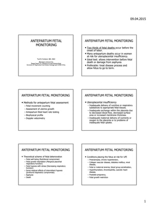
1. 09.04.2015
1
ANTEPARTUM FETALANTEPARTUM FETAL
MONITORINGMONITORING
Tevfik Yoldemir MD, BBA
Marmara University
Department of Obstetrics and Gynecology
Division of Reproductive Endocrinology and Infertility
ANTEPARTUM FETAL MONITORINGANTEPARTUM FETAL MONITORING
•• Two thirds of fetal deathsTwo thirds of fetal deaths occur before theoccur before the
onset of labor.onset of labor.
•• ManyMany antepartumantepartum deaths occur in womendeaths occur in women
at risk forat risk for uteroplacentaluteroplacental insufficiency.insufficiency.
•• Ideal test: allows intervention before fetalIdeal test: allows intervention before fetal
death or damage from asphyxia.death or damage from asphyxia.
•• Preferable: treat disease process andPreferable: treat disease process and
allow fetus to go to term.allow fetus to go to term.
ANTEPARTUM FETAL MONITORINGANTEPARTUM FETAL MONITORING
•• Methods forMethods for antepartumantepartum fetal assessmentfetal assessment
–– Fetal movement countingFetal movement counting
–– Assessment of uterine growthAssessment of uterine growth
–– AntepartumAntepartum fetal heart rate testingfetal heart rate testing
–– Biophysical profileBiophysical profile
–– DopplerDoppler velocimetryvelocimetry
ANTEPARTUM FETAL MONITORINGANTEPARTUM FETAL MONITORING
•• Uteroplacental insufficiencyUteroplacental insufficiency
–– Inadequate delivery of nutritive or respiratoryInadequate delivery of nutritive or respiratory
substances to appropriate fetal tissues.substances to appropriate fetal tissues.
–– Inadequate exchange within the placenta dueInadequate exchange within the placenta due
to decreased blood flow, decreased surfaceto decreased blood flow, decreased surface
area or increased membrane thickness.area or increased membrane thickness.
–– Inadequate maternal delivery of nutrients orInadequate maternal delivery of nutrients or
oxygen to the placenta or to problems ofoxygen to the placenta or to problems of
inadequate fetal uptake.inadequate fetal uptake.
ANTEPARTUM FETAL MONITORINGANTEPARTUM FETAL MONITORING
•• Theoretical scheme of fetal deteriorationTheoretical scheme of fetal deterioration
–– Fetal well being (Nutritional compromise)Fetal well being (Nutritional compromise)
–– Fetal growth retardation (Marginal placentalFetal growth retardation (Marginal placental
respiratory function)respiratory function)
–– Fetal hypoxia with stress (Decreasing respiratoryFetal hypoxia with stress (Decreasing respiratory
function)function)
–– Some residual effects of intermittent hypoxiaSome residual effects of intermittent hypoxia
(profound respiratory compromise)(profound respiratory compromise)
–– AsphyxiaAsphyxia
–– DeathDeath
ANTEPARTUM FETAL MONITORINGANTEPARTUM FETAL MONITORING
•• Conditions placing the fetus at risk for UPIConditions placing the fetus at risk for UPI
–– Preeclampsia, chronic hypertension,Preeclampsia, chronic hypertension,
–– Collagen vascular disease, diabetes mellitus, renalCollagen vascular disease, diabetes mellitus, renal
disease,disease,
–– Fetal or maternal anemia, blood group sensitization,Fetal or maternal anemia, blood group sensitization,
–– Hyperthyroidism, thrombophilia, cyanotic heartHyperthyroidism, thrombophilia, cyanotic heart
disease,disease,
–– Postdate pregnancy,Postdate pregnancy,
–– Fetal growth restrictionFetal growth restriction
2. 09.04.2015
2
ANTEPARTUM FETAL MONITORINGANTEPARTUM FETAL MONITORING
•• Fetal movement countingFetal movement counting
–– Maternal perception of a decrease in fetalMaternal perception of a decrease in fetal
movements may be a sign of impending fetalmovements may be a sign of impending fetal
death.death.
–– It costs nothing.It costs nothing.
–– In a systematic fashion, especially in low riskIn a systematic fashion, especially in low risk
populations, may detect unsuspected fetalpopulations, may detect unsuspected fetal
jeopardy.jeopardy.
ANTEPARTUM FETAL MONITORINGANTEPARTUM FETAL MONITORING
•• Fetal movement countingFetal movement counting
–– 3 movements in 30 minutes (Sadovsky).3 movements in 30 minutes (Sadovsky).
–– Elapsed time to register 10 fetal movementsElapsed time to register 10 fetal movements
(Moore and Piacquadio).(Moore and Piacquadio).
ANTEPARTUM FETAL MONITORINGANTEPARTUM FETAL MONITORING
•• Assessment of uterine growthAssessment of uterine growth
–– General rule: fundal height in centimeters will equalGeneral rule: fundal height in centimeters will equal
the weeks of gestation.the weeks of gestation.
–– Exceptions: maternal obesity, multiple gestation,Exceptions: maternal obesity, multiple gestation,
polyhydramnios, abnormal fetal lie, oligohydramnios,polyhydramnios, abnormal fetal lie, oligohydramnios,
low fetal station, and fetal growth restriction.low fetal station, and fetal growth restriction.
–– Abnormalities of fundal height should lead to furtherAbnormalities of fundal height should lead to further
investigation.investigation.
–– Accuracy: poor?Accuracy: poor?
ANTEPARTUM FETAL MONITORINGANTEPARTUM FETAL MONITORING
•• When to begin testingWhen to begin testing
–– Single factors with minimal to moderateSingle factors with minimal to moderate
increased risk forincreased risk for antepartumantepartum fetal death:fetal death: 3232
weeksweeks..
–– Highest maternal risk factors: 26 weeks.Highest maternal risk factors: 26 weeks.
–– When estimated fetal maturity is sufficient toWhen estimated fetal maturity is sufficient to
expect a reasonable chance of survival shouldexpect a reasonable chance of survival should
intervention be necessary.intervention be necessary.
ANTEPARTUM FETAL MONITORINGANTEPARTUM FETAL MONITORING
•• Which test to use?Which test to use?
–– Contraction stress testContraction stress test
•• Low incidence of unexpected fetal deathLow incidence of unexpected fetal death
•• Increase in time, cost and inconvenienceIncrease in time, cost and inconvenience
–– Nonstress testNonstress test
–– Biophysical profile, modified biophysicalBiophysical profile, modified biophysical
profileprofile
–– Doppler velocimetryDoppler velocimetry
ANTEPARTUM FETAL MONITORINGANTEPARTUM FETAL MONITORING
•• Nonstress test (NST)Nonstress test (NST)
–– Healthy fetuses display normal oscillations andHealthy fetuses display normal oscillations and
fluctuations of the baseline FHR (Hammacher, 1966;fluctuations of the baseline FHR (Hammacher, 1966;
Kubli, 1969).Kubli, 1969).
–– Absence of these patterns was associated withAbsence of these patterns was associated with
increase in neonatal depression and perinatalincrease in neonatal depression and perinatal
mortality.mortality.
–– Accelerations of the FHR during stress testingAccelerations of the FHR during stress testing
correlated with fetal well being (Trierweiler, 1976).correlated with fetal well being (Trierweiler, 1976).
3. 09.04.2015
3
ANTEPARTUM FETAL MONITORINGANTEPARTUM FETAL MONITORING
•• Nonstress test (NST)Nonstress test (NST)
–– Accelerations of the FHR occur with fetalAccelerations of the FHR occur with fetal
movement, uterine contractions, or inmovement, uterine contractions, or in
response to external stimuli.response to external stimuli.
–– FHR accelerations appear to be a reflection ofFHR accelerations appear to be a reflection of
CNS alertness and activity.CNS alertness and activity.
–– Absence of FHR accelerations seems to depictAbsence of FHR accelerations seems to depict
CNS depression caused by hypoxia, drugs,CNS depression caused by hypoxia, drugs,
fetal sleep, or congenital anomalies.fetal sleep, or congenital anomalies.
ANTEPARTUM FETAL MONITORINGANTEPARTUM FETAL MONITORING
•• Nonstress test (NST)Nonstress test (NST)
–– The endpoint of the NST is the presence or absenceThe endpoint of the NST is the presence or absence
of FHR accelerations within a specified period of time.of FHR accelerations within a specified period of time.
–– Most clinicians use 2 accelerations of 15 beats perMost clinicians use 2 accelerations of 15 beats per
minute (BPM) for 15 seconds in a 20minute (BPM) for 15 seconds in a 20--minute period.minute period.
–– A healthy fetus < 32 weeks’ gestation may not haveA healthy fetus < 32 weeks’ gestation may not have
the reactivity or the accelerations that meet thethe reactivity or the accelerations that meet the
criteria of 15 BPM for 15 seconds.criteria of 15 BPM for 15 seconds.
–– The more remote from term, the more likely thatThe more remote from term, the more likely that
nonreactivity will be due to fetal prematurity.nonreactivity will be due to fetal prematurity.
Basic Features of FH TraceBasic Features of FH Trace
Baseline variability CTGBaseline variability CTG
Baseline variabilityBaseline variability
ANTEPARTUM FETAL MONITORINGANTEPARTUM FETAL MONITORING
•• Performing the NSTPerforming the NST
–– External monitors for contraction and FHRExternal monitors for contraction and FHR
measurement applied.measurement applied.
–– Patient in semiPatient in semi--fowler position or left lateralfowler position or left lateral
tilt (to minimize supine hypotension).tilt (to minimize supine hypotension).
–– Fetal movement is recorded.Fetal movement is recorded.
ANTEPARTUM FETAL MONITORINGANTEPARTUM FETAL MONITORING
•• Interpreting the NSTInterpreting the NST
–– Reactive: 2 or more accelerations in 20Reactive: 2 or more accelerations in 20
minutes.minutes.
•• Accelerations: an increase of at least 15 BPMAccelerations: an increase of at least 15 BPM
above the baseline lasting at least 15 seconds.above the baseline lasting at least 15 seconds.
–– Fetal sound stimulation may be used to elicitFetal sound stimulation may be used to elicit
a response.a response.
4. 09.04.2015
4
ANTEPARTUM FETAL MONITORINGANTEPARTUM FETAL MONITORING
•• Interpreting the NSTInterpreting the NST
–– Non reactive: Less than 2 accelerations in a 20Non reactive: Less than 2 accelerations in a 20--
minute period.minute period.
•• May extend the testing period to 40 minutes or perform aMay extend the testing period to 40 minutes or perform a
backback--up test.up test.
–– There is no universal agreement on the number ofThere is no universal agreement on the number of
accelerations required to consider the test reactive.accelerations required to consider the test reactive.
–– Reactive/Nonreactive with decelerations: individualizeReactive/Nonreactive with decelerations: individualize
managementmanagement
ANTEPARTUM FETAL MONITORINGANTEPARTUM FETAL MONITORING
•• Nonstress testNonstress test
–– Perinatal mortality: 6.2/1000Perinatal mortality: 6.2/1000
–– False positive rate: 50%False positive rate: 50%
–– False negative rate: 3.2 / 1000False negative rate: 3.2 / 1000
www.yoldemir.comwww.yoldemir.com
ANTEPARTUM FETAL MONITORINGANTEPARTUM FETAL MONITORING
•• Contraction stress test (CST)Contraction stress test (CST)
–– Uterine contractions producing an intraUterine contractions producing an intra--amnioticamniotic
pressure in excess of 30 mm Hg create an intrapressure in excess of 30 mm Hg create an intra--
myometrialmyometrial pressure that exceeds mean intrapressure that exceeds mean intra--arterialarterial
pressure, therefore temporarily halting uterine bloodpressure, therefore temporarily halting uterine blood
flow.flow.
–– A hypoxic fetus will manifestA hypoxic fetus will manifest late decelerationslate decelerations..
–– Late decelerations correlate with stillbirth, IUGR, andLate decelerations correlate with stillbirth, IUGR, and
lowlow ApgarApgar scores.scores.
–– OxytocinOxytocin challenge test (OCT)challenge test (OCT)
–– Breast (nipple) stimulationBreast (nipple) stimulation
ANTEPARTUM FETAL MONITORINGANTEPARTUM FETAL MONITORING
•• How to perform the CSTHow to perform the CST
–– External monitors for contraction and FHRExternal monitors for contraction and FHR
measurement applied.measurement applied.
–– Patient inPatient in semisemi--fowler position or left lateralfowler position or left lateral
tilt (to minimize supine hypotension).tilt (to minimize supine hypotension).
–– Protocol forProtocol for oxytocinoxytocin infusion or breastinfusion or breast
stimulation.stimulation.
–– Goal:Goal: three contractions in ten minutesthree contractions in ten minutes..
5. 09.04.2015
5
ANTEPARTUM FETAL MONITORINGANTEPARTUM FETAL MONITORING
•• Interpretation of the CSTInterpretation of the CST
–– Negative:Negative: no late decelerationsno late decelerations and adequateand adequate
FHR recordingFHR recording
–– Positive: Late decelerations present with thePositive: Late decelerations present with the
majority of contractions (without excessivemajority of contractions (without excessive
uterine activity)uterine activity)
–– Equivocal test results: Suspicious,Equivocal test results: Suspicious,
hyperstimulationhyperstimulation, unsatisfactory., unsatisfactory.
www.yoldemir.comwww.yoldemir.com
Early Decelerations
www.yoldemir.comwww.yoldemir.com
Late Decelerations
ANTEPARTUM FETAL MONITORINGANTEPARTUM FETAL MONITORING
•• Interpretation of the CSTInterpretation of the CST
–– Suspicious: Late decelerations are present with lessSuspicious: Late decelerations are present with less
than half of the contractions.than half of the contractions.
–– Hyperstimulation: Decelerations after contractionsHyperstimulation: Decelerations after contractions
lasting more than 90 seconds, or with contractionlasting more than 90 seconds, or with contraction
frequency greater than every 2 minutes.frequency greater than every 2 minutes.
–– Unsatisfactory: Cannot induce adequate contractionsUnsatisfactory: Cannot induce adequate contractions
or FHR recording is of poor quality.or FHR recording is of poor quality.
ANTEPARTUM FETAL MONITORINGANTEPARTUM FETAL MONITORING
•• Other patternsOther patterns
–– Variable decelerations: considerVariable decelerations: consider
oligohydramnios or cord entrapment.oligohydramnios or cord entrapment.
–– Loss of variability and blunting ofLoss of variability and blunting of
decelerations: ominous sign.decelerations: ominous sign.
–– Sinusoidal pattern: ominous pattern. FetalSinusoidal pattern: ominous pattern. Fetal
anemia or fetalanemia or fetal--maternal hemorrhage.maternal hemorrhage.
–– Nonreactive negative CST: should not occur,Nonreactive negative CST: should not occur,
preexisting CNS abnormality?preexisting CNS abnormality?
6. 09.04.2015
6
www.yoldemir.comwww.yoldemir.com
Variable Decelerations
ANTEPARTUM FETAL MONITORINGANTEPARTUM FETAL MONITORING
•• Management of CSTManagement of CST
–– Negative test: repeated weeklyNegative test: repeated weekly
–– Positive test: acted on according to clinicalPositive test: acted on according to clinical
conditioncondition
–– Equivocal test: repeat test the next dayEquivocal test: repeat test the next day
ANTEPARTUM FETAL MONITORINGANTEPARTUM FETAL MONITORING
•• When to shorten the interval betweenWhen to shorten the interval between
testingtesting
–– Deterioration in diabetic controlDeterioration in diabetic control
–– Worsening hypertensionWorsening hypertension
–– Need to introduce antihypertensiveNeed to introduce antihypertensive
medicationmedication
–– Decreased fetal movementDecreased fetal movement
ANTEPARTUM FETAL MONITORINGANTEPARTUM FETAL MONITORING
•• Contraindications to CSTContraindications to CST
–– PROMPROM
–– Previous classical cesarean deliveryPrevious classical cesarean delivery
–– Placenta previaPlacenta previa
–– Incompetent cervixIncompetent cervix
–– History of premature labor in this pregnancyHistory of premature labor in this pregnancy
–– Multiple gestationMultiple gestation
7. 09.04.2015
7
ANTEPARTUM FETAL MONITORINGANTEPARTUM FETAL MONITORING
•• Biophysical profile (BPP)Biophysical profile (BPP)
–– Described by Manning (1980)Described by Manning (1980)
–– The number of biophysical activities thatThe number of biophysical activities that
could be recorded increased with real timecould be recorded increased with real time
ultrasound:ultrasound:
•• Fetal movement (FM)Fetal movement (FM)
•• Fetal tone (FT)Fetal tone (FT)
•• Fetal breathing movements (FB)Fetal breathing movements (FB)
•• Amniotic fluid volume (AFV)Amniotic fluid volume (AFV)
ANTEPARTUM FETAL MONITORINGANTEPARTUM FETAL MONITORING
•• Biophysical profile (BPP)Biophysical profile (BPP) –– variablesvariables
–– NST: reactiveNST: reactive –– as described earlier.as described earlier.
–– FBM: presentFBM: present -- at least 1 episode of at least 30at least 1 episode of at least 30
seconds duration (within a 30 minute period).seconds duration (within a 30 minute period).
–– FM: presentFM: present -- at least 3 discrete episodes.at least 3 discrete episodes.
–– FT: normalFT: normal -- at least 1 episode of extension ofat least 1 episode of extension of
extremities or spine with return to flexion.extremities or spine with return to flexion.
–– AFV: normalAFV: normal –– largest pocket of fluid greater than 1largest pocket of fluid greater than 1
cm in vertical diameter.cm in vertical diameter.
ANTEPARTUM FETAL MONITORINGANTEPARTUM FETAL MONITORING
•• Biophysical profile (BPP)Biophysical profile (BPP)
–– Each variableEach variable
•• When normal: 2When normal: 2
•• When abnormal: 0When abnormal: 0
–– Highest Score: 10, Lowest Score: 0Highest Score: 10, Lowest Score: 0
–– Accuracy improved by increasing the numberAccuracy improved by increasing the number
of variables assessed.of variables assessed.
–– Overall false negative rate: 0.6/1000Overall false negative rate: 0.6/1000
ANTEPARTUM FETAL MONITORINGANTEPARTUM FETAL MONITORING
•• Biophysical profile (BPP)Biophysical profile (BPP)
–– Acute markers of fetal compromise: NST, FT,Acute markers of fetal compromise: NST, FT,
FBM, FMFBM, FM
–– Chronic marker of fetal compromise: AFVChronic marker of fetal compromise: AFV
–– Nervous impulses that initiate fetal biophysicalNervous impulses that initiate fetal biophysical
activities arise from different anatomic sitesactivities arise from different anatomic sites
within the brain.within the brain.
ANTEPARTUM FETAL MONITORINGANTEPARTUM FETAL MONITORING
•• Biophysical profile (BPP)Biophysical profile (BPP)
–– Activities that become active first in fetalActivities that become active first in fetal
development (development (FTFT,, FMFM) are the last to) are the last to
disappear when asphyxia arrests all activities.disappear when asphyxia arrests all activities.
–– Activities that become active later in gestationActivities that become active later in gestation
((NSTNST,,FBMFBM) will be abolished 1) will be abolished 1stst in cases ofin cases of
hypoxia and acidosis.hypoxia and acidosis.
8. 09.04.2015
8
ANTEPARTUM FETAL MONITORINGANTEPARTUM FETAL MONITORING
•• Biophysical profile (BPP)Biophysical profile (BPP)
–– Fetal tone: 7.5 to 8.5 weeksFetal tone: 7.5 to 8.5 weeks
–– Fetal movement: 9 weeksFetal movement: 9 weeks
–– Fetal breathing: 20 to 21 weeksFetal breathing: 20 to 21 weeks
–– NST: 24 to 28 weeksNST: 24 to 28 weeks
ANTEPARTUM FETAL MONITORINGANTEPARTUM FETAL MONITORING
•• Biophysical profile (BPP)Biophysical profile (BPP)
–– When hypoxia and acidosisWhen hypoxia and acidosis
•• Late decelerations appear (CST)Late decelerations appear (CST)
•• Accelerations disappear (CST, NST, BPP)Accelerations disappear (CST, NST, BPP)
•• Fetal breathing stops (BPP)Fetal breathing stops (BPP)
•• Fetal movement ceases (BPP, FMC)Fetal movement ceases (BPP, FMC)
•• Fetal tone absent (BPP)Fetal tone absent (BPP)
–– Assessment of fetal wellAssessment of fetal well--being in high riskbeing in high risk
pregnanciespregnancies
•• Reduced perinatal mortality rate from 65/1000 to 5/1000Reduced perinatal mortality rate from 65/1000 to 5/1000
ANTEPARTUM FETAL MONITORINGANTEPARTUM FETAL MONITORING
•• BPP and perinatal mortality (PNMR)BPP and perinatal mortality (PNMR)
–– 12,000 pregnancies (Manning, 1985)12,000 pregnancies (Manning, 1985)
–– BPP Score Corrected PNMRBPP Score Corrected PNMR
•• 88--10 0.610 0.6
•• 6 0.06 0.0
•• 4 22.04 22.0
•• 2 42.62 42.6
•• 0 187.00 187.0
ANTEPARTUM FETAL MONITORINGANTEPARTUM FETAL MONITORING
•• Biophysical profile (BPP)Biophysical profile (BPP)
–– When the FHR accelerates, there is virtuallyWhen the FHR accelerates, there is virtually
always fetal movement (FM)always fetal movement (FM)
–– If the NST is reactive, there is fetal movementIf the NST is reactive, there is fetal movement
(FM) and tone (FT)(FM) and tone (FT)
–– If the NST is reactive, do not need theIf the NST is reactive, do not need the
ultrasound parameters of the BPPultrasound parameters of the BPP
–– Only the AFV would add additionalOnly the AFV would add additional
informationinformation
ANTEPARTUM FETAL MONITORINGANTEPARTUM FETAL MONITORING
•• Modified biophysical profile (BPP)Modified biophysical profile (BPP)
–– A standard NST is combined with an amniotic fluidA standard NST is combined with an amniotic fluid
index (AFI)index (AFI)
–– Negative: Reactive NST / AFI > 5.0 cmNegative: Reactive NST / AFI > 5.0 cm
–– If NST is nonreactive or has decelerations, or if theIf NST is nonreactive or has decelerations, or if the
AFI isAFI is << 5.0 cm, then a BPP is performed.5.0 cm, then a BPP is performed.
–– Negative results are repeated every 3 to 4 days.Negative results are repeated every 3 to 4 days.
–– If the AFI > 5.0 cm, a repeat AFI may be done in oneIf the AFI > 5.0 cm, a repeat AFI may be done in one
week.week.
9. 09.04.2015
9
ANTEPARTUM FETAL MONITORINGANTEPARTUM FETAL MONITORING
•• Primary fetal surveillancePrimary fetal surveillance
–– There have been no adequate prospectiveThere have been no adequate prospective
randomized studies comparing the various testingrandomized studies comparing the various testing
modalities.modalities.
–– The final decision regarding choice of fetalThe final decision regarding choice of fetal
surveillance test is most often determined bysurveillance test is most often determined by
institutional preference and experience.institutional preference and experience.
–– All forms of fetal testing are valuable and need to beAll forms of fetal testing are valuable and need to be
interpreted cautiously with full knowledge of theinterpreted cautiously with full knowledge of the
specific test limitations.specific test limitations.
ANTEPARTUM FETAL MONITORINGANTEPARTUM FETAL MONITORING
•• Primary fetal surveillancePrimary fetal surveillance
–– NST: The most popular methodNST: The most popular method
•• Easy to perform, easy to interpret, has fewerEasy to perform, easy to interpret, has fewer
equivocal results, has excellent patient andequivocal results, has excellent patient and
physician acceptance.physician acceptance.
•• BPP as a back up test.BPP as a back up test.
–– BPP:BPP:
•• Can identify oligohydramnios and anomalousCan identify oligohydramnios and anomalous
babies.babies.
•• Antepartum death rate is less than with the NST.Antepartum death rate is less than with the NST.
ANTEPARTUM FETAL MONITORINGANTEPARTUM FETAL MONITORING
•• Doppler velocimetry of the umbilical arteriesDoppler velocimetry of the umbilical arteries
–– 40% of combined ventricular output is directed to the40% of combined ventricular output is directed to the
placenta by umbilical arteries.placenta by umbilical arteries.
–– Assessment of umbilical blood flow providesAssessment of umbilical blood flow provides
information on blood perfusion of the fetoplacentalinformation on blood perfusion of the fetoplacental
unit.unit.
–– Volume of flow increases and vascular impedanceVolume of flow increases and vascular impedance
decreases with advancing gestational age.decreases with advancing gestational age.
–– Low vascular impedance allows a continuous forwardLow vascular impedance allows a continuous forward
blood flow throughout the cardiac cycle.blood flow throughout the cardiac cycle.
ANTEPARTUM FETAL MONITORINGANTEPARTUM FETAL MONITORING
•• Doppler velocimetryDoppler velocimetry
–– An increase in the vascular resistance of theAn increase in the vascular resistance of the
fetoplacental unit leads to a decrease in end diastolicfetoplacental unit leads to a decrease in end diastolic
flow velocity or its absence in the flow velocityflow velocity or its absence in the flow velocity
waveform.waveform.
–– Abnormal waveforms reflect the presence of aAbnormal waveforms reflect the presence of a
structural placental lesion.structural placental lesion.
–– Abnormal Doppler results require specificAbnormal Doppler results require specific
management protocols and intensive fetalmanagement protocols and intensive fetal
surveillance.surveillance.
ANTEPARTUM FETAL MONITORINGANTEPARTUM FETAL MONITORING
•• Doppler velocimetryDoppler velocimetry
–– A poor indicator of fetal compromise orA poor indicator of fetal compromise or
adaptation to the placental abnormality butadaptation to the placental abnormality but
does identify patients at risk for increaseddoes identify patients at risk for increased
perinatal mortality.perinatal mortality.
–– Strong association between high systolic toStrong association between high systolic to
diastolic ratios and IUGR.diastolic ratios and IUGR.
ThankThank youyou forfor youryour attentionattention..