
More Related Content
What's hot
What's hot (20)
Viewers also liked
Viewers also liked (12)
Similar to Ankle Syndesmosis TightRope
Similar to Ankle Syndesmosis TightRope (20)
Recently uploaded
Recently uploaded (20)
Ankle Syndesmosis TightRope
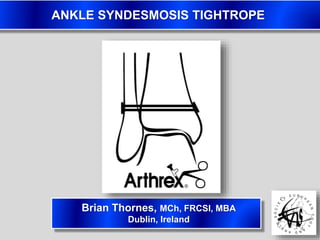
- 1. ANKLE SYNDESMOSIS TIGHTROPE Brian Thornes, MCh, FRCSI, MBA Dublin, Ireland
- 3. LOSS OF SCREW FIXATION
- 4. COMPLICATIONS REMOVING SCREWS 2011: Schepers T, et al Complications of Syndesmosis Screw Removal 76 patients N=7 (9%) wound infection N=5 (7%) recurrent diastasis N=5 (7%) occult broken screw TOTAL: 22% complication rate
- 5. COMPLICATIONS REMOVING SCREWS 2007: S. Hakkalamani, et al Syndesmotic screw removal in Weber ‘C’ ankle fractures 42 patients N=6 wound infection (14%) N=4 instability pain (10%) N=1 DVT N=1 occult broken screw TOTAL: 26% complication rate
- 6. LATE DIASTASIS FOLLOWING REMOVAL 2007: Wahlquist M. Late Diastasis of the Syndesmosis following Syndesmotic Screw Removal (podium presentation) 21 patients Average 2mm widening of tibio-fibular clear space 38% of patients symptomatic
- 9. BROKEN SCREWS BEST ??!! 2009 Hamid N, et al Outcome after fixation of ankle fractures with an injury to the syndesmosis. The effect of a syndesmosis screw 52 patients 27 intact screws (AOFAS score 83) 15 elective removal (AOFAS score 86) 10 broken screws (AOFAS score 92) Average 30 (12-56) month follow-up
- 10. SYNDESMOSIS SCREWS Old Debate / Controversies 1. What size/number of screws to use (3.5mm / 4.5mm)? 2. How many cortices to engage (3 or 4 cortices)? 3. If/when to remove before screw breakage?
- 11. SYNDESMOSIS INJURIES Better Questions: 1. What is the healing time for syndesmosis ligaments?
- 12. SYNDESMOSIS INJURIES Better Questions: 1. What is the healing time for syndesmosis ligaments? 2. Is rigid fixation the correct environment to promote healing?
- 13. SYNDESMOSIS INJURIES Better Questions: 1. What is the healing time for syndesmosis ligaments? 2. Is rigid fixation the correct environment to promote healing? 3. How to hold & maintain reduction, with physiological movement?
- 14. GENESIS OF THE TIGHTROPE 2003: Thornes B, Walsh A, Hislop M, Murray P, O’Brien M Suture-Endobutton Fixation of Ankle Tibio-Fibular Diastasis: A Cadaver Study 2005: Thornes B, Shannon F, Guiney AM, Masterson E Suture-Button Syndesmosis Fixation. Accelerated Rehabilitation and Improved Outcomes 2006: Thornes B, McCartan D Ankle Syndesmosis Injuries Treated with the TightRope Suture-Button Kit
- 17. TIGHTROPE
- 18. TIGHTROPE
- 19. 18YR OLD, 120KG WEIGHT...?NWB
- 20. CLINICAL SERIES 2009: Cottom JM Transosseous fixation of the syndesmosis: Comparison of suture-button to traditional screw fixation in 50 cases 25 Tightrope vs 25 Screw cohorts Similar ankle outcome scores 68% removal rate with screws 0% removal rate with TightRope
- 21. CLINICAL SERIES 2009: Coetzee JC Treatment of syndesmoses disruptions: A prospective, randomized study of screw fixation vs TightRope® 12 TightRope vs 12 Screw cases 12 month AOFAS score: 85 (TightRope) vs 76 (screw) Significantly better range of motion in TightRope group
- 22. CLINICAL SERIES 2011: DeGroot H, et al Outcomes of Suture Button Repair of the Distal Tibiofibular Syndesmosis 24 TightRope cases AOFAS score: 94 (71-100) at 18 months 6 cases: local irritation from button/suture knot... elective removal without difficulty
- 23. CLINICAL SERIES 2011: DeGroot H, et al Outcomes of Suture Button Repair of the Distal Tibiofibular Syndesmosis DISCUSSION “In summary, we believe the suture button device represents a viable alternative to screw fixation for syndesmosis injuries. The disrupted syndesmotic relationships were normalised by the application of the suture button and remained within normal limits through the study period in all cases. Because of the ease of use of the device and the ability to allow full weightbearing without concerns about implant breakage, we feel that suture-button fixation is superior to conventional metallic screws.”
- 24. CLINICAL SERIES 2012 (in press): Naqvi GA, Shafqat A, Awan N Tightrope fixation of ankle syndesmosis injuries: Clinical outcome, complications and technique modification 49 TightRope cases AOFAS score 86 (78-93) at 6 months 3 cases of implant removal (irritation/infection) Senior author recommends burying lateral suture tails sub-perisoteally
- 25. CURRENT CONTOVERSIES 1. Mal-Reduction 2. Mid-diaphyseal Fibular Fractures 3. Osteoporotic bone
- 26. MAL-REDUCTION 25 screw patients 52% incongruity of fibula within incisura on postop CT scan 2006: Gardner M, et al Malareduction of the Tibiofibular Syndesmosis in Ankle Fractures
- 28. MID-SHAFT FIBULA FRACTURE 2008. Ho JY et al. Mid-Diaphyseal Fibular Fractures with Syndesmotic Disruption: Should We Plate the Fibula? Cadaver study, 8 paired samples • Rotational stability • Load-to-failure • Stiffness All better with additional fibular plating versus syndesmosis (screw) fixation alone Therefore: if you can, FIX THE FIBULA
- 29. OSTEOPOROTIC ANKLE #’S INCREASING
- 30. OSTEOPOROTIC ANKLE #’S INCREASING
- 31. OSTEOPOROTIC ANKLE #’S INCREASING
- 32. 81YR OLD, OSTEOPOROTIC LADY...?NWB
- 33. NEXT TIME...