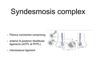
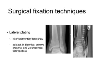
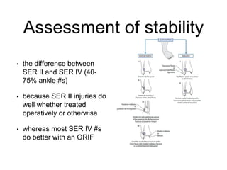
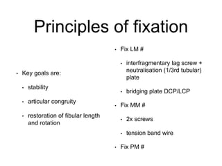
Ankle fracture fixation
Derek Park
Consultant Trauma & Orthopaedic Surgeon
Barnet & Chase Farm Hospitals
Outline
• Surgical anatomy and
approaches
• Surgical fixation techniques
Ankle fractures
• Why does this ankle
fracture…
Ankle fractures
• End up like this?
Ankle fractures
• And why do Orthopaedic
surgeons do things like
this?
Ankle fractures
• Or this?
Ankle fractures
• Or even this?
Why do people get it wrong?
• Underestimation of the fracture
• Lack of understanding of ankle
fracture patterns
• Lack of understanding of the
basic principles of fracture
reduction & stabilisation
Surgical and functional anatomy of
the ankle joint
• Ankle joint = 3 bone articulation
between tibia, fibula and talus
• 80% load transmitted through
the tibial plafond of the talar
dome in single leg stance
• Talar dome is wider anteriorly
than posteriorly = section of a
cone, with apex medial, fully
congruous
Surgical and functional anatomy of
the ankle joint
• Strong ligaments support
the ankle Medially:
• superficial deltoid ligament
• deep deltoid ligament
• Deep deltoid acts as a
check-rein to abnormal
talar movement
• Draws in and stabilizes the
talus on axial loading
Surgical and functional anatomy of
the ankle joint
• Lateral ligament complex
comprises the:
• anterior talofibular ligament - ATFL
• calcaneofibular ligament- CFL
• posterior talofibular ligament - PTFL
Syndesmosis complex
• Fibrous connection comprising:
• anterior & posterior tibiofibular
ligaments (AITFL & PITFL)
• interosseous ligament
Surgical approaches
• Direct lateral
• Posterolateral approach
• Peroneus & FHL
Surgical fixation techniques
• Lateral plating
• Interfragmentary lag screw
• at least 2x bicortical screws
proximal and 2x unicortical
screws distal
Surgical fixation techniques
• Posterior (antiglide) plating
Surgical fixation techniques
• Distal fibular locked plating
• decreased bone stock
• severely comminuted fx
Treatment rationale
• What is stable and what
isn’t?
• How do you assess
stability?
Weber classification
• Level of fibular fracture
relative to the syndesmosis
• A = below syndesmosis
• B = level of syndesmosis
• C = above level of
syndesmosis
1. Position of the foot
2. Deforming force
Assessment of stability
• the difference between
SER II and SER IV (40-
75% ankle #s)
• because SER II injuries do
well whether treated
operatively or otherwise
• whereas most SER IV #s
do better with an ORIF
Supination/Adduction
Pronation/External rotation
• Is it a PER injury?
• If “high” fibular fracture (Weber
C) = Yes
• ORIF
Principles of fixation
• Key goals are:
• stability
• articular congruity
• restoration of fibular length
and rotation
• Fix LM #
• interfragmentary lag screw +
neutralisation (1/3rd tubular)
plate
• bridging plate DCP/LCP
• Fix MM #
• 2x screws
• tension band wire
• Fix PM #
Diastasis screw - how
• Primary issues
• clamp reduction
• position 2-3cm above joint line
• position screw not compression
screw
• through plate if low enough
• Secondary issues
• 1 vs 2 screws
• 3.5mm vs 4.5mm screws
• 3 vs 4 cortices
• ankle position at time of
insertion
• post-op regimen
In Summary
• Isolated Weber B LM #s, no talar shift, no medial
signs = (probably) Stable
• Isolated Weber B LM #s, no talar shift, +ve medial
signs = Assess for stability
• Bimalleolar #s = ORIF
• Isolated Weber B LM #s with talar shift = ORIF
• Weber C LM #s = ORIF

Anke fx fixation - Derek Park

  • 1.
    Ankle fracture fixation DerekPark Consultant Trauma & Orthopaedic Surgeon Barnet & Chase Farm Hospitals
  • 2.
    Outline • Surgical anatomyand approaches • Surgical fixation techniques
  • 3.
    Ankle fractures • Whydoes this ankle fracture…
  • 4.
  • 5.
    Ankle fractures • Andwhy do Orthopaedic surgeons do things like this?
  • 6.
  • 7.
  • 8.
    Why do peopleget it wrong? • Underestimation of the fracture • Lack of understanding of ankle fracture patterns • Lack of understanding of the basic principles of fracture reduction & stabilisation
  • 9.
    Surgical and functionalanatomy of the ankle joint • Ankle joint = 3 bone articulation between tibia, fibula and talus • 80% load transmitted through the tibial plafond of the talar dome in single leg stance • Talar dome is wider anteriorly than posteriorly = section of a cone, with apex medial, fully congruous
  • 10.
    Surgical and functionalanatomy of the ankle joint • Strong ligaments support the ankle Medially: • superficial deltoid ligament • deep deltoid ligament • Deep deltoid acts as a check-rein to abnormal talar movement • Draws in and stabilizes the talus on axial loading
  • 11.
    Surgical and functionalanatomy of the ankle joint • Lateral ligament complex comprises the: • anterior talofibular ligament - ATFL • calcaneofibular ligament- CFL • posterior talofibular ligament - PTFL
  • 12.
    Syndesmosis complex • Fibrousconnection comprising: • anterior & posterior tibiofibular ligaments (AITFL & PITFL) • interosseous ligament
  • 13.
    Surgical approaches • Directlateral • Posterolateral approach • Peroneus & FHL
  • 14.
    Surgical fixation techniques •Lateral plating • Interfragmentary lag screw • at least 2x bicortical screws proximal and 2x unicortical screws distal
  • 15.
  • 16.
    Surgical fixation techniques •Posterior (antiglide) plating
  • 17.
    Surgical fixation techniques •Distal fibular locked plating • decreased bone stock • severely comminuted fx
  • 18.
    Treatment rationale • Whatis stable and what isn’t? • How do you assess stability?
  • 19.
    Weber classification • Levelof fibular fracture relative to the syndesmosis • A = below syndesmosis • B = level of syndesmosis • C = above level of syndesmosis
  • 20.
    1. Position ofthe foot 2. Deforming force
  • 21.
    Assessment of stability •the difference between SER II and SER IV (40- 75% ankle #s) • because SER II injuries do well whether treated operatively or otherwise • whereas most SER IV #s do better with an ORIF
  • 22.
  • 23.
    Pronation/External rotation • Isit a PER injury? • If “high” fibular fracture (Weber C) = Yes • ORIF
  • 24.
    Principles of fixation •Key goals are: • stability • articular congruity • restoration of fibular length and rotation • Fix LM # • interfragmentary lag screw + neutralisation (1/3rd tubular) plate • bridging plate DCP/LCP • Fix MM # • 2x screws • tension band wire • Fix PM #
  • 25.
    Diastasis screw -how • Primary issues • clamp reduction • position 2-3cm above joint line • position screw not compression screw • through plate if low enough • Secondary issues • 1 vs 2 screws • 3.5mm vs 4.5mm screws • 3 vs 4 cortices • ankle position at time of insertion • post-op regimen
  • 31.
    In Summary • IsolatedWeber B LM #s, no talar shift, no medial signs = (probably) Stable • Isolated Weber B LM #s, no talar shift, +ve medial signs = Assess for stability • Bimalleolar #s = ORIF • Isolated Weber B LM #s with talar shift = ORIF • Weber C LM #s = ORIF