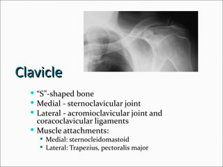
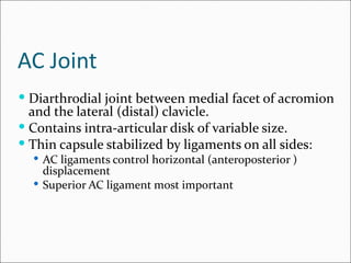
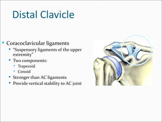
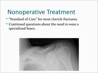
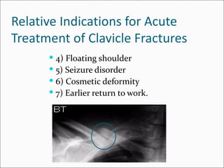
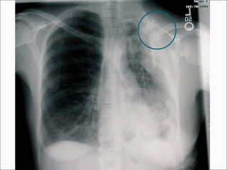
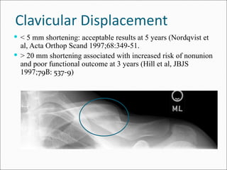
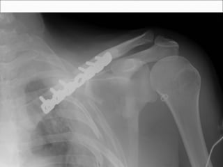
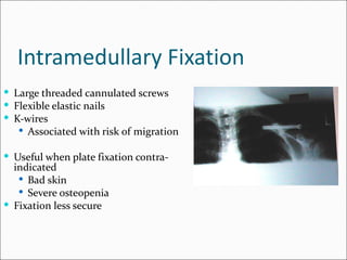
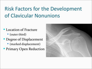
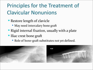
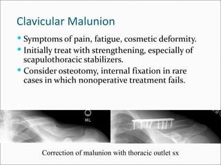
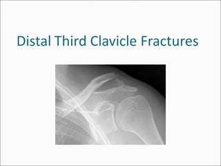
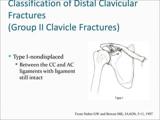
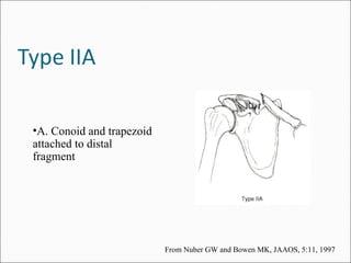
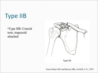
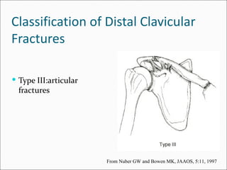
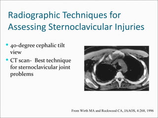
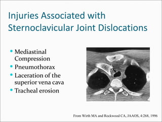
Clavicle
   “S”-shaped bone
   Medial - sternoclavicular joint
   Lateral - acromioclavicular joint and
    coracoclavicular ligaments
   Muscle attachments:
     Medial: sternocleidomastoid
     Lateral: Trapezius, pectoralis major
AC Joint
 Diarthrodial joint between medial facet of acromion
  and the lateral (distal) clavicle.
 Contains intra-articular disk of variable size.
 Thin capsule stabilized by ligaments on all sides:
   AC ligaments control horizontal (anteroposterior )
    displacement
   Superior AC ligament most important
Distal Clavicle

 Coracoclavicular ligaments
   “Suspensory ligaments of the upper
    extremity”
   Two components:
       Trapezoid
       Conoid
   Stronger than AC ligaments
   Provide vertical stability to AC joint
Mechanism of Injury
 Moderate or high-energy traumatic impacts to the
    shoulder
    1.   Fall from height
    2.   Motor vehicle accident
    3.   Sports injury
    4.   Blow to the point of the shoulder
    5.   Rarely a direct injury to the clavicle
Physical Examination
 Inspection
    Evaluate deformity and/or
     displacement
    Beware of rare inferior or
     posterior displacement of distal
     or medial ends of clavicle
    Compare to opposite side.
Physical Examination

   Palpation
      Evaluate pain
      Look for instability with stress
Physical Examination

 Neurovascular examination
 Evaluate upper extremity motor and sensation
 Measure shoulder range-of-motion
Radiographic Evaluation
 of the Clavicle

 Anteroposterior
 View


 30-degree Cephalic
 Tilt View
Clavicle Fractures
Classification of
Clavicle Fractures

     Group I : Middle third
       Most common (80% of clavicle fractures)
     Group II: Distal third
       10-15% of clavicle injuries
     Group III: Medial third
       Least common (approx. 5%)
Treatment Options
      Nonoperative
         Sling
         Brace
      Surgical
        Plate Fixation
        Screw or Pin Fixation
Nonoperative Treatment
 “Standard of Care” for most clavicle fractures.
 Continued questions about the need to wear a
 specialized brace.
Simple Sling vs.
Figure-of-8 Bandage
  Prospective randomized trial of 61 patients
  Simple sling
    Less discomfort
  Functional and cosmetic results identical
  Alignment of healed fractures unchanged from the
  initial displacement in both groups


             Andersen et al., Acta Orthop Scand 58: 71-4, 1987.
Nonoperative Treatment
 It is difficult to reduce clavicle fractures by closed
  means.
 Most clavicle fractures unite rapidly despite
  displacement
 Significantly displaced mid-shaft and distal-third
  injuries have a higher incidence of nonunion.
Nonoperative Treatment
 There is new evidence that the outcome of
 nonoperative management of displaced middle-third
 clavicle fractures is not as good as traditionally
 thought, with many patients having significant
 functional problems.
Deficits following nonoperative treatment of
displaced midshaft clavicular fractures
  A patient-based outcome questionnaire and muscle-
   strength testing were used to evaluate 30 patients after
   nonoperative care of a displaced midshaft fracture of
   the clavicle.
  At a minimum of twelve months (mean 55 mos),
   outcomes were measured with the Constant shoulder
   score and the DASH patient questionnaire. In addition,
   shoulder muscle-strength testing was performed with
   the Baltimore Therapeutic Equipment Work Simulator,
   with the uninjured arm serving as a control.


McKee et al. J Bone Joint Surg Am 2006;88-A:35-40.
Deficits following nonoperative treatment of
displaced midshaft clavicular fractures
  The strength of the injured shoulder was 81%
   for maximum flexion, 75% for endurance of
   flexion, 82% for maximum abduction, 67% for
   endurance of abduction, 81% for maximum
   external rotation, 82% for endurance of external
   rotation, 85% for maximum internal rotation,
   and 78% for endurance of internal rotation (p <
   0.05 for all).
  The mean Constant score was 71 points, and the
   mean DASH score was 24.6 points, indicating
   substantial residual disability.

McKee et al. J Bone Joint Surg Am 2006;88-A:35-40.
 Displaced midshaft clavicle fractures can cause
 significant, persistent disability, even if they heal
 uneventfully.
Definite Indications for Surgical
Treatment of Clavicle Fractures
   1) Open fractures
   2) Associated neurovascular injury
Relative Indications for Acute
Treatment of Clavicle Fractures
    1) Widely displaced fractures
    2) Multiple trauma
    3) Displaced distal-third fractures
Relative Indications for Acute
Treatment of Clavicle Fractures
       4) Floating shoulder
       5) Seizure disorder
       6) Cosmetic deformity
       7) Earlier return to work.
Clavicular Displacement
 < 5 mm shortening: acceptable results at 5 years (Nordqvist et
  al, Acta Orthop Scand 1997;68:349-51.
 > 20 mm shortening associated with increased risk of nonunion
  and poor functional outcome at 3 years (Hill et al, JBJS
  1997;79B: 537-9)
Plate Fixation
 Traditional means of ORIF
 Plate applied superiorly or inferiorly
   Inferior plating associated with lower risk of hardware
    prominence
 Used for acute displaced fractures and nonunions.
Intramedullary Fixation
 Large threaded cannulated screws
 Flexible elastic nails
 K-wires
    Associated with risk of migration

 Useful when plate fixation contra-
  indicated
    Bad skin
    Severe osteopenia
 Fixation less secure
Complications of Clavicular
Fractures and its Treatment
      Nonunion
      Malunion
      Neurovascular Sequelae
      Post-Traumatic Arthritis
Risk Factors for the Development
of Clavicular Nonunions

 Location of Fracture
   (outer third)
 Degree of Displacement
   (marked displacement)
 Primary Open Reduction
Principles for the Treatment of
Clavicular Nonunions
 Restore length of clavicle
   May need intercalary bone graft
 Rigid internal fixation, usually with a plate
 Iliac crest bone graft
   Role of bone-graft substitutes not yet defined.
Clavicular Malunion
 Symptoms of pain, fatigue, cosmetic deformity.
 Initially treat with strengthening, especially of
  scapulothoracic stabilizers.
 Consider osteotomy, internal fixation in rare
  cases in which nonoperative treatment fails.


       Correction of malunion with thoracic outlet sx
Neurologic Sequelae
 Occasionally, fracture fragments or abundant callus
  can cause brachial plexus symptoms.
 Treatment is reduction and fixation of the fracture, or
  resection of callus with or without osteotomy and
  fixation for malunions.
Classification of Distal Clavicular
Fractures
(Group II Clavicle Fractures)

 Type I-nondisplaced
    Between the CC and AC
     ligaments with ligament
     still intact


                           From Nuber GW and Bowen MK, JAAOS, 5:11, 1997
Classification of Distal Clavicular
Fractures
    Type II
      Typically displaced secondary to a
       fracture medial to the coracoclavicular
       ligaments, keeping the distal fragment
       reduced while allowing the medial
       fragmetn to displace superiorly
      Highest rate of nonunion (up to 30%)
      Two Types
•A. Conoid and trapezoid
attached to distal
fragment


                           From Nuber GW and Bowen MK, JAAOS, 5:11, 1997
•Type IIB: Conoid
torn, trapezoid
attached


                    From Nuber GW and Bowen MK, JAAOS, 5:11, 1997
Classification of Distal Clavicular
Fractures

 Type III:articular
  fractures


                       From Nuber GW and Bowen MK, JAAOS, 5:11, 1997
Treatment of Distal-Third (Type II)
Clavicle Fractures
        Nonoperative treatment
          22 to 33% failed to unite
          45 to 67% took more than three months to heal

        Operative treatment
          100% of fractures healed within 6 to 10 weeks after
           surgery
 Displaced Type II fractures of the distal clavicle are
  often treated more aggressively because of the
  increased risk of nonunion with nonoperative
  treatment
Techniques for Acute Operative
Treatment of Distal Clavicle Fractures

 Kirschner wires inserted into the distal
  fragment
 Dorsal plate fixation
 CC screw fixation
 Tension-band wire or suture
 Transfer of coracoid process to the clavicle
 Clavicular Hook Plate
 For most techniques of clavicular fixation,
 coracoclavicular fixation is also needed to prevent
 redisplacement of the medial clavicle.
• The Hook Plate (Synthes USA, Paoli, PA)
  was specifically designed to avoid this
  problem of redisplacement.
Hook Plate - Results
 Recent series of distal clavicle fractuers treated with
  the Hook Plate document high union rates of 88% -
  100%. Complications are rare but potentially
  significant, including new fracture about the implant,
  rotator cuff tear, and frequent subacromial
  impingement.
Indications for Late Surgery for
Distal Clavicle Fractures

        Pain
        Weakness
        Deformity
Radiographic Evaluation of the
 Acromioclavicular Joint
 Proper exposure of the AC joint requires one-third
  to one-half the x-ray penetration of routine
  shoulder views
 Initial Views:
   Anteroposterior view
 Other views:
   Axillary: demonstrates anterior-posterior displacement
   Stress views: not generally relevant for treatment
    decisions.
Type I
 Sprain of
  acromioclavicular
  ligament
 AC joint intact
 Coracoclavicular
  ligaments intact
 Deltoid and trapezius
  muscles intact


                          From Nuber GW and Bowen MK, JAAOS, 5:11, 1997
Type II
 AC joint disrupted
 < 50% Vertical
  displacement
 Sprain of the
  coracoclavicular
  ligaments
 CC ligaments intact
 Deltoid and trapezius
  muscles intact

                          From Nuber GW and Bowen MK, JAAOS, 5:11, 1997
Type III
 AC ligaments and CC
  ligaments all disrupted
 AC joint dislocated and
  the shoulder complex
  displaced inferiorly
 CC interspace greater
  than the normal
  shoulder(25-100%)
 Deltoid and trapezius
  muscles usually detached
  from the distal clavicle

                             From Nuber GW and Bowen MK, JAAOS, 5:11, 1997
Type III Variants
    “Pseudodislocation” through an intact
     periosteal sleeve
    Physeal injury
    Coracoid process fracture
Type IV
 AC and CC ligaments
  disrupted
 AC joint dislocated and
  clavicle displaced
  posteriorly into or
  through the trapezius
  muscle
 Deltoid and trapezius
  muscles detached from
  the distal clavicle
                        From Nuber GW and Bowen MK, JAAOS, 5:11, 1997
Type V
 AC ligaments disrupted
 CC ligaments disrupted
 AC joint dislocated and
  gross disparity between
  the clavicle and the
  scapula (100-300%)
 Deltoid and trapezius
  muscles detached from
  the distal half of clavicle

                            From Nuber GW and Bowen MK, JAAOS, 5:11, 1997
Type VI
 AC joint dislocated and
  clavicle displaced inferior
  to the acromion or the
  coracoid process
 AC and CC ligaments
  disrupted
 Deltoid and trapezius
  muscles detached from
  the distal clavicle


                            From Nuber GW and Bowen MK, JAAOS, 5:11, 1997
Treatment Options for Types I - II
Acromioclavicular Joint Injuries

   Nonoperative: Ice and protection until pain
    subsides (7 to 10 days).
   Return to sports as pain allows (1-2 weeks)
   No apparent benefit to the use of specialized
    braces.
 Type II operative treatment
    Generally reserved only for the patient with chronic
     pain.
    Treatment is resection of the distal clavicle and
     reconstruction of the coracoclavicular ligaments.
Treatment Options for Type III-VI
Acromioclavicular Joint Injuries
  Nonoperative treatment
    Closed reduction and application of a sling and harness
     to maintain reduction of the clavicle
    Short-term sling and early range of motion
  Operative treatment
    Primary AC joint fixation
    Primary CC ligament fixation
    Excision of the distal clavicle
    Dynamic muscle transfers
 Type III Injuries: Need for acute surgical treatment
  remains very controversial.
 Most surgeons recommend conservative treatment
  except in the throwing athlete or overhead worker.
 Repair generally avoided in contact athletes because of
  the risk of reinjury.
Indications for Acute Surgical
Treatment of Acromioclavicular
Injuries

 Type III injuries in highly
  active patients

 Type IV, V, and VI injuries
Surgical Options for AC Joint
Instability
 Coracoid process transfer to distal transfer (Dynamic
  muscle transfer)
 Primary AC joint fixation
 Primary Coracoclavicular Fixation
 Distal Clavicle Excision with CC ligament
  reconstruction.
Weaver-Dunn Procedure
 The distal clavicle is excised.
 The CA ligament is transferred
  to the distal clavicle.
 The CC ligaments are repaired
  and/or augmented with a
  coracoclavicular screw or suture.
 Repair of deltotrapezial fascia


                                    From Nuber GW and Bowen MK, JAAOS, 5:11, 1997
Indications for Late Surgical
Treatment of Acromioclavicular
Injuries

          Pain
          Weakness
          Deformity
Techniques for Late Surgical
Treatment of Acromioclavicular
Injuries

  Reduction of AC joint and repair of AC and CC
   ligaments
  Resection of distal clavicle and reconstruction of
   CC ligaments (Weaver-Dunn Procedure)
The Anatomy of the Sternoclavicular Joint


   Diarthrodial Joint
   “Saddle shaped”
   Poor congruence
   Intra-articular disc
    ligament. Divides SC joint
    into two separate joint
    spaces.
   Costoclavicular ligament-
    (rhomboid ligament) Short
    and strong and consist of
    an anterior and posterior
    fasciculus
 Interclavicular ligament- Connects the
  superomedial aspects of each clavicle with the
  capsular ligaments and the upper sternum
 Capsular ligament- Covers the anterior and
  posterior aspects of the joint and represents
  thickenings of the joint capsule. The anterior
  portion of the ligament is heavier and stronger
  than the posterior portion.
Epiphysis of the Medial Clavicle
 Medial Physis- Last of the ossification centers to
  appear in the body and the last epiphysis to close.
 Does not ossify until 18th to 20th year
 Does not unite with the clavicle until the 23rd to 25th
  year
Radiographic Techniques for
 Assessing Sternoclavicular Injuries
 40-degree cephalic tilt
  view
 CT scan- Best technique
  for sternoclavicular joint
  problems


                               From Wirth MA and Rockwood CA, JAAOS, 4:268, 1996
Injuries Associated with
Sternoclavicular Joint Dislocations

 Mediastinal
  Compression
 Pneumothorax
 Laceration of the
  superior vena cava
 Tracheal erosion


                       From Wirth MA and Rockwood CA, JAAOS, 4:268, 1996
Treatment of Anterior
Sternoclavicular Dislocations
 Nonoperative treatment
     Analgesics and immobilization
     Functional outcome usually good
 Closed reduction
     Often not successful
     Direct pressure over the medial end of
      the clavicle may reduce the joint
Treatment of Posterior
Sternoclavicular Dislocations
 Careful examination of the patient is extremely
  important to rule out vascular compromise.
 Consider CT to rule out mediastinal compression
 Attempt closed reduction - it is often successful and
  remains stable.
Closed Reduction Techniques
  Abduction traction
  Adduction traction
  “Towel Clip” - anterior force applied to clavicle by
   percutaneously applied towel clip
Operative Techniques
 Resection arthroplasty
   May result in instability of remaining
    clavicle unless stabilization is done.
   Suggest minimal resection of bone and
    fixation of medial clavicle to first rib.
 Sternoclavicular reconstruction with suture, tendon
 graft.

U01 clavicle ac_sc_joints1

  • 2.
    Clavicle “S”-shaped bone  Medial - sternoclavicular joint  Lateral - acromioclavicular joint and coracoclavicular ligaments  Muscle attachments:  Medial: sternocleidomastoid  Lateral: Trapezius, pectoralis major
  • 3.
    AC Joint  Diarthrodialjoint between medial facet of acromion and the lateral (distal) clavicle.  Contains intra-articular disk of variable size.  Thin capsule stabilized by ligaments on all sides:  AC ligaments control horizontal (anteroposterior ) displacement  Superior AC ligament most important
  • 4.
    Distal Clavicle  Coracoclavicularligaments  “Suspensory ligaments of the upper extremity”  Two components:  Trapezoid  Conoid  Stronger than AC ligaments  Provide vertical stability to AC joint
  • 5.
    Mechanism of Injury Moderate or high-energy traumatic impacts to the shoulder 1. Fall from height 2. Motor vehicle accident 3. Sports injury 4. Blow to the point of the shoulder 5. Rarely a direct injury to the clavicle
  • 6.
    Physical Examination  Inspection  Evaluate deformity and/or displacement  Beware of rare inferior or posterior displacement of distal or medial ends of clavicle  Compare to opposite side.
  • 7.
    Physical Examination  Palpation  Evaluate pain  Look for instability with stress
  • 8.
    Physical Examination  Neurovascularexamination  Evaluate upper extremity motor and sensation  Measure shoulder range-of-motion
  • 9.
    Radiographic Evaluation ofthe Clavicle  Anteroposterior View  30-degree Cephalic Tilt View
  • 10.
  • 11.
    Classification of Clavicle Fractures  Group I : Middle third  Most common (80% of clavicle fractures)  Group II: Distal third  10-15% of clavicle injuries  Group III: Medial third  Least common (approx. 5%)
  • 12.
    Treatment Options  Nonoperative  Sling  Brace  Surgical  Plate Fixation  Screw or Pin Fixation
  • 13.
    Nonoperative Treatment  “Standardof Care” for most clavicle fractures.  Continued questions about the need to wear a specialized brace.
  • 14.
    Simple Sling vs. Figure-of-8Bandage  Prospective randomized trial of 61 patients  Simple sling  Less discomfort  Functional and cosmetic results identical  Alignment of healed fractures unchanged from the initial displacement in both groups Andersen et al., Acta Orthop Scand 58: 71-4, 1987.
  • 15.
    Nonoperative Treatment  Itis difficult to reduce clavicle fractures by closed means.  Most clavicle fractures unite rapidly despite displacement  Significantly displaced mid-shaft and distal-third injuries have a higher incidence of nonunion.
  • 16.
    Nonoperative Treatment  Thereis new evidence that the outcome of nonoperative management of displaced middle-third clavicle fractures is not as good as traditionally thought, with many patients having significant functional problems.
  • 17.
    Deficits following nonoperativetreatment of displaced midshaft clavicular fractures  A patient-based outcome questionnaire and muscle- strength testing were used to evaluate 30 patients after nonoperative care of a displaced midshaft fracture of the clavicle.  At a minimum of twelve months (mean 55 mos), outcomes were measured with the Constant shoulder score and the DASH patient questionnaire. In addition, shoulder muscle-strength testing was performed with the Baltimore Therapeutic Equipment Work Simulator, with the uninjured arm serving as a control. McKee et al. J Bone Joint Surg Am 2006;88-A:35-40.
  • 18.
    Deficits following nonoperativetreatment of displaced midshaft clavicular fractures  The strength of the injured shoulder was 81% for maximum flexion, 75% for endurance of flexion, 82% for maximum abduction, 67% for endurance of abduction, 81% for maximum external rotation, 82% for endurance of external rotation, 85% for maximum internal rotation, and 78% for endurance of internal rotation (p < 0.05 for all).  The mean Constant score was 71 points, and the mean DASH score was 24.6 points, indicating substantial residual disability. McKee et al. J Bone Joint Surg Am 2006;88-A:35-40.
  • 19.
     Displaced midshaftclavicle fractures can cause significant, persistent disability, even if they heal uneventfully.
  • 20.
    Definite Indications forSurgical Treatment of Clavicle Fractures  1) Open fractures  2) Associated neurovascular injury
  • 21.
    Relative Indications forAcute Treatment of Clavicle Fractures  1) Widely displaced fractures  2) Multiple trauma  3) Displaced distal-third fractures
  • 22.
    Relative Indications forAcute Treatment of Clavicle Fractures  4) Floating shoulder  5) Seizure disorder  6) Cosmetic deformity  7) Earlier return to work.
  • 24.
    Clavicular Displacement  <5 mm shortening: acceptable results at 5 years (Nordqvist et al, Acta Orthop Scand 1997;68:349-51.  > 20 mm shortening associated with increased risk of nonunion and poor functional outcome at 3 years (Hill et al, JBJS 1997;79B: 537-9)
  • 25.
    Plate Fixation  Traditionalmeans of ORIF  Plate applied superiorly or inferiorly  Inferior plating associated with lower risk of hardware prominence  Used for acute displaced fractures and nonunions.
  • 27.
    Intramedullary Fixation  Largethreaded cannulated screws  Flexible elastic nails  K-wires  Associated with risk of migration  Useful when plate fixation contra- indicated  Bad skin  Severe osteopenia  Fixation less secure
  • 28.
    Complications of Clavicular Fracturesand its Treatment  Nonunion  Malunion  Neurovascular Sequelae  Post-Traumatic Arthritis
  • 29.
    Risk Factors forthe Development of Clavicular Nonunions  Location of Fracture  (outer third)  Degree of Displacement  (marked displacement)  Primary Open Reduction
  • 30.
    Principles for theTreatment of Clavicular Nonunions  Restore length of clavicle  May need intercalary bone graft  Rigid internal fixation, usually with a plate  Iliac crest bone graft  Role of bone-graft substitutes not yet defined.
  • 31.
    Clavicular Malunion  Symptomsof pain, fatigue, cosmetic deformity.  Initially treat with strengthening, especially of scapulothoracic stabilizers.  Consider osteotomy, internal fixation in rare cases in which nonoperative treatment fails. Correction of malunion with thoracic outlet sx
  • 32.
    Neurologic Sequelae  Occasionally,fracture fragments or abundant callus can cause brachial plexus symptoms.  Treatment is reduction and fixation of the fracture, or resection of callus with or without osteotomy and fixation for malunions.
  • 34.
    Classification of DistalClavicular Fractures (Group II Clavicle Fractures)  Type I-nondisplaced  Between the CC and AC ligaments with ligament still intact From Nuber GW and Bowen MK, JAAOS, 5:11, 1997
  • 35.
    Classification of DistalClavicular Fractures  Type II  Typically displaced secondary to a fracture medial to the coracoclavicular ligaments, keeping the distal fragment reduced while allowing the medial fragmetn to displace superiorly  Highest rate of nonunion (up to 30%)  Two Types
  • 36.
    •A. Conoid andtrapezoid attached to distal fragment From Nuber GW and Bowen MK, JAAOS, 5:11, 1997
  • 37.
    •Type IIB: Conoid torn,trapezoid attached From Nuber GW and Bowen MK, JAAOS, 5:11, 1997
  • 38.
    Classification of DistalClavicular Fractures  Type III:articular fractures From Nuber GW and Bowen MK, JAAOS, 5:11, 1997
  • 39.
    Treatment of Distal-Third(Type II) Clavicle Fractures  Nonoperative treatment  22 to 33% failed to unite  45 to 67% took more than three months to heal  Operative treatment  100% of fractures healed within 6 to 10 weeks after surgery
  • 40.
     Displaced TypeII fractures of the distal clavicle are often treated more aggressively because of the increased risk of nonunion with nonoperative treatment
  • 41.
    Techniques for AcuteOperative Treatment of Distal Clavicle Fractures  Kirschner wires inserted into the distal fragment  Dorsal plate fixation  CC screw fixation  Tension-band wire or suture  Transfer of coracoid process to the clavicle  Clavicular Hook Plate
  • 42.
     For mosttechniques of clavicular fixation, coracoclavicular fixation is also needed to prevent redisplacement of the medial clavicle.
  • 43.
    • The HookPlate (Synthes USA, Paoli, PA) was specifically designed to avoid this problem of redisplacement.
  • 45.
    Hook Plate -Results  Recent series of distal clavicle fractuers treated with the Hook Plate document high union rates of 88% - 100%. Complications are rare but potentially significant, including new fracture about the implant, rotator cuff tear, and frequent subacromial impingement.
  • 46.
    Indications for LateSurgery for Distal Clavicle Fractures  Pain  Weakness  Deformity
  • 47.
    Radiographic Evaluation ofthe Acromioclavicular Joint  Proper exposure of the AC joint requires one-third to one-half the x-ray penetration of routine shoulder views  Initial Views:  Anteroposterior view  Other views:  Axillary: demonstrates anterior-posterior displacement  Stress views: not generally relevant for treatment decisions.
  • 48.
    Type I  Sprainof acromioclavicular ligament  AC joint intact  Coracoclavicular ligaments intact  Deltoid and trapezius muscles intact From Nuber GW and Bowen MK, JAAOS, 5:11, 1997
  • 49.
    Type II  ACjoint disrupted  < 50% Vertical displacement  Sprain of the coracoclavicular ligaments  CC ligaments intact  Deltoid and trapezius muscles intact From Nuber GW and Bowen MK, JAAOS, 5:11, 1997
  • 50.
    Type III  ACligaments and CC ligaments all disrupted  AC joint dislocated and the shoulder complex displaced inferiorly  CC interspace greater than the normal shoulder(25-100%)  Deltoid and trapezius muscles usually detached from the distal clavicle From Nuber GW and Bowen MK, JAAOS, 5:11, 1997
  • 51.
    Type III Variants  “Pseudodislocation” through an intact periosteal sleeve  Physeal injury  Coracoid process fracture
  • 52.
    Type IV  ACand CC ligaments disrupted  AC joint dislocated and clavicle displaced posteriorly into or through the trapezius muscle  Deltoid and trapezius muscles detached from the distal clavicle From Nuber GW and Bowen MK, JAAOS, 5:11, 1997
  • 53.
    Type V  ACligaments disrupted  CC ligaments disrupted  AC joint dislocated and gross disparity between the clavicle and the scapula (100-300%)  Deltoid and trapezius muscles detached from the distal half of clavicle From Nuber GW and Bowen MK, JAAOS, 5:11, 1997
  • 54.
    Type VI  ACjoint dislocated and clavicle displaced inferior to the acromion or the coracoid process  AC and CC ligaments disrupted  Deltoid and trapezius muscles detached from the distal clavicle From Nuber GW and Bowen MK, JAAOS, 5:11, 1997
  • 55.
    Treatment Options forTypes I - II Acromioclavicular Joint Injuries  Nonoperative: Ice and protection until pain subsides (7 to 10 days).  Return to sports as pain allows (1-2 weeks)  No apparent benefit to the use of specialized braces.
  • 56.
     Type IIoperative treatment  Generally reserved only for the patient with chronic pain.  Treatment is resection of the distal clavicle and reconstruction of the coracoclavicular ligaments.
  • 57.
    Treatment Options forType III-VI Acromioclavicular Joint Injuries  Nonoperative treatment  Closed reduction and application of a sling and harness to maintain reduction of the clavicle  Short-term sling and early range of motion  Operative treatment  Primary AC joint fixation  Primary CC ligament fixation  Excision of the distal clavicle  Dynamic muscle transfers
  • 58.
     Type IIIInjuries: Need for acute surgical treatment remains very controversial.  Most surgeons recommend conservative treatment except in the throwing athlete or overhead worker.  Repair generally avoided in contact athletes because of the risk of reinjury.
  • 59.
    Indications for AcuteSurgical Treatment of Acromioclavicular Injuries  Type III injuries in highly active patients  Type IV, V, and VI injuries
  • 60.
    Surgical Options forAC Joint Instability  Coracoid process transfer to distal transfer (Dynamic muscle transfer)  Primary AC joint fixation  Primary Coracoclavicular Fixation  Distal Clavicle Excision with CC ligament reconstruction.
  • 61.
    Weaver-Dunn Procedure  Thedistal clavicle is excised.  The CA ligament is transferred to the distal clavicle.  The CC ligaments are repaired and/or augmented with a coracoclavicular screw or suture.  Repair of deltotrapezial fascia From Nuber GW and Bowen MK, JAAOS, 5:11, 1997
  • 62.
    Indications for LateSurgical Treatment of Acromioclavicular Injuries  Pain  Weakness  Deformity
  • 63.
    Techniques for LateSurgical Treatment of Acromioclavicular Injuries  Reduction of AC joint and repair of AC and CC ligaments  Resection of distal clavicle and reconstruction of CC ligaments (Weaver-Dunn Procedure)
  • 65.
    The Anatomy ofthe Sternoclavicular Joint  Diarthrodial Joint  “Saddle shaped”  Poor congruence  Intra-articular disc ligament. Divides SC joint into two separate joint spaces.  Costoclavicular ligament- (rhomboid ligament) Short and strong and consist of an anterior and posterior fasciculus
  • 66.
     Interclavicular ligament-Connects the superomedial aspects of each clavicle with the capsular ligaments and the upper sternum  Capsular ligament- Covers the anterior and posterior aspects of the joint and represents thickenings of the joint capsule. The anterior portion of the ligament is heavier and stronger than the posterior portion.
  • 67.
    Epiphysis of theMedial Clavicle  Medial Physis- Last of the ossification centers to appear in the body and the last epiphysis to close.  Does not ossify until 18th to 20th year  Does not unite with the clavicle until the 23rd to 25th year
  • 68.
    Radiographic Techniques for Assessing Sternoclavicular Injuries  40-degree cephalic tilt view  CT scan- Best technique for sternoclavicular joint problems From Wirth MA and Rockwood CA, JAAOS, 4:268, 1996
  • 69.
    Injuries Associated with SternoclavicularJoint Dislocations  Mediastinal Compression  Pneumothorax  Laceration of the superior vena cava  Tracheal erosion From Wirth MA and Rockwood CA, JAAOS, 4:268, 1996
  • 70.
    Treatment of Anterior SternoclavicularDislocations  Nonoperative treatment  Analgesics and immobilization  Functional outcome usually good  Closed reduction  Often not successful  Direct pressure over the medial end of the clavicle may reduce the joint
  • 71.
    Treatment of Posterior SternoclavicularDislocations  Careful examination of the patient is extremely important to rule out vascular compromise.  Consider CT to rule out mediastinal compression  Attempt closed reduction - it is often successful and remains stable.
  • 72.
    Closed Reduction Techniques  Abduction traction  Adduction traction  “Towel Clip” - anterior force applied to clavicle by percutaneously applied towel clip
  • 73.
    Operative Techniques  Resectionarthroplasty  May result in instability of remaining clavicle unless stabilization is done.  Suggest minimal resection of bone and fixation of medial clavicle to first rib.  Sternoclavicular reconstruction with suture, tendon graft.