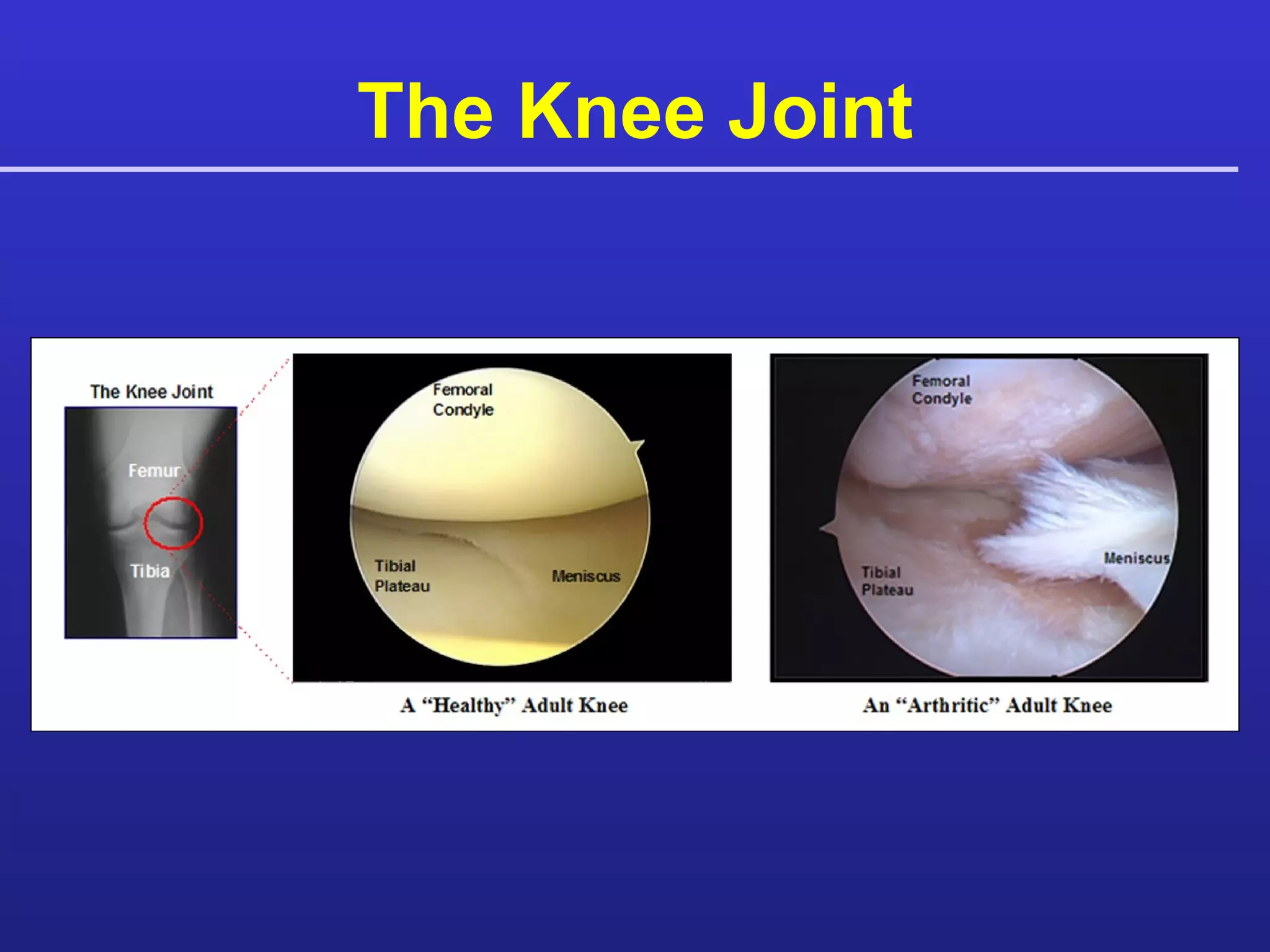
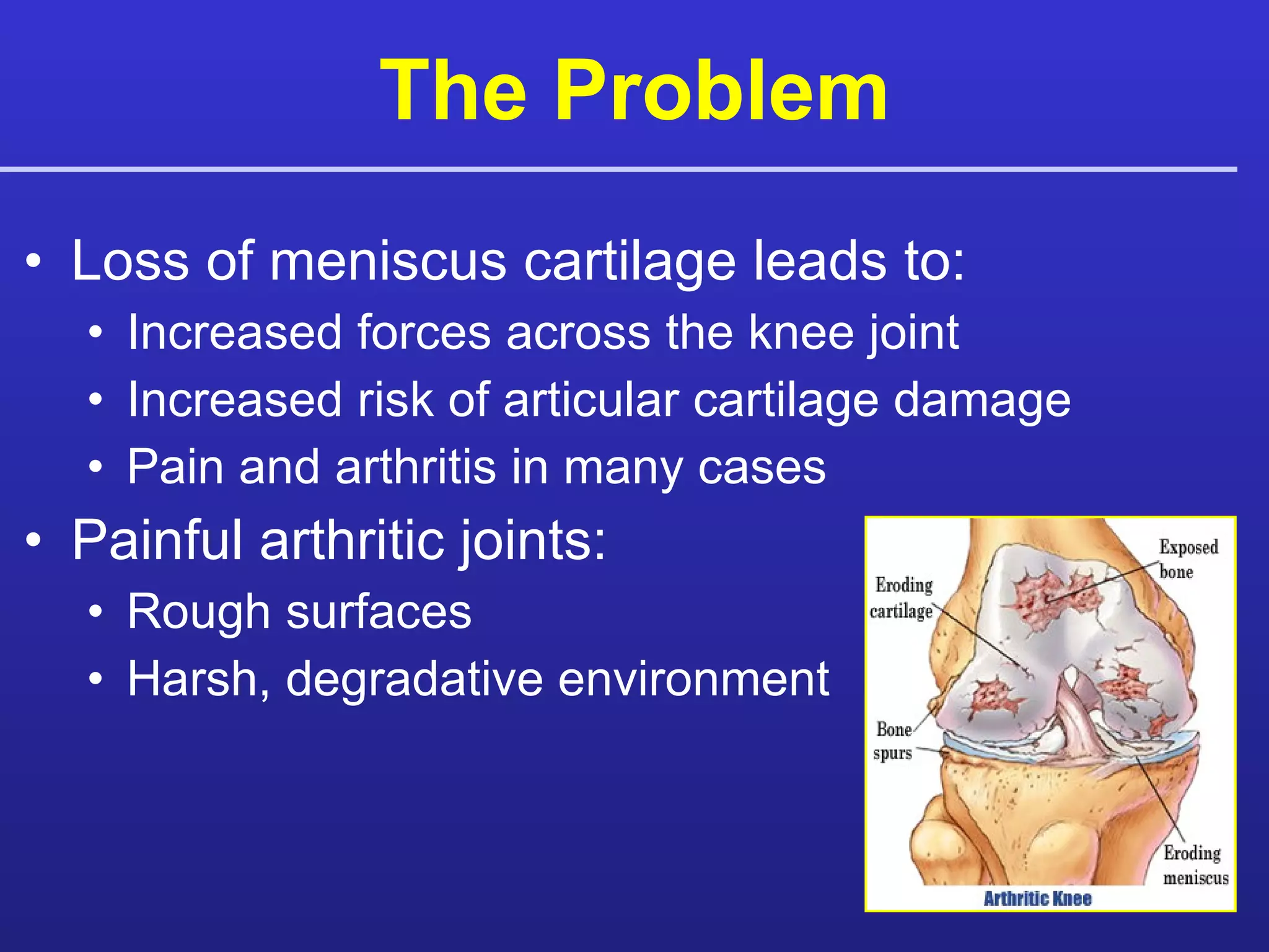
Meniscus Allograft: State of the Art Kevin R. Stone, MD Ann W. Walgenbach, RNNP  Wendy S. Adelson, MS Jonathan R. Pelsis, MHS Meniskus – Ersatz: Collagen Meniskus & Allograft 15. Janur 2010 Stone Research Foundation San Francisco
The Aging Knee Pediatric  Normal Adult  OA Adult
The Knee Joint
Meniscus  Key shock absorber in the knee Torn 1.5M times annually US Minimal healing No spontaneous regeneration template
Loss of meniscus cartilage leads to: Increased forces across the knee joint Increased risk of articular cartilage damage Pain and arthritis in many cases Painful arthritic joints: Rough surfaces Harsh, degradative environment The Problem
Reduce pain and improve function Preserve the biology of the knee Restore a biomechanically favorable environment Provide a buffer to prevent  bone-on-bone contact and pain The Goal
The Alignment Controversy Is osteotomy a two plane crude correction of a multiplanar deformed geometry? Is osteotomy really a correction? Are the complications worth it? “ Bad biomechanics ruins good biology any day of the week…” … However, biology lasts decades even in mechanically disadvantaged knees.
A Solution ? Biologic Joint Replacement Smooth, repair, replace, or regenerate damaged articular cartilage Meniscus reconstruction Meniscus allograft transplantation Fibrous interpostional joint arthroplasty Reduce pain and improve function Increase success of cartilage grafts
Outerbridge Grading System  For Cartilaginous Degeneration   Outerbridge RE. The etiology of chondromalacia patellae.  J Bone Joint Surg Br,  1961;43: 752-7.  Grade I Soft discolored superficial fibrillation Grade II Fragmentation < 1.3 cm  2 Grade III Fragmentation > 1.3 cm  2 Grade IV Erosion to subchondral bone (eburnation)
Meniscus Allograft Transplantation:  Indications? Traditional thought:   Meniscus Transplantation does not work in arthritic knees  ( Noyes & Barber-Westin 1995, Stollsteimer 2000, Rath 2001) Current thought:   Meniscus Transplantation  does  work in arthritic knees if damaged articular cartilage is treated as well  (van Arkel 2002, Noyes 2004, Verdonk 2005, Cole 2006, Stone 2006, Farr 2007, Rue 2008)
Sizing: Surgeon Concerns “It takes me 6 months to get a properly sized meniscus.” “My measurements do not match the bank’s measurements.” “Is there an easier, more accurate method for sizing?”
Meniscus Allograft: Sizing Success rate may be dependant on accurate sizing Image-based sizing measures bony landmarks and insertion points however:  Contrast limitations  Identification of soft versus mineralized tissue interface Magnification errors Schaffer B, Kennedy S, Flimkiewicz J, Yao L.  Preoperative Sizing of the Meniscal Allografts in Meniscal transplantation.  Am Journal of Sports Med. Vol. 28, No. 4, 2000.
Supporting Studies: Sizing 148 heights and weights compared to MRI meniscus size Pearson’s Correlations (r):  Height vs Total Tibial Plateau (TTP) r = 0.7194 Weight vs TTP r = 0.5470  TTP vs Medial and Lateral Meniscal Width r = 0.7386, r = 0.7209 TTP vs Medial and Lateral Meniscal Length r =  0.7040, r = 0.7209 Stone KR, Freyer A, Turek T, Walgenbach AW, Wadhwa S, Crues J. Meniscal sizing based on gender, height, and weight. Arthroscopy 2007;23-5:503-8 Meniscal Sizing Based on Gender, Height, and Weight
The Three-Tunnel Technique Replacing the Meniscus Stone KR, Walgenbach AW. “Meniscal Allografting: the Three-Tunnel Technique.”  Arthroscopy – The Journal of Arthroscopic and Related Surgery. 2003, 19(4):426-30.
Articular Cartilage Paste Graft  Procedure Step 1 Step 5 Step 4 Step 3 Step 2
Meniscus Transplantation 173 patients since 1997 Clinical Exam + Patient Reported Subjective Outcome  (1, 2, 3, 5, 7, 10, 15+ yrs) IKDC WOMAC TEGNER The Stone Clinic Experience
Current Study: Long-Term Survival of Concurrent  Meniscus Allograft Transplantation  and Articular Cartilage Repair:  A Prospective 12-Year  Follow-Up Evaluation  Pre-Allograft Allograft in place Transplantation OB IV
Study Design Study Inclusion Irreparable injury of the meniscus Or Loss of the meniscus More than 50% OB III/IV  ROM  ≥  90° Study Exclusion Rheumatoid Arthritis Tri-compartment arthritis Total loss of joint space Simultaneous med/lat meniscus allograft transplantation
Patient Selection Young patients with cartilage loss and pain Older patients with cartilage loss and focal pain who want to remain athletic and delay or avoid a knee arthroplasty. “Doc, isn’t there a shock absorber you can put in my knee?”
Patient Selection Identify the EXACT location of the patient’s pain “ It hurts right here”  ->   Biologic Surgery = Effective VS “ It hurts all over”  ->  Biologic Surgery = Worrisome
Surgical Technique Medial Meniscus Allograft Transplantation:  Performed utilizing periosteum, but not bone blocks, at the meniscus horns.  Lateral Meniscus Allograft Transplantation:  Preformed by preserving the bony block between the horns and inserting it into a bone trough.
119 Meniscus Allograft Transplant Cases Mean age = 46.9 years  (14.1 – 73.2 yrs) Mean follow-up = 5.8 years  (2.1 mo – 12.3 yrs) 118 patients ≥ 3 months from injury to time of surgery  (Mean = 14.2 years) Patient Population
Patient Population  (N = 119) Neutral / Varus / Valgus Moderate ( 5 – 7°) / Severe ( > 7°) Grade III / Grade IV Medial / Lateral Male / Female None / Mild–Moderate / Severe (Kellgren-Lawrence)
Review of Literature Mixed Patient Studies N = 119 N = 100 N = 29 N = 31 N = 44
Results Procedure failure: Removal of allograft without revision  (N = 7) , or progression to knee arthroplasty  [N = 18 (TKA or UNI)].   94/119 allograft cases successful (79%) Of 25 failures, Mean time-to-failure:  4.65 ± 2.99 years Range: 2.1 months – 10.37 years Kaplan-Meier estimated mean survival time was 9.93 ± 0.40 years  [95%CI: 9.14,10.72] 13 patients were lost to follow-up
Complications 4 Early Postoperative Infections 3 Deep (1 Staphphylococcus Aures, 2 negative serologies) 1 Superficial (Staphylococcus Epidemis) All cases were treated arthroscopically with irrigation and debridement and IV antibiotics.  All cases resolved, but one deep infection case ultimately failed, with the allograft being removed 12.5 months later.
Subsequent Surgeries – 1 4 2 Meniscus Allograft Revision – 2 1 12 Meniscus Allograft Repair – 1 9 22 Meniscectomy 1 1 – 4 Microfracture / Articular Cartilage Paste Grafting 1 4 6 20 Chondroplasty / Debridement – 1 1 2 Other 4 th N = 2 3 rd N = 10 2 nd N = 21 1 st N = 62 Subsequent Surgeries Primary Procedure
Kaplan-Meier Survival Analysis In Patients OB III/IV Time-to-failure analysis with continuous enrollment over 12-yrs Takes into account remaining patients (still intact / lost to follow-up  (N=13) ) Intact/Lost To Follow-Up 94% 92% 84% 79% 67%
Cox Proportional Hazards Model What is it? A Cox model provides an estimate of a variable’s effect on survival after adjustment for other explanatory variables. In addition, it allows us to estimate the hazard (or risk) of procedure failure, given their prognostic variables.
What factors affect survival? Cox Proportional Hazards Model was used to explore the relationship between procedure failure and several covariates. Age (p = 0.026) Number of Previous Surgeries (p = 0.006)  Number of Additional Surgeries Osteotomy performed concomitantly Number of concomitant procedures Outerbridge Grade (III or IV) Medial v. Lateral Allograft Joint Space Narrowing Malalignment Severity Alignment Type Sex NOT RELATED RELATED
Cox Model - Related Hazards Independent of actual time-to-failure, increased number of previous surgeries (p = 0.026) and increased age at time of surgery (p = 0.006) increases the risk of meniscus allograft transplantation failure.
Effect of Age 53 patients over 50 (Mean = 56 yrs) KM mean survival = 8.84 years  [95% CI: 7.51,10.17] 71.7% (38/53) Success Rate 1 allograft removed 2 mo. post-op 14 progressed to Joint Arthroplasty @ mean 5.1 years 66 patients under 50 (Mean = 39 yrs) KM mean survival = 10.67 years  [95% CI: 9.76,11.58]  84.8% (56/66) Success Rate 6 allografts removed @ mean 4.0 years 4 Progressed to Joint Arthroplasty @ mean 5.2 years
Medial v. Lateral Allografts Non Significant Hazard (p = 0.848) Medial (N = 85) KM mean survival: 9.91 ± 0.46 years Lateral (N = 34) KM mean survival: 10.17 ± 0.78 years
Malalignment Severity of Mal-Alignment  (p = 0.535) Severe Malalignment (>7 º)  (N = 10) Moderate Malalignment (5 – 7 º)  (N = 39) 7 Osteotomies –  71.4% Success Rate (5/7) –  2 UNI 3 NO Osteotomy –  66.7% Success Rate (2/3) –  1 UNI –  50% Success Rate (4/8) –  2 TKA, 1 UNI, 1 Removed 8  Osteotomies –  80.6% Success Rate (25/31) –  2 TKA, 2 UNI, 2 Removed 31 NO Osteotomy
Subjective Outcome Scores IKDC, WOMAC, and TEGNER questionnaire follow-up schedule was preoperatively and at 2, 3, 5, 7, 10, 15 year post-op. Tegner Index was used to normalize return to activity across a diverse population * * Rodkey et al. Comparison of the collagen meniscus implant with partial meniscectomy. A prospective randomized trial. J Bone Joint Surg Am 2008;90-7:1413-26. Current Tegner activity score Highest reported pre-injury score = Tegner Index Score
Subjective Outcome Scores 1.00 0.90 0.80 0.70 0.60 0.50 0.40 0.30 0.20 0.10 0.00 Mean Tegner Index Score
Patient Example: BK 27 year old male Torn lateral meniscus in high school wrestling 1996 Partial lateral meniscectomy 2/96, 8/04 Pre-Operative X-Rays
BK: Pre-Op MRI MRI documents degenerative changes   to LTP and loss of lateral meniscus
Patient Example: BK Lateral Meniscus Allograft Transplantation
Patient Example: BK 8 months post  Arthroscopy for suprapatellar pouch and anterolateral swelling Lateral meniscus allograft transplant had healed
BK MRI 4 Years Post Op Lateral meniscus allograft appears normal and   well positioned Patient reports no pain - “It feels really good”
Patient Example: JL 35 Year Old Female Right Knee   1984 - Lateral Meniscectomy 1988 - Lateral release 2003 - Knee locked, total meniscectomy Valgus Alignment
Patient Example: JL OB III/IV far-posterior aspect LFC, Microfracture LFC
JL: 4 months Post-Op Flexion contracture, debridement, closed manipulation, notchplasty No evidence of   meniscal impingement Healed, intact lateral   meniscus
JL: 6 years Post-Op Lateral Meniscus repair, chondroplasty, debridement, notchplasty
Patient Example: JA 37 Year old female Meniscectomy at age 20 R-Lateral Meniscus missing OB III chondral defect Microfracture, Chondroplasty LFC Long-Leg AP
JA: Preoperative X-ray Lateral AP
JA: Preoperative MRI Lateral meniscus: Absent posterior horn Articular Cartilage: Chondral damage to LFC
JA Operative Images A B C Deficient Lateral Meniscus Chondral Lesion of LFC Microfracture of Lesion
JA Operative Images A B C Absent Meniscus Lateral Meniscus Allograft Allograft Placement
JA: 5 Months Post-Op  Full ROM with smooth articulation
JA: 2Yr Postoperative X-ray PA Flexion AP
JA: 2yr Post-operative MRI Healed lateral meniscal allograft
JA: 5Yr Postoperative X-Ray PA Flexion AP
JA: 5Yr Postoperative MRI Virtually unchanged meniscal allograft
Patient Example: GC 7 o  varus L-knee Medial joint space narrowing Active 53 y.o. male. Meniscectomy: 1986, 1996 Medial meniscus-allograft 3/99  Paste Graft MFC & MTP High medial tibial osteotomy (Bionx wedge and allograft bone)
GC: Preoperative Images Sagittal MRI Loss of cartilage MFC PA Flexion Medial joint space narrowing
GC: Operative Images A B Bipolar lesions Morselization of MFC & MTP Loss of medial meniscus
GC: Operative Images A B C Placement of medial meniscal allograft Impaction of paste graft Paste Grafted Lesion
GC: Postoperative X-Ray Long-leg AP
GC: 3Yr Postoperative X-ray AP Long-leg
GC: 3Yr Postoperative Images 3 Years post-op L-medial allograft, osteotomy, & paste graft
GC: Comparison of healing 3-Years post-op allograft and paste graft to MFC Operative 3 yrs Post-op 3 yrs Post-op
Patient Example: SC 39 y.o. male Injury: 1970s playing hockey  Meniscectomy (1999) Pre-op:  Varus  Joint space narrowing Right Medial Meniscus  Allograft (2000)
SC: Preoperative MRI Bucket-handle tear with bipolar cartilage lesions on MFC & MTP Coronal Sagittal
SC: Operative Images Right knee bucket-handle tear displaced into intercondylar notch A B
SC: Operative Images Eburnated bone MFC Eburnated bone MTP Microfracture MTP Microfracture MTP
SC: Placement of Allograft Right Knee Placement of Medial Meniscal Allograft
SC: Comparison of healing   Return to full activity Intermittent catching and pain 17 Mo Post-op Pre-op Note improved joint space compared to pre-op
SC: 17 Mo Post Op MRI Coronal Sagittal
SC: 2nd Surgery Movie 17 mo. Post  Paste graft MFC +  Meniscus  Allograft Initial Surgery 17 mo. Post-op Meniscectomy
SC: 5 yr Post Op Images AP Allograft present with maturing degenerative changes Coronal
Patient Example DB 47 YO Male Skier R Knee:  Chronic Pain Moderate to Severe Bilateral Pain
DB: Right Knee Right Knee:  09/91:  Medial Meniscectomy, Drilling MFC, Chondroplasty 12/97: (triple) Medial Meniscus Allograft, Osteotomy, Art Cart MFC, MFx LFC 05/98: Revision Osteotomy, Medial Meniscectomy, Debridement, MFx MTP 10/2000: Ilizarov, Meniscectomy, Chondroplasty Pre-Op XRAY
DB: Right Knee 10 Yr PostOp MRI
DB: 10 Yr Post Op XRAY
DB: 10 Yr PostOp 63 YO, Tegner = 6, Skis 30+ days/yr, Snow skis 50+ days/yr.
47 YO Female Beach volleyball injury (11/03) Failed debridement (11/03) Clinical exam: Pain at rest = 8/10 Severe swelling  Giving way Meniscus Allograft, ACL reconstruction, Chondroplasty (3/05) Patient Example: RT
RT: Pre-Operative MRI
Torn medial meniscus MFC chondral lesion LFC chondral lesion Torn ACL Patient Example: RT
Medial meniscus Allograft Allograft Insertion Allograft placement ACL BTB allograft Patient Example: RT
Intact meniscus allograft ACL hardware removal due to   prominence of fixation screw RT: 3 Months Post
Excellent joint space, intact meniscus allograft and ACL, but right knee clicking and catching RT: 18 Months Post
Intact meniscus allograft and ACL with diffuse thinning of patellofemoral cartilage RT: 18 Months Post
Surgery for catching due to chondral flap at patellofemoral joint Intact meniscus allograft and ACL RT: 18 Months Post
Patient Example: RM 57 Male Long-leg MRI Injury: Football tackle 1978  Previous Meniscectomy: 1978, 1993 Moderate varus mal alignment ( ≤ 7°) MFC OCD lesion
RM: Operative Images Severe Articular Cartilage Damage
RM: Operative Images  Allograft insertion Articular Cartilage  Paste Grafting Repair
RM: Operative Images  High tibial opening wedge osteotomy
RM: 3.5 Years Post-Allograft  Movie  Primary Surgery Second Look, 3.5 years later
RM: Histology
RM: 3.5 Years Post Operative Long-leg
RM: 5 Years Post Operative MRI Sagittal Coronal
RM: 6 Year Post Operative X-Ray Long-leg Lateral
Patient Example: HM 5-7 o  varus L-knee 18 Yrs Post meniscectomy  2 Meniscectomies (‘86, ’96) Pain >1 year Varus deformity Medial joint space narrowing L-medial meniscus-allograft (3/1999)  Paste graft MFC & MTP High medial tibial osteotomy  (Bionx wedge and allograft bone) Chondroplasty LFC Partial lateral meniscectomy Notchplasty
HM  4 Yrs 9 Mo Post Paste Graft Debrided lesion Healed paste graft
HM   4 Yrs 9 Mo Post Paste Graft Biopsy Histology
Biologic Knee Revision Surgery
Patient Example: TA 48 y.o. world-class female marathoner 86 marathons,  12 Ironmans 3 Double Ironman   Triathalons Neutral alignment / mild medial joint space narrowing Meniscectomy: 4/2001 and 1/2002 AP X-ray R Medial Meniscus Allograft + Microfracture (bipolar lesions)
TA: Preoperative MRI Tear at horn of medial meniscus Osteoarthritis: medial compartment Saggital Coronal
4/22/02: Right medial meniscus rim before allograft TA: Operative Images
TA: Meniscus Allograft Placement Preparation of medial meniscal allograft Placement of medial meniscal allograft Relationship of lesion to meniscus A B Movie
TA: Injury C Injury: 2 Mo. Post-op  Swam in pool for 2 hours  Developed immediate swelling A Movie
TA: Revision C A Movie Revision: 8 Months Post-allograft
TA: Revision: Operative Images Insertion of Meniscus Allograft with Articular Cartilage Paste Grafting Joint Arthroplasty 3/2006 (38 Mo. Post Op) A B C D
Patient Example: RT 34 YO male Partial meniscectomy for torn lateral meniscus (9/91), debridement 2006 Lateral joint line pain Severe pain and swelling with activities Positive Apley’s, McMurray’s, and hyperextension tests
RT: Pre-Op Imaging X-rays: Collapse of lateral joint space. Mild patellar spurring.
RT: Pre-Op MRI Loss of articular cartilage on posterior aspect of LFC Loss of posterior and central aspects of lateral   meniscus PD SAG PD COR
RT:  Surgery 11/2009 Lateral Meniscus Allograft transplantation Microfracture LFC and LTP (too far posterior for Articular Cartilage Paste Grafting) Removal of anvil osteophyte
RT: MRI 2 days Post Op Allograft intact without evidence of tear Anterior subluxation of the posterior aspect of the lateral meniscus with anterior displacement of the bone block (12 mm). FSE T2 SAG FSE PD SAG
RT: Revision Surgery Interval repositioning of the lateral meniscus 5 days post index procedure. Re-microfracture of distal femoral condyle to ensure good blood clot.
RT: MRI 1 Day Post Revision Repositioned Lateral Meniscus Allograft FSE T2 SAG FSE PD SAG
Conclusions Our research represents the largest and longest prospective study of meniscus replacement patients with severe chondral damage. Meniscus replacement can improve symptoms,  even in severe OA . Meniscus replacement should not be limited to young patients without articular cartilage damage. Axial malalignment does not affect outcome.
Conclusions Height and weight can be used to size meniscal allograft tissue. Three-tunnel Technique is necessary to fix meniscus allograft to tibial plateau, not the surrounding tissue, to avoid meniscus subluxation
Conclusions Repair of severe articular cartilage damage combined with meniscus replacement provides significant improvements in activity, pain, and function. Improvements are maintained over the course of follow-up (2 – 12 yrs).
Conclusions  The number of TKA surgeries is predicted to increase to 3.4 million by 2030 * , with increasing costs † . 18/119(15%) cases in our study progressed to knee arthroplasty 4.8 years after meniscus replacement  (range: 1.3 – 10.4 yrs) . Average age at time of knee arthroplasty was 61 years  (range: 52 – 72 yrs) .  * Kurtz, AAOS Chicago, 2006 † Kurtz, JBJS, 2007
Conclusions Biologic joint reconstruction, rather than bionic (artificial) replacement, may be an appropriate first step for many people with knee joint arthritis.
Acknowledgements Thomas Turek Mark Coleman Abhi Freyer Ann Walgenbach Jonathan Pelsis Wendy Adelson Sharon Bobrow Meniscus Allograft Transplantation: 1997 – 2010 Articular Cartilage Paste Grafting: 1991 – 2010
2009 Team 2005 Team

Meniscus Transplant and Replacement

  • 1.
    Meniscus Allograft: Stateof the Art Kevin R. Stone, MD Ann W. Walgenbach, RNNP Wendy S. Adelson, MS Jonathan R. Pelsis, MHS Meniskus – Ersatz: Collagen Meniskus & Allograft 15. Janur 2010 Stone Research Foundation San Francisco
  • 2.
    The Aging KneePediatric Normal Adult OA Adult
  • 3.
  • 4.
    Meniscus Keyshock absorber in the knee Torn 1.5M times annually US Minimal healing No spontaneous regeneration template
  • 5.
    Loss of meniscuscartilage leads to: Increased forces across the knee joint Increased risk of articular cartilage damage Pain and arthritis in many cases Painful arthritic joints: Rough surfaces Harsh, degradative environment The Problem
  • 6.
    Reduce pain andimprove function Preserve the biology of the knee Restore a biomechanically favorable environment Provide a buffer to prevent bone-on-bone contact and pain The Goal
  • 7.
    The Alignment ControversyIs osteotomy a two plane crude correction of a multiplanar deformed geometry? Is osteotomy really a correction? Are the complications worth it? “ Bad biomechanics ruins good biology any day of the week…” … However, biology lasts decades even in mechanically disadvantaged knees.
  • 8.
    A Solution ?Biologic Joint Replacement Smooth, repair, replace, or regenerate damaged articular cartilage Meniscus reconstruction Meniscus allograft transplantation Fibrous interpostional joint arthroplasty Reduce pain and improve function Increase success of cartilage grafts
  • 9.
    Outerbridge Grading System For Cartilaginous Degeneration Outerbridge RE. The etiology of chondromalacia patellae. J Bone Joint Surg Br, 1961;43: 752-7. Grade I Soft discolored superficial fibrillation Grade II Fragmentation < 1.3 cm 2 Grade III Fragmentation > 1.3 cm 2 Grade IV Erosion to subchondral bone (eburnation)
  • 10.
    Meniscus Allograft Transplantation: Indications? Traditional thought: Meniscus Transplantation does not work in arthritic knees ( Noyes & Barber-Westin 1995, Stollsteimer 2000, Rath 2001) Current thought: Meniscus Transplantation does work in arthritic knees if damaged articular cartilage is treated as well (van Arkel 2002, Noyes 2004, Verdonk 2005, Cole 2006, Stone 2006, Farr 2007, Rue 2008)
  • 11.
    Sizing: Surgeon Concerns“It takes me 6 months to get a properly sized meniscus.” “My measurements do not match the bank’s measurements.” “Is there an easier, more accurate method for sizing?”
  • 12.
    Meniscus Allograft: SizingSuccess rate may be dependant on accurate sizing Image-based sizing measures bony landmarks and insertion points however: Contrast limitations Identification of soft versus mineralized tissue interface Magnification errors Schaffer B, Kennedy S, Flimkiewicz J, Yao L. Preoperative Sizing of the Meniscal Allografts in Meniscal transplantation. Am Journal of Sports Med. Vol. 28, No. 4, 2000.
  • 13.
    Supporting Studies: Sizing148 heights and weights compared to MRI meniscus size Pearson’s Correlations (r): Height vs Total Tibial Plateau (TTP) r = 0.7194 Weight vs TTP r = 0.5470 TTP vs Medial and Lateral Meniscal Width r = 0.7386, r = 0.7209 TTP vs Medial and Lateral Meniscal Length r = 0.7040, r = 0.7209 Stone KR, Freyer A, Turek T, Walgenbach AW, Wadhwa S, Crues J. Meniscal sizing based on gender, height, and weight. Arthroscopy 2007;23-5:503-8 Meniscal Sizing Based on Gender, Height, and Weight
  • 14.
    The Three-Tunnel TechniqueReplacing the Meniscus Stone KR, Walgenbach AW. “Meniscal Allografting: the Three-Tunnel Technique.” Arthroscopy – The Journal of Arthroscopic and Related Surgery. 2003, 19(4):426-30.
  • 15.
    Articular Cartilage PasteGraft Procedure Step 1 Step 5 Step 4 Step 3 Step 2
  • 16.
    Meniscus Transplantation 173patients since 1997 Clinical Exam + Patient Reported Subjective Outcome (1, 2, 3, 5, 7, 10, 15+ yrs) IKDC WOMAC TEGNER The Stone Clinic Experience
  • 17.
    Current Study: Long-TermSurvival of Concurrent Meniscus Allograft Transplantation and Articular Cartilage Repair: A Prospective 12-Year Follow-Up Evaluation Pre-Allograft Allograft in place Transplantation OB IV
  • 18.
    Study Design StudyInclusion Irreparable injury of the meniscus Or Loss of the meniscus More than 50% OB III/IV ROM ≥ 90° Study Exclusion Rheumatoid Arthritis Tri-compartment arthritis Total loss of joint space Simultaneous med/lat meniscus allograft transplantation
  • 19.
    Patient Selection Youngpatients with cartilage loss and pain Older patients with cartilage loss and focal pain who want to remain athletic and delay or avoid a knee arthroplasty. “Doc, isn’t there a shock absorber you can put in my knee?”
  • 20.
    Patient Selection Identifythe EXACT location of the patient’s pain “ It hurts right here” -> Biologic Surgery = Effective VS “ It hurts all over” -> Biologic Surgery = Worrisome
  • 21.
    Surgical Technique MedialMeniscus Allograft Transplantation: Performed utilizing periosteum, but not bone blocks, at the meniscus horns. Lateral Meniscus Allograft Transplantation: Preformed by preserving the bony block between the horns and inserting it into a bone trough.
  • 22.
    119 Meniscus AllograftTransplant Cases Mean age = 46.9 years (14.1 – 73.2 yrs) Mean follow-up = 5.8 years (2.1 mo – 12.3 yrs) 118 patients ≥ 3 months from injury to time of surgery (Mean = 14.2 years) Patient Population
  • 23.
    Patient Population (N = 119) Neutral / Varus / Valgus Moderate ( 5 – 7°) / Severe ( > 7°) Grade III / Grade IV Medial / Lateral Male / Female None / Mild–Moderate / Severe (Kellgren-Lawrence)
  • 24.
    Review of LiteratureMixed Patient Studies N = 119 N = 100 N = 29 N = 31 N = 44
  • 25.
    Results Procedure failure:Removal of allograft without revision (N = 7) , or progression to knee arthroplasty [N = 18 (TKA or UNI)]. 94/119 allograft cases successful (79%) Of 25 failures, Mean time-to-failure: 4.65 ± 2.99 years Range: 2.1 months – 10.37 years Kaplan-Meier estimated mean survival time was 9.93 ± 0.40 years [95%CI: 9.14,10.72] 13 patients were lost to follow-up
  • 26.
    Complications 4 EarlyPostoperative Infections 3 Deep (1 Staphphylococcus Aures, 2 negative serologies) 1 Superficial (Staphylococcus Epidemis) All cases were treated arthroscopically with irrigation and debridement and IV antibiotics. All cases resolved, but one deep infection case ultimately failed, with the allograft being removed 12.5 months later.
  • 27.
    Subsequent Surgeries –1 4 2 Meniscus Allograft Revision – 2 1 12 Meniscus Allograft Repair – 1 9 22 Meniscectomy 1 1 – 4 Microfracture / Articular Cartilage Paste Grafting 1 4 6 20 Chondroplasty / Debridement – 1 1 2 Other 4 th N = 2 3 rd N = 10 2 nd N = 21 1 st N = 62 Subsequent Surgeries Primary Procedure
  • 28.
    Kaplan-Meier Survival AnalysisIn Patients OB III/IV Time-to-failure analysis with continuous enrollment over 12-yrs Takes into account remaining patients (still intact / lost to follow-up (N=13) ) Intact/Lost To Follow-Up 94% 92% 84% 79% 67%
  • 29.
    Cox Proportional HazardsModel What is it? A Cox model provides an estimate of a variable’s effect on survival after adjustment for other explanatory variables. In addition, it allows us to estimate the hazard (or risk) of procedure failure, given their prognostic variables.
  • 30.
    What factors affectsurvival? Cox Proportional Hazards Model was used to explore the relationship between procedure failure and several covariates. Age (p = 0.026) Number of Previous Surgeries (p = 0.006) Number of Additional Surgeries Osteotomy performed concomitantly Number of concomitant procedures Outerbridge Grade (III or IV) Medial v. Lateral Allograft Joint Space Narrowing Malalignment Severity Alignment Type Sex NOT RELATED RELATED
  • 31.
    Cox Model -Related Hazards Independent of actual time-to-failure, increased number of previous surgeries (p = 0.026) and increased age at time of surgery (p = 0.006) increases the risk of meniscus allograft transplantation failure.
  • 32.
    Effect of Age53 patients over 50 (Mean = 56 yrs) KM mean survival = 8.84 years [95% CI: 7.51,10.17] 71.7% (38/53) Success Rate 1 allograft removed 2 mo. post-op 14 progressed to Joint Arthroplasty @ mean 5.1 years 66 patients under 50 (Mean = 39 yrs) KM mean survival = 10.67 years [95% CI: 9.76,11.58] 84.8% (56/66) Success Rate 6 allografts removed @ mean 4.0 years 4 Progressed to Joint Arthroplasty @ mean 5.2 years
  • 33.
    Medial v. LateralAllografts Non Significant Hazard (p = 0.848) Medial (N = 85) KM mean survival: 9.91 ± 0.46 years Lateral (N = 34) KM mean survival: 10.17 ± 0.78 years
  • 34.
    Malalignment Severity ofMal-Alignment (p = 0.535) Severe Malalignment (>7 º) (N = 10) Moderate Malalignment (5 – 7 º) (N = 39) 7 Osteotomies – 71.4% Success Rate (5/7) – 2 UNI 3 NO Osteotomy – 66.7% Success Rate (2/3) – 1 UNI – 50% Success Rate (4/8) – 2 TKA, 1 UNI, 1 Removed 8 Osteotomies – 80.6% Success Rate (25/31) – 2 TKA, 2 UNI, 2 Removed 31 NO Osteotomy
  • 35.
    Subjective Outcome ScoresIKDC, WOMAC, and TEGNER questionnaire follow-up schedule was preoperatively and at 2, 3, 5, 7, 10, 15 year post-op. Tegner Index was used to normalize return to activity across a diverse population * * Rodkey et al. Comparison of the collagen meniscus implant with partial meniscectomy. A prospective randomized trial. J Bone Joint Surg Am 2008;90-7:1413-26. Current Tegner activity score Highest reported pre-injury score = Tegner Index Score
  • 36.
    Subjective Outcome Scores1.00 0.90 0.80 0.70 0.60 0.50 0.40 0.30 0.20 0.10 0.00 Mean Tegner Index Score
  • 37.
    Patient Example: BK27 year old male Torn lateral meniscus in high school wrestling 1996 Partial lateral meniscectomy 2/96, 8/04 Pre-Operative X-Rays
  • 38.
    BK: Pre-Op MRIMRI documents degenerative changes to LTP and loss of lateral meniscus
  • 39.
    Patient Example: BKLateral Meniscus Allograft Transplantation
  • 40.
    Patient Example: BK8 months post Arthroscopy for suprapatellar pouch and anterolateral swelling Lateral meniscus allograft transplant had healed
  • 41.
    BK MRI 4Years Post Op Lateral meniscus allograft appears normal and well positioned Patient reports no pain - “It feels really good”
  • 42.
    Patient Example: JL35 Year Old Female Right Knee 1984 - Lateral Meniscectomy 1988 - Lateral release 2003 - Knee locked, total meniscectomy Valgus Alignment
  • 43.
    Patient Example: JLOB III/IV far-posterior aspect LFC, Microfracture LFC
  • 44.
    JL: 4 monthsPost-Op Flexion contracture, debridement, closed manipulation, notchplasty No evidence of meniscal impingement Healed, intact lateral meniscus
  • 45.
    JL: 6 yearsPost-Op Lateral Meniscus repair, chondroplasty, debridement, notchplasty
  • 46.
    Patient Example: JA37 Year old female Meniscectomy at age 20 R-Lateral Meniscus missing OB III chondral defect Microfracture, Chondroplasty LFC Long-Leg AP
  • 47.
  • 48.
    JA: Preoperative MRILateral meniscus: Absent posterior horn Articular Cartilage: Chondral damage to LFC
  • 49.
    JA Operative ImagesA B C Deficient Lateral Meniscus Chondral Lesion of LFC Microfracture of Lesion
  • 50.
    JA Operative ImagesA B C Absent Meniscus Lateral Meniscus Allograft Allograft Placement
  • 51.
    JA: 5 MonthsPost-Op Full ROM with smooth articulation
  • 52.
    JA: 2Yr PostoperativeX-ray PA Flexion AP
  • 53.
    JA: 2yr Post-operativeMRI Healed lateral meniscal allograft
  • 54.
    JA: 5Yr PostoperativeX-Ray PA Flexion AP
  • 55.
    JA: 5Yr PostoperativeMRI Virtually unchanged meniscal allograft
  • 56.
    Patient Example: GC7 o varus L-knee Medial joint space narrowing Active 53 y.o. male. Meniscectomy: 1986, 1996 Medial meniscus-allograft 3/99 Paste Graft MFC & MTP High medial tibial osteotomy (Bionx wedge and allograft bone)
  • 57.
    GC: Preoperative ImagesSagittal MRI Loss of cartilage MFC PA Flexion Medial joint space narrowing
  • 58.
    GC: Operative ImagesA B Bipolar lesions Morselization of MFC & MTP Loss of medial meniscus
  • 59.
    GC: Operative ImagesA B C Placement of medial meniscal allograft Impaction of paste graft Paste Grafted Lesion
  • 60.
  • 61.
    GC: 3Yr PostoperativeX-ray AP Long-leg
  • 62.
    GC: 3Yr PostoperativeImages 3 Years post-op L-medial allograft, osteotomy, & paste graft
  • 63.
    GC: Comparison ofhealing 3-Years post-op allograft and paste graft to MFC Operative 3 yrs Post-op 3 yrs Post-op
  • 64.
    Patient Example: SC39 y.o. male Injury: 1970s playing hockey Meniscectomy (1999) Pre-op: Varus Joint space narrowing Right Medial Meniscus Allograft (2000)
  • 65.
    SC: Preoperative MRIBucket-handle tear with bipolar cartilage lesions on MFC & MTP Coronal Sagittal
  • 66.
    SC: Operative ImagesRight knee bucket-handle tear displaced into intercondylar notch A B
  • 67.
    SC: Operative ImagesEburnated bone MFC Eburnated bone MTP Microfracture MTP Microfracture MTP
  • 68.
    SC: Placement ofAllograft Right Knee Placement of Medial Meniscal Allograft
  • 69.
    SC: Comparison ofhealing Return to full activity Intermittent catching and pain 17 Mo Post-op Pre-op Note improved joint space compared to pre-op
  • 70.
    SC: 17 MoPost Op MRI Coronal Sagittal
  • 71.
    SC: 2nd SurgeryMovie 17 mo. Post Paste graft MFC + Meniscus Allograft Initial Surgery 17 mo. Post-op Meniscectomy
  • 72.
    SC: 5 yrPost Op Images AP Allograft present with maturing degenerative changes Coronal
  • 73.
    Patient Example DB47 YO Male Skier R Knee: Chronic Pain Moderate to Severe Bilateral Pain
  • 74.
    DB: Right KneeRight Knee: 09/91: Medial Meniscectomy, Drilling MFC, Chondroplasty 12/97: (triple) Medial Meniscus Allograft, Osteotomy, Art Cart MFC, MFx LFC 05/98: Revision Osteotomy, Medial Meniscectomy, Debridement, MFx MTP 10/2000: Ilizarov, Meniscectomy, Chondroplasty Pre-Op XRAY
  • 75.
    DB: Right Knee10 Yr PostOp MRI
  • 76.
    DB: 10 YrPost Op XRAY
  • 77.
    DB: 10 YrPostOp 63 YO, Tegner = 6, Skis 30+ days/yr, Snow skis 50+ days/yr.
  • 78.
    47 YO FemaleBeach volleyball injury (11/03) Failed debridement (11/03) Clinical exam: Pain at rest = 8/10 Severe swelling Giving way Meniscus Allograft, ACL reconstruction, Chondroplasty (3/05) Patient Example: RT
  • 79.
  • 80.
    Torn medial meniscusMFC chondral lesion LFC chondral lesion Torn ACL Patient Example: RT
  • 81.
    Medial meniscus AllograftAllograft Insertion Allograft placement ACL BTB allograft Patient Example: RT
  • 82.
    Intact meniscus allograftACL hardware removal due to prominence of fixation screw RT: 3 Months Post
  • 83.
    Excellent joint space,intact meniscus allograft and ACL, but right knee clicking and catching RT: 18 Months Post
  • 84.
    Intact meniscus allograftand ACL with diffuse thinning of patellofemoral cartilage RT: 18 Months Post
  • 85.
    Surgery for catchingdue to chondral flap at patellofemoral joint Intact meniscus allograft and ACL RT: 18 Months Post
  • 86.
    Patient Example: RM57 Male Long-leg MRI Injury: Football tackle 1978 Previous Meniscectomy: 1978, 1993 Moderate varus mal alignment ( ≤ 7°) MFC OCD lesion
  • 87.
    RM: Operative ImagesSevere Articular Cartilage Damage
  • 88.
    RM: Operative Images Allograft insertion Articular Cartilage Paste Grafting Repair
  • 89.
    RM: Operative Images High tibial opening wedge osteotomy
  • 90.
    RM: 3.5 YearsPost-Allograft Movie Primary Surgery Second Look, 3.5 years later
  • 91.
  • 92.
    RM: 3.5 YearsPost Operative Long-leg
  • 93.
    RM: 5 YearsPost Operative MRI Sagittal Coronal
  • 94.
    RM: 6 YearPost Operative X-Ray Long-leg Lateral
  • 95.
    Patient Example: HM5-7 o varus L-knee 18 Yrs Post meniscectomy 2 Meniscectomies (‘86, ’96) Pain >1 year Varus deformity Medial joint space narrowing L-medial meniscus-allograft (3/1999) Paste graft MFC & MTP High medial tibial osteotomy (Bionx wedge and allograft bone) Chondroplasty LFC Partial lateral meniscectomy Notchplasty
  • 96.
    HM 4Yrs 9 Mo Post Paste Graft Debrided lesion Healed paste graft
  • 97.
    HM 4 Yrs 9 Mo Post Paste Graft Biopsy Histology
  • 98.
  • 99.
    Patient Example: TA48 y.o. world-class female marathoner 86 marathons, 12 Ironmans 3 Double Ironman Triathalons Neutral alignment / mild medial joint space narrowing Meniscectomy: 4/2001 and 1/2002 AP X-ray R Medial Meniscus Allograft + Microfracture (bipolar lesions)
  • 100.
    TA: Preoperative MRITear at horn of medial meniscus Osteoarthritis: medial compartment Saggital Coronal
  • 101.
    4/22/02: Right medialmeniscus rim before allograft TA: Operative Images
  • 102.
    TA: Meniscus AllograftPlacement Preparation of medial meniscal allograft Placement of medial meniscal allograft Relationship of lesion to meniscus A B Movie
  • 103.
    TA: Injury CInjury: 2 Mo. Post-op Swam in pool for 2 hours Developed immediate swelling A Movie
  • 104.
    TA: Revision CA Movie Revision: 8 Months Post-allograft
  • 105.
    TA: Revision: OperativeImages Insertion of Meniscus Allograft with Articular Cartilage Paste Grafting Joint Arthroplasty 3/2006 (38 Mo. Post Op) A B C D
  • 106.
    Patient Example: RT34 YO male Partial meniscectomy for torn lateral meniscus (9/91), debridement 2006 Lateral joint line pain Severe pain and swelling with activities Positive Apley’s, McMurray’s, and hyperextension tests
  • 107.
    RT: Pre-Op ImagingX-rays: Collapse of lateral joint space. Mild patellar spurring.
  • 108.
    RT: Pre-Op MRILoss of articular cartilage on posterior aspect of LFC Loss of posterior and central aspects of lateral meniscus PD SAG PD COR
  • 109.
    RT: Surgery11/2009 Lateral Meniscus Allograft transplantation Microfracture LFC and LTP (too far posterior for Articular Cartilage Paste Grafting) Removal of anvil osteophyte
  • 110.
    RT: MRI 2days Post Op Allograft intact without evidence of tear Anterior subluxation of the posterior aspect of the lateral meniscus with anterior displacement of the bone block (12 mm). FSE T2 SAG FSE PD SAG
  • 111.
    RT: Revision SurgeryInterval repositioning of the lateral meniscus 5 days post index procedure. Re-microfracture of distal femoral condyle to ensure good blood clot.
  • 112.
    RT: MRI 1Day Post Revision Repositioned Lateral Meniscus Allograft FSE T2 SAG FSE PD SAG
  • 113.
    Conclusions Our researchrepresents the largest and longest prospective study of meniscus replacement patients with severe chondral damage. Meniscus replacement can improve symptoms, even in severe OA . Meniscus replacement should not be limited to young patients without articular cartilage damage. Axial malalignment does not affect outcome.
  • 114.
    Conclusions Height andweight can be used to size meniscal allograft tissue. Three-tunnel Technique is necessary to fix meniscus allograft to tibial plateau, not the surrounding tissue, to avoid meniscus subluxation
  • 115.
    Conclusions Repair ofsevere articular cartilage damage combined with meniscus replacement provides significant improvements in activity, pain, and function. Improvements are maintained over the course of follow-up (2 – 12 yrs).
  • 116.
    Conclusions Thenumber of TKA surgeries is predicted to increase to 3.4 million by 2030 * , with increasing costs † . 18/119(15%) cases in our study progressed to knee arthroplasty 4.8 years after meniscus replacement (range: 1.3 – 10.4 yrs) . Average age at time of knee arthroplasty was 61 years (range: 52 – 72 yrs) . * Kurtz, AAOS Chicago, 2006 † Kurtz, JBJS, 2007
  • 117.
    Conclusions Biologic jointreconstruction, rather than bionic (artificial) replacement, may be an appropriate first step for many people with knee joint arthritis.
  • 118.
    Acknowledgements Thomas TurekMark Coleman Abhi Freyer Ann Walgenbach Jonathan Pelsis Wendy Adelson Sharon Bobrow Meniscus Allograft Transplantation: 1997 – 2010 Articular Cartilage Paste Grafting: 1991 – 2010
  • 119.

Editor's Notes

  • #2 Kevin R. Stone, MD
  • #3 Kevin R. Stone, MD
  • #4 Kevin R. Stone, MD
  • #6 Kevin R. Stone, MD
  • #7 Kevin R. Stone, MD
  • #9 Kevin R. Stone, MD
  • #10 Kevin R. Stone, MD
  • #11 Kevin R. Stone, MD Rath = severe arthritis excluded
  • #12 Kevin R. Stone, MD
  • #13 Kevin R. Stone, MD
  • #14 Kevin R. Stone, MD
  • #15 Kevin R. Stone, MD
  • #16 Kevin R. Stone, MD The procedure step by step.
  • #17 Kevin R. Stone, MD
  • #30 Examine the coefficients for each explanatory variable. Positive Coefficient means that the hazard is higher WORSE PROGNOSIS Negative Coefficient implies a lower hazard BETTER PROGNOSIS
  • #38 Kevin R. Stone, MD Bryan Kelly
  • #47 Kevin R. Stone, MD
  • #48 Kevin R. Stone, MD
  • #49 Kevin R. Stone, MD
  • #50 Kevin R. Stone, MD
  • #51 Kevin R. Stone, MD
  • #52 Kevin R. Stone, MD
  • #53 Kevin R. Stone, MD
  • #54 Kevin R. Stone, MD
  • #55 Kevin R. Stone, MD
  • #56 Kevin R. Stone, MD
  • #57 Kevin R. Stone, MD
  • #58 Kevin R. Stone, MD A= MRI confirming articular cartilage loss of the MFC B= Long leg x-ray demonstrating varus deformity of (L-knee??? I think it should be the Right knee: see x-rays and chart notes ) of about 5-7 degrees C= PA Flexion view demonstrating medial joint space narrowing bialterally L worse than R (nearly bone on bone on the Left). 51 yo ♂ real estate broker both knees w/ problems L worse than R. He has a long hx/o degenerative changes in the medial compartment, loss of the medial meniscus and previous efforts at surgical debridement in order to relieve his medial compartment pain. Pre-operative x-rays revealed medial joint space narrowing and loss of articular cartilage. Pre-operative MRI confirmed loss of the medial meniscus and loss of the artircular cartilage of the medial compartment. He stood in varus. In view of his young age and atheletic activities he requested an effort at biological reconstruction of the medial compartment.   03/10/1999 L-med-Allo/ ArtCart-MFC &amp; MTP/ Open high tib med wedge opening osteotomy using BionX implants and allograft bone/ chon-LFC/ debridement/   Sx: developed a “clicking soreness” on upper MFC thought to be scar tissue requested an effort at operative debridement   03/20/2002 L-knee arthros/ chon-troch/ partial (M)ectomy of Allo where at the posterior 1/3 there was a small flap tear
  • #59 Kevin R. Stone, MD A= Kissing lesion, MFC, MTP w/ loss of medial meniscus B= Morcellation of the MFC &amp; MTP lesions and loss of medial meniscus
  • #60 Kevin R. Stone, MD A= Placement of medial meniscal allograft B&amp;C= Articular cartilage paste grafting MFC.
  • #61 Kevin R. Stone, MD
  • #62 Kevin R. Stone, MD A= MRI (03/18/02) documenting site of medial meniscus allograft and cartilage paste graft B= Long-leg x-ray (03/14/02) demonstrating post-op alignment C= PA Flexion view (03/14/02) documenting previous osteotomy and preservation of some joint space.   03/14/02 Patient seen 3 years post-op. He noted that before surgery he was unable to do certain activities that he would like to do, and he noted that the knee just pops w/ squatting. He is otherwise quite happy. Px: He had 2 prominent bumps at the medial side of his femoral condyle that he is complaining about. He had patellofemoral crepitus. His pain level is minimal, and his activity level is high. Dx: Arthrofibrosis and bursitis of L-knee. Sx: developed a “clicking soreness” on upper MFC thought to be scar tissue requested an effort at operative debridement   03/20/2002 L-knee arthroscopy/ chond-troch/ partial (M)ectomy of Allo where at the posterior 1/3 there was a small flap tear
  • #63 Kevin R. Stone, MD
  • #64 Kevin R. Stone, MD A= Medial meniscus allograft 3 years S/P transplantation B= Medial meniscus allograft 3 years S/P transplantation C= Biopsy MFC 3 years S/P ArtCart
  • #65 Kevin R. Stone, MD 11-06-2000 R-leg = 4 o varus L leg = 2 o varus Steve Cousins 04-23-2002 R-leg = 5 o varus L leg = 2.5 o varus 39 yo ♂ owner of a “Spicy Sports” company with a long history of injuries playing hockey and lacrosse. Symptoms since 1977 w/ knee locking on one occasion (1982) but spontaneously released without surgery. Eventually came to surgery 1999 but after skiing for 4 months pain recurred. Symptoms at time of xam: R-knee pain, swelling, instability.   11/07/2000 R-med-Allo/ ArtCart- MFC/ Mfx-MTP/ removal bucket-handle tear   Developed intermittent anterior knee catching and pain for which HE requested a repeat arthroscopic evaluation and again requested that osteotomy be delayed. Physical exam: lacked final few degrees of extension – excellent flexion and stability. MRI – intact meniscus, damage on the articular cartilage surface, and anterior arthrofibrosis. X-rays- well preserved joint space.   04/02/2002 R-partial med-meniscus/ chondroplasty – trochlea/ debridement
  • #66 Kevin R. Stone, MD
  • #67 Kevin R. Stone, MD A= Bucket-handle tear medial meniscus, displacing into the intercondylar notch. B= Bucket-handle tear medial meniscus, displacing into the intercondylar notch.
  • #68 Kevin R. Stone, MD
  • #69 Kevin R. Stone, MD Placement of the medial meniscal allograft in relation to ArtCart of MFC The only other picture of this meniscus is washed out and less distinct in demonstrating the implanted meniscus.
  • #70 Kevin R. Stone, MD R-lat R-med L-med L-lat 11-6-2000 8.31 mm 0.70 mm 3.89 mm 6.91 mm 04-23-2002 7.28 mm 1.83 mm 4.85 mm 6.85 mm
  • #71 Kevin R. Stone, MD A= Torn posterior medial meniscus B= S/P partial medial meniscectomy Slide “C” is a movie slide – demonstrating the allograft in relation to the healed MFC ArtCart 1.5 years post-op. Developed intermittent anterior knee catching and pain for which HE requested a repeat arthroscopic evaluation and again requested that osteotomy be delayed. Px: lacked final few degrees of extension – excellent flexion and stability. MRI – intact meniscus/ damage on the articular cartilage surface, and anterior arthrofibrosis. X-rays- well preserved joint space.   04/02/2002 R-partial medial meniscectomy/ chondroplasty – trochlea/ debride
  • #72 Kevin R. Stone, MD A= Torn posterior medial meniscus B= S/P partial medial meniscectomy Slide “C” is a movie slide – demonstrating the allograft in relation to the healed MFC ArtCart 1.5 years post-op. Developed intermittent anterior knee catching and pain for which HE requested a repeat arthroscopic evaluation and again requested that osteotomy be delayed. Px: lacked final few degrees of extension – excellent flexion and stability. MRI – intact meniscus/ damage on the articular cartilage surface, and anterior arthrofibrosis. X-rays- well preserved joint space.   04/02/2002 R-partial medial meniscectomy/ chondroplasty – trochlea/ debride
  • #73 Kevin R. Stone, MD A= Torn posterior medial meniscus B= S/P partial medial meniscectomy Slide “C” is a movie slide – demonstrating the allograft in relation to the healed MFC ArtCart 1.5 years post-op. Developed intermittent anterior knee catching and pain for which HE requested a repeat arthroscopic evaluation and again requested that osteotomy be delayed. Px: lacked final few degrees of extension – excellent flexion and stability. MRI – intact meniscus/ damage on the articular cartilage surface, and anterior arthrofibrosis. X-rays- well preserved joint space.   04/02/2002 R-partial medial meniscectomy/ chondroplasty – trochlea/ debride
  • #74 Kevin R. Stone, MD Kevin R. Stone, Biological Knee Reconstruction Annual Joint Preserving Meeting, Johns Hopkins 2004
  • #75 Kevin R. Stone, MD Kevin R. Stone, Biological Knee Reconstruction Annual Joint Preserving Meeting, Johns Hopkins 2004
  • #76 Kevin R. Stone, MD Kevin R. Stone, Biological Knee Reconstruction Annual Joint Preserving Meeting, Johns Hopkins 2004
  • #77 Kevin R. Stone, MD Kevin R. Stone, Biological Knee Reconstruction Annual Joint Preserving Meeting, Johns Hopkins 2004
  • #78 Kevin R. Stone, MD Kevin R. Stone, Biological Knee Reconstruction Annual Joint Preserving Meeting, Johns Hopkins 2004
  • #79 Kevin R. Stone, MD Rhonda Topple
  • #81 Kevin R. Stone, MD Kevin R. Stone, Biological Knee Reconstruction Annual Joint Preserving Meeting, Johns Hopkins 2004 RT
  • #82 Kevin R. Stone, MD Kevin R. Stone, Biological Knee Reconstruction Annual Joint Preserving Meeting, Johns Hopkins 2004 RT
  • #83 Kevin R. Stone, MD Kevin R. Stone, Biological Knee Reconstruction Annual Joint Preserving Meeting, Johns Hopkins 2004 RT
  • #84 Kevin R. Stone, MD Kevin R. Stone, Biological Knee Reconstruction Annual Joint Preserving Meeting, Johns Hopkins 2004 RT
  • #85 Kevin R. Stone, MD Kevin R. Stone, Biological Knee Reconstruction Annual Joint Preserving Meeting, Johns Hopkins 2004 RT
  • #86 Kevin R. Stone, MD Kevin R. Stone, Biological Knee Reconstruction Annual Joint Preserving Meeting, Johns Hopkins 2004 RT
  • #87 Kevin R. Stone, MD
  • #88 Kevin R. Stone, MD
  • #89 Kevin R. Stone, MD
  • #90 Kevin R. Stone, MD
  • #91 Kevin R. Stone, MD
  • #92 Kevin R. Stone, MD
  • #93 Kevin R. Stone, MD
  • #94 Kevin R. Stone, MD
  • #95 Kevin R. Stone, MD
  • #96 Kevin R. Stone, MD
  • #97 Kevin R. Stone, MD Test
  • #98 Kevin R. Stone, MD TEST
  • #100 Kevin R. Stone, MD Tracy Achiles A= MRI demonstrating full-thickness MFC defect B= MRI demonstrating loss of medial meniscus C= MRI demonstrating loss of articular cartilage 48 yo ♀ fitness manager and triathlon/ iron-man competitor who tripped over her dog while walking it down the driveway landing on both knees. Subsequently saw local orthop surgeon who found a R-med meniscal tear and she underwent partial (M)ectomy Apr 27, 2001. Able to return to running but Sx pain/ swelling recurred. 2 nd Surgery Jan 2002   04/02/2002 – Right knee Surg: R-med-Allo/ R-MFC-ArtCart/ R-MFC|MTP|-Mfx/ Chon – troch   Two weeks post-op she swam in a pool for two hours with her legs kicking and developed immediate swelling. It was presumed that she most likely had re-torn her meniscus allograft. However, she was treated conservatively to see whether or not it would heal on its own. It failed to do so. She had recurrent swelling w/ activities and not responsive to a single effort of cortisone injection . 06/26/2002 - Right knee Surg: R- med-Allo repair/ Mfx-MFC /Chon -MTP
  • #101 Kevin R. Stone, MD
  • #102 Kevin R. Stone, MD A= Full thickness chondral defect MFC and loss of medial meniscus B= Full thickness chondral defect MFC and loss of medial meniscus
  • #103 Kevin R. Stone, MD A= Preparation and placement of medial meniscal allograft B= Placement of medial meniscal allograft Slide “C” is a movie clip – demonstrates relationship of lesion to meniscus
  • #104 Kevin R. Stone, MD A= Retained medial meniscal allograft. C= Refixation of medial meniscal allograft. Repair of the “unstable junction of meniscal capsule w/ medial meniscus allograft. Slide “B*” is a movie - demonstrates the instability of the junction of the junction of the capsule w/ the allograft
  • #105 Kevin R. Stone, MD A= Retained medial meniscal allograft. C= Refixation of medial meniscal allograft. Repair of the “unstable junction of meniscal capsule w/ medial meniscus allograft. Slide “B*” is a movie - demonstrates the instability of the junction of the junction of the capsule w/ the allograft
  • #106 Kevin R. Stone, MD A= Retained medial meniscal allograft. C= Refixation of medial meniscal allograft. Repair of the “unstable junction of meniscal capsule w/ medial meniscus allograft. Slide “B*” is a movie - demonstrates the instability of the junction of the junction of the capsule w/ the allograft
  • #107 Kevin R. Stone, MD Ryan Timbrook
  • #114 Kevin R. Stone, MD
  • #115 Kevin R. Stone, MD
  • #116 Kevin R. Stone, MD
  • #117 Kevin R. Stone, MD
  • #118 Kevin R. Stone, MD
  • #119 Kevin R. Stone, MD
  • #120 Kevin R. Stone, MD