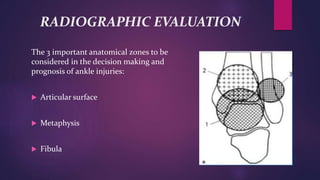
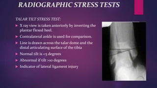
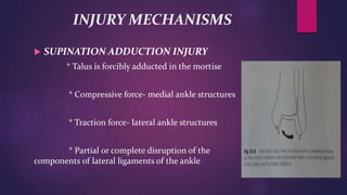
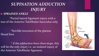
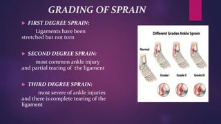
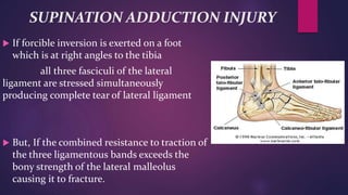
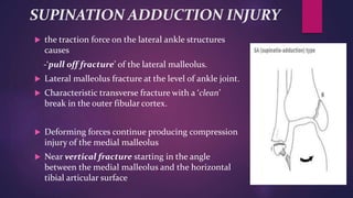
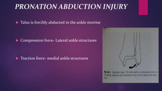
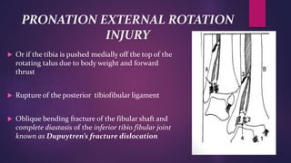
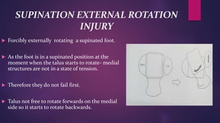
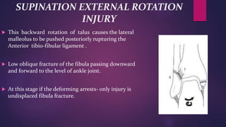
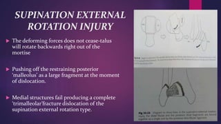
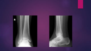
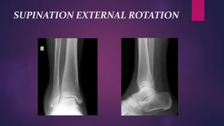
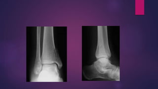
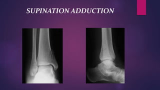
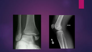
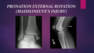
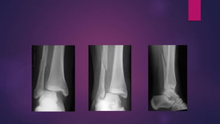
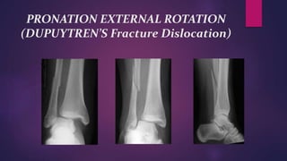
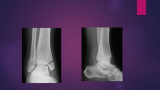
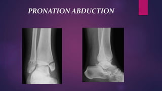
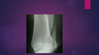
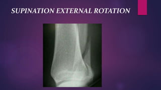
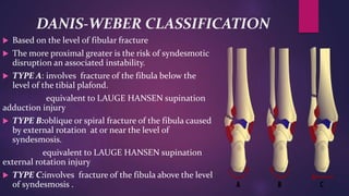
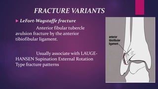
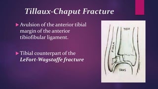
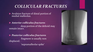
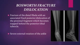
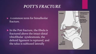
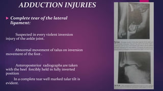
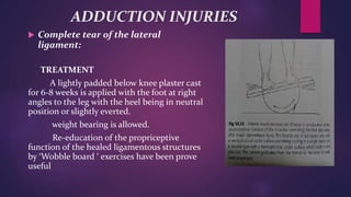
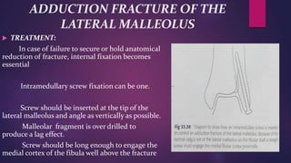
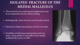
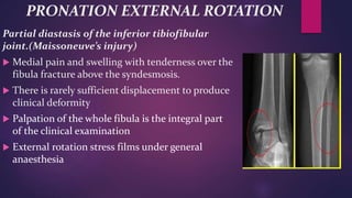
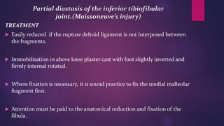
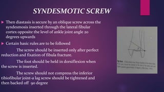
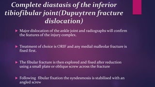
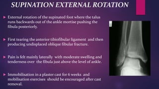
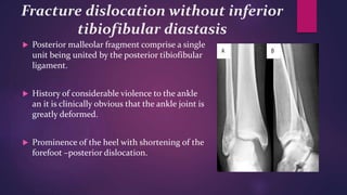
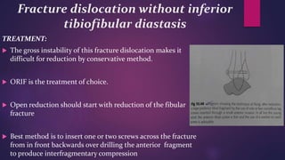
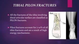
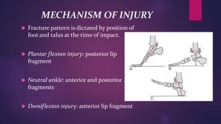
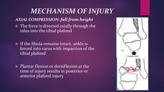
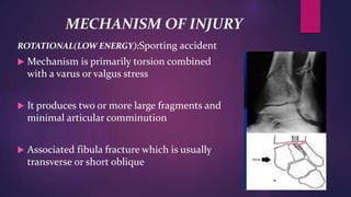
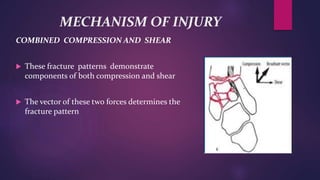
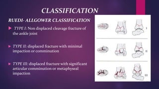
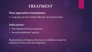
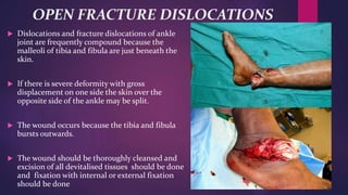
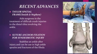
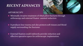
SlideShare a Scribd company logo
VARIOUS ANKLE
INJURIES AND
IT’S MANAGEMENT
PRESENTER:
DR. AMOL GAIKWAD
PG TRAINEE
DEPARTMENT OF ORTHOPAEDICS
MODERATOR:
DR. P. S. CHAKRABARTY
ASSOCIATE PROFESSOR
DEPARTMENT OF ORTHOPAEDICS
INTRODUCTION
 Ankle injury refers to disruption of any component
or components of the ankle joint following trauma.
 Ankle injuries occur frequently, and have high
propensity for complications.
ANATOMY
 Ankle joint is a synovial joint of hinge variety
 ARTICULAR SURFACES:
*Upper articular surface:
lower end of the tibia including the medial
malleolus
lateral malleolus of fibula
inferior transverse tibiofibular ligament
*Inferior articular surface:
upper medial and lateral aspects of Talus
ANATOMY
 LIGAMENTS
* Fibrous capsule
surrounds the joint and it is attached all around
the articular margins.
* Deltoid or Medial ligament
very strong triangular ligament divided into
superficial and deep part
superficial part- anterior fibres/tibionavicular
middle fibres/tibiocalcaneal
posterior fibres/posterior tibiotalar
deep part or anterior tibiotalar
ANATOMY
 LIGAMENTS
* Lateral ligament
- anterior talofibular ligament
flat band which passes from the
anterior margin of the lateral malleolus to the neck of the
talus.
- posterior talofibular ligament
passes from the lower part of the
malleolar fossa of the fibula to the lateral tubercle of the
talus
- calcaneofibular ligament
long rounded cord which passes from
the notch on the lower border of the lateral malleolus to
the tubercle on the lateral surface of the calcaneum
ANATOMY
 SYNDESMOTIC LIGAMENT COMPLEX
resists the axial, rotational and translational forces to
maintain the structural integrity of the mortise
4 ligaments-
*Anterior inferior tibio fibular ligament
*Posterior inferior tibio fibular ligament
*Inferior Transverse tibio fibular ligament
*Interosseous ligament
BIOMECHANICS
CLINICAL EVALUATION
 Variable presentation from a limp to nonambulatory in
significant pain and discomfort.
 Neurovascular status should be carefully documented.
 Extent of soft tissue injury should be evaluated.
 Entire length of fibula should be palpated.
 A dislocated ankle should be reduced and splinted
immediately.
RADIOGRAPHIC EVALUATION
 Anteroposterior(AP), Lateral and Mortise views of the
ankle should be obtained.
 AP view
*Tibiofibula overlap of <10 mm is abnormal and
implies syndesmotic injury
*Tibiofibula clear space of >5mm is abnormal and
implies syndesmotic injury
 Lateral view
*dome of the talus should be centered under the tibia.
*posterior tibial tuberosity fractures can be identified
and direction of fibular injury
*avulsion fracture of talus.
RADIOGRAPHIC EVALUATION
 MORTISE VIEW
* view taken with the foot in 15-20 degrees of
internal rotation to offset the intermalleolar axis
* medial clear space >4-5mm is abnormal and
indicates lateral talar shift
* Talocrural angle- is the angle subtended
between intermalleolar line and a line passing to the
distal tibial articular surface.
should be approx. 83degrees and should be within
2-3 degrees of the uninjured ankle.
RADIOGRAPHIC EVALUATION
The 3 important anatomical zones to be
considered in the decision making and
prognosis of ankle injuries:
 Articular surface
 Metaphysis
 Fibula
RADIOGRAPHIC STRESS TESTS
TALAR TILT STRESS TEST:
 X ray view is taken anteriorly by inverting the
plantar flexed heel.
 Contralateral ankle is used for comparison.
 Line is drawn across the talar dome and the
distal articulating surface of the tibia
 Normal tilt is <5 degrees
 Abnormal if tilt >10 degrees
 Indicator of lateral ligament injury
RADIOGRAPHIC STRESS TESTS
STANDING TALAR TILT STRESS TEST:
 More sensitive
 Patient stands on inverted stress platform
 Foot and ankle in 40 degrees of plantar flexion 50 degrees
of inversion
EXTERNAL ROTATION STRESS TEST:
 Evaluates the syndesmotic and the deep deltoid ligaments
 External rotation of the foot on a stabilised leg with ankle
in dorsiflexion
 Difference in width of superior clear space between medial
and lateral side of the joint should be <2mm
RADIOGRAPHIC EVALUATION
 Computed Tomography(CT) scans help to delineate bony
anatomy, especially in tibial plafond fractures.
 Magnetic Resonance Imaging(MRI) may be used for assessing
occult cartilaginous, ligamentous or tendinous injuries
CLASSIFICATION OF ANKLE
INJURIES
 LAUGE- HANSEN CLASSIFICATION
*end product of a sequence of bony and
ligamentous failures resulting from a deforming
force
* for a given deforming force the failure
sequence occurs in the same order to produce a
complex injury pattern which is pathognomonic
of that deforming force
INJURY MECHANISMS
 SUPINATION ADDUCTION INJURY
* Talus is forcibly adducted in the mortise
* Compressive force- medial ankle structures
* Traction force- lateral ankle structures
* Partial or complete disruption of the
components of lateral ligaments of the ankle
SUPINATION ADDUCTION
INJURY
 SPRAINED ANKLE
*Partial lateral ligament injury with a
tear of the Anterior Talofibular fasciculus only.
*forcible inversion of the plantar
flexed foot
* if the adduction force then stops, this
will be the only injury i.e. an isolated injury of
the Anterior Talofibular ligament.
GRADING OF SPRAIN
 FIRST DEGREE SPRAIN:
Ligaments have been
stretched but not torn
 SECOND DEGREE SPRAIN:
most common ankle injury
and partial tearing of the ligament
 THIRD DEGREE SPRAIN:
most severe of ankle injuries
and there is complete tearing of the
ligament
SUPINATION ADDUCTION INJURY
 If forcible inversion is exerted on a foot
which is at right angles to the tibia
all three fasciculi of the lateral
ligament are stressed simultaneously
producing complete tear of lateral ligament
 But, If the combined resistance to traction of
the three ligamentous bands exceeds the
bony strength of the lateral malleolus
causing it to fracture.
SUPINATION ADDUCTION INJURY
 the traction force on the lateral ankle structures
causes
-‘pull off fracture’ of the lateral malleolus.
 Lateral malleolus fracture at the level of ankle joint.
 Characteristic transverse fracture with a ‘clean’
break in the outer fibular cortex.
 Deforming forces continue producing compression
injury of the medial malleolus
 Near vertical fracture starting in the angle
between the medial malleolus and the horizontal
tibial articular surface
PRONATION ABDUCTION INJURY
 Talus is forcibly abducted in the ankle mortise
 Compression force- Lateral ankle structures
 Traction force- medial ankle structures
PRONATION ABDUCTION INJURY
 Medial traction force may cause either a complete
tear of the deltoid ligament or ‘pull off fracture’ of
the medial malleolus.
 Lateral compression force produces a lower fibular
fracture angling slightly upwards from the level of
the ankle joint always with a communition of
lateral fibular cortex.
PRONATION EXTERNAL ROTATION
INJURY
 With the foot pronated the deltoid ligament
is under tension.
 The talus starts to rotate externally in the
ankle mortise- so the medial structures
which fail first.
 Talus is then free of its medial tether and
swings forwards out of the inner side of the
ankle mortise about a lateral axis.
PRONATION EXTERNAL
ROTATION INJURY
 This imparts a torsional force to the fibula which
first tears the anterior tibiofibular ligament
 Followed by the rupture of the interosseous
ligament of the inferior tibiofibular joint
 At this point only the posterior tibiofibular
ligament of the syndesmosis is intact
PRONATION EXTERNAL ROTATION
INJURY
 If the deforming force continues to rotate the
fibula it will relax the posterior tibiofibular
ligament
 Either resulting in the spiral fracture of the
fibula just above the level of the syndesmosis
or may be as high as the neck of fibula-
Partial diastasis of the inferior tibio fibular
joint which is known as Maisonneuve’s
injury.
PRONATION EXTERNAL ROTATION
INJURY
 Or if the tibia is pushed medially off the top of the
rotating talus due to body weight and forward
thrust
 Rupture of the posterior tibiofibular ligament
 Oblique bending fracture of the fibular shaft and
complete diastasis of the inferior tibio fibular joint
known as Dupuytren’s fracture dislocation
SUPINATION EXTERNAL ROTATION
INJURY
 Forcibly externally rotating a supinated foot.
 As the foot is in a supinated position at the
moment when the talus starts to rotate- medial
structures are not in a state of tension.
 Therefore they do not fail first.
 Talus not free to rotate forwards on the medial
side so it starts to rotate backwards.
SUPINATION EXTERNAL ROTATION
INJURY
 This backward rotation of talus causes the lateral
malleolus to be pushed posteriorly rupturing the
Anterior tibio-fibular ligament .
 Low oblique fracture of the fibula passing downward
and forward to the level of ankle joint.
 At this stage if the deforming arrests- only injury is
undisplaced fibula fracture.
SUPINATION EXTERNAL
ROTATION INJURY
 The deforming forces does not cease-talus
will rotate backwards right out of the
mortise
 Pushing off the restraining posterior
‘malleolus’ as a large fragment at the moment
of dislocation.
 Medial structures fail producing a complete
‘trimalleolar’fracture dislocation of the
supination external rotation type.
SUPINATION EXTERNAL ROTATION
SUPINATION ADDUCTION
PRONATION EXTERNAL ROTATION
(MAISSONEUVE’S INJURY)
PRONATION EXTERNAL ROTATION
(DUPUYTREN’S Fracture Dislocation)
PRONATION ABDUCTION
SUPINATION EXTERNAL ROTATION
DANIS-WEBER CLASSIFICATION
 Based on the level of fibular fracture
 The more proximal greater is the risk of syndesmotic
disruption an associated instability.
 TYPE A: involves fracture of the fibula below the
level of the tibial plafond.
equivalent to LAUGE HANSEN supination
adduction injury
 TYPE B:oblique or spiral fracture of the fibula caused
by external rotation at or near the level of
syndesmosis.
equivalent to LAUGE HANSEN supination
external rotation injury
 TYPE C:involves fracture of the fibula above the level
of syndesmosis .
FRACTURE VARIANTS
 LeFort-Wagstaffe fracture
Anterior fibular tubercle
avulsion fracture by the anterior
tibiofibular ligament.
Usually associate with LAUGE-
HANSEN Supination External Rotation
Type fracture patterns
Tillaux-Chaput Fracture
 Avulsion of the anterior tibial
margin of the anterior
tibiofibular ligament.
 Tibial counterpart of the
LeFort-Wagstaffe fracture
COLLICULAR FRACTURES
 Avulsion fracture of distal portion of
medial malleolus.
 Anterior colliculus fractures
deep portion of the deltoid may
remain intact.
 Posterior colliculus fractures
fragment is usually non
displaced.
‘supramalleolar spike’
BOSWORTH FRACTURE
DISLOCATION
 Fracture of the distal fibula with an
associated fixed posterior dislocation of
the proximal fragment which becomes
trapped behind the posterior tibial
tubercle.
 Severe external rotation of the ankle
POTT’S FRACTURE
 A common term for bimalleolar
fracture.
 In the Pott fracture, the fibula is
fractured above the intact distal
tibiofibular syndesmosis, the
deltoid ligament is ruptured, and
the talus is subluxed laterally
DIAGNOSIS AND TREATMENT
 ADDUCTION INJURIES
SPRAINED ANKLE: common injury caused by the inversion twist of
plantar flexed foot
DIAGNOSIS: characteristic swelling over the lateral aspect of ankle
joint.
* inverting the heel with foot at right angle produces little pain
* while in plantar flexed foot it produces severe pain in front of
lateral malleolus- pathognomonic of isolated anterior talofibular
ligament injury.
SPRAINED ANKLE
 TREATMENT
RICE
 Rest- stay off the Ankle, crutches may be needed
 Ice- 15 minutes - 4 to 5 times a day
 Compression- bandaging to keep swelling down
 Elevation- keep ankle above heart level when
possible, allows gravity to pump out swelling
Use of EVERSION STIRRUP
ADDUCTION INJURIES
 Complete tear of the lateral
ligament:
Suspected in every violent inversion
injury of the ankle joint.
Abnormal movement of talus on inversion
movement of the foot .
Anteroposterior radiographs are taken
with the heel forcibly held in fully inverted
position
In a complete tear well marked talar tilt is
evident.
ADDUCTION INJURIES
 Complete tear of the lateral
ligament:
TREATMENT
A lightly padded below knee plaster cast
for 6-8 weeks is applied with the foot at right
angles to the leg with the heel being in neutral
position or slightly everted.
weight bearing is allowed.
Re-education of the propriceptive
function of the healed ligamentous structures
by ‘Wobble board ‘ exercises have been prove
useful
ADDUCTION FRACTURE OF THE
LATERAL MALLEOLUS
 Pain and gross swelling over the outer aspect of ankle
 Tenderness is present at the base of lateral malleolus and fracture
gap is felt to open up if gentle attempts are made to open up the
heel.
TREATMENT:
 Conservative management usually gives excellent functional
results.
 Lightly padded below knee cast is applied and the heel is
moulded into slight eversion
ADDUCTION FRACTURE OF THE
LATERAL MALLEOLUS
 TREATMENT:
In case of failure to secure or hold anatomical
reduction of fracture, internal fixation becomes
essential
Intramedullary screw fixation can be one.
Screw should be inserted at the tip of the
lateral malleolus and angle as vertically as possible.
Malleolar fragment is over drilled to
produce a lag effect.
Screw should be long enough to engage the
medial cortex of the fibula well above the fracture
BIMALLEOLAR ADDUCTION
FRACTURES
 Bimalleolar fractures when widely displaced are very
unstable injuries and reduction cannot be achieved by
conservative methods.
 Internal fixation is the treatment of choice.
 Lateral malleolus should be fixed by intramedullary
screw or TBW
 Medial malleolar fragment is secure with one or two
almost horizontal screws.
 Immobilisation for atleast 10 weeks and non weight
bearing for first 6 weeks
ABDUCTION INJURIES
 Foot is in valgus deformity with swelling both
medially and laterally
 Medial tenderness may be directly over the
medial malleolus and lateral swelling with
tenderness is at the base of lateral malleolus.
 Radiographs reveal the typical fibular fracture
with varying degrees of comminution at the
lateral cortex and separation of the medial
malleolar fracture
ABDUCTION INJURIES
 TREATMENT
Undisplaced isolated abduction fracture of the
medial malleolus unites if immobilised in a below knee
plaster cast.
If the original manipulation fails to achieve
anatomical reduction, there may be soft tissue
interposition between the fragments.
A flap, attached to the distal fragment and
consisting of superficial fibres of the deltoid ligament,
may tuck itself into the fracture gap.
Such a flap needs to be excise and the malleolus is
reduced and fixed with a screw to produce
interfragmentary compression.
TENSION BAND WIRING
 A method of fixation, especially useful for smaller
fragments is the tension band wiring described by
Weber and Vasey
 Reduced fragments are fixed using two parallel K-
wires at right angle to the fracture plane.
 A horizontal through and through drill hole is then
made about 3cms proximal to the fracture and a
length of 20 G SS wire is passed through the hole.
 The ends of the wire are crossed over and the two ends
of the loop tightened and twisted.
PRONATION EXTERNAL ROTATION
 External rotation being applied to the pronated foot.
 Medial structures fail first.
 Sequential tearing of the anterior tibiofibular and
interosseous ligament.
 Torsional force may continue causing spiral fibula
fracture leaving the posterior tibiofibular ligment
intact.
 Torsional force may further continue tearing the
posterior tibiofibular ligment causing the oblique
comminuted bending fracture of the fibula.
ISOLATED FRACTURE OF THE
MEDIAL MALLEOLUS
 These fractures are rarely grossly displaced an may
be accompanied only by a little swelling.
 Radiologically ,these fractures can be easily missed.
 Treated in a below knee plaster cast for 6-8 weeks
 Possibility of soft tissue interposition and non
union many prefer to internally fix the medial
malleolus by a screw or TBW.
PRONATION EXTERNAL ROTATION
Partial diastasis of the inferior tibiofibular
joint.(Maissoneuve’s injury)
 Medial pain and swelling with tenderness over the
fibula fracture above the syndesmosis.
 There is rarely sufficient displacement to produce
clinical deformity
 Palpation of the whole fibula is the integral part
of the clinical examination
 External rotation stress films under general
anaesthesia
Partial diastasis of the inferior tibiofibular
joint.(Maissoneuve’s injury)
TREATMENT
 Easily reduced if the rupture deltoid ligament is not interposed between
the fragments.
 Immobilisation in above knee plaster cast with foot slightly inverted and
firmly internal rotated.
 Where fixation is necessary, it is sound practice to fix the medial malleolar
fragment first.
 Attention must be paid to the anatomical reduction and fixation of the
fibula.
SYNDESMOTIC SCREW
 Then diastasis is secure by an oblique screw across the
syndesmosis inserted through the lateral fibular
cortex opposite the level of ankle joint angle 20
degrees upwards
 Certain basic rules are to be followed
The screw should be inserted only after perfect
reduction and fixation of fibula fracture.
The foot should be held in dorsiflexion when
the screw is inserted.
The screw should not compress the inferior
tibiofibular joint-a lag screw should be tightened and
then backed off 90 degree
Complete diastasis of the inferior
tibiofibular joint(Dupuytren fracture
dislocation)
 Major dislocation of the ankle joint and radiographs will confirm
the features of the injury complex.
 Treatment of choice is ORIF and any medial malleolar fracture is
fixed first.
 The fibular fracture is then explored and fixed after reduction
using a small plate or oblique screw across the fracture
 Following fibular fixation the syndesmosis is stabilised with an
angled screw
SUPINATION EXTERNAL ROTATION
 External rotation of the supinated foot where the talus
runs backwards out of the ankle mortise pushing the
fibula posteriorly.
 First tearing the anterior tibiofibular ligament and then
producing undisplaced oblique fibular fracture.
 Pain is felt mainly laterally with moderate swelling and
tenderness over the fibula just above the level of ankle.
 Immobilisation in a plaster cast for 6 weeks and
mobilisation exercises should be encouraged after cast
removal.
Fracture dislocation without inferior
tibiofibular diastasis
 Posterior malleolar fragment comprise a single
unit being united by the posterior tibiofibular
ligament.
 History of considerable violence to the ankle
an it is clinically obvious that the ankle joint is
greatly deformed.
 Prominence of the heel with shortening of the
forefoot –posterior dislocation.
Fracture dislocation without inferior
tibiofibular diastasis
TREATMENT:
 The gross instability of this fracture dislocation makes it
difficult for reduction by conservative method.
 ORIF is the treatment of choice.
 Open reduction should start with reduction of the fibular
fracture
 Best method is to insert one or two screws across the fracture
from in front backwards over drilling the anterior fragment
to produce interfragmentary compression
TIBIAL PILON FRACTURES
 All the fractures of the tibia involving
distal articular surface are classified as
PILON fractures.
 Pilon fractures account for 7-10% of all
tibia fractures and are a result of high
energy mechanisms .
MECHANISM OF INJURY
 Fracture pattern is dictated by position of
foot and talus at the time of impact.
 Plantar flexion injury: posterior lip
fragment
 Neutral ankle: anterior and posterior
fragments
 Dorsiflexion injury: anterior lip fragment
MECHANISM OF INJURY
AXIAL COMPRESSION: fall from height
 The force is directed axially through the
talus into the tibial plafond
 If the fibula remains intact, ankle is
forced into varus with impaction of the
tibial plafond
 Plantar flexion or dorsiflexion at the
time of injury results in posterior or
anterior plafond injury
MECHANISM OF INJURY
ROTATIONAL(LOW ENERGY):Sporting accident
 Mechanism is primarily torsion combined
with a varus or valgus stress
 It produces two or more large fragments and
minimal articular comminution
 Associated fibula fracture which is usually
transverse or short oblique
MECHANISM OF INJURY
COMBINED COMPRESSION AND SHEAR
 These fracture patterns demonstrate
components of both compression and shear
 The vector of these two forces determines the
fracture pattern
CLASSIFICATION
RUEDI- ALLGOWER CLASSIFICATION
 TYPE I: Non displaced cleavage fracture of
the ankle joint
 TYPE II: displaced fracture with minimal
impaction or comminution
 TYPE III: displaced fracture with significant
articular comminution or metaphyseal
impaction
TREATMENT OPTIONS
 Conservative treatment with cast
 Open reduction and internal fixation
 Combination of different types of external fixators with
or without internal fixation
TREATMENT
Non operative treatment:
 Long leg cast for 6 weeks followed by fracture brace.
Indications:
 Non displaced fracture patterns
 Severely debilitated patient
Manipulation of displaced fractures is unlikely to result in
reduction of intra articular fragments
OPERATIVE TREATMENT
 Displaced pilon fractures are usually treated surgically
TIMING OF SURGERY:
 Surgery may be delayed for several days (7-10 days average) to
allow for optimization of soft tissue status, diminution of
swelling about the ankle, resolution of fracture blisters and
sloughing of compromise soft tissues
 High energy injuries can be treated with spanning external
fixator
Goals of operative treatment
 Maintenance of fibula length and stability
 Restoration of tibial articular surface
 Bone grafting of metaphyseal defects
 Stabilisation of distal tibia
METHODS OF FIXATION
JOINT SPANNING EXTERNAL FIXATION
 Used in patients with significant soft tissue
compromise or open fractures
 Reduction is maintained via distraction and
ligamentotaxis
 Non articulating (rigid) external fixation is commonly
used theoretically allowing no ankle motion
 Articulating external fixation allows motion in the
sagittal plane, preventing varus and shortening
Hybrid external fixation
 Non spanning external fixator
 Fracture reduction is enhanced using
thin wires with or without olives to
restore the articular surface and
maintain bony stability.
 Useful when internal fixation of any
kind is contra indicated
 3% deep wound infection
INTERNAL FIXATION
 Open reduction and plate fixation may be the best way to achieve a
precisely reduced articular surface
 To minimise complications of plating, following techniques have been
recommended.
surgical delay until definitive surgical treatment
use of small, precontoured, low profile implants and mini
fragment screws
use of indirect reduction techniques to minimise soft tissue
stripping
percutaneous techniques for plate fixation
ORIF OF TIBIAL PLAFOND
FRACTURES
 First step is to reduce and fix the fibular
fracture.
 This locates a major lateral tibial fragment.
 The tibial fragments are then exposed
 Reconstruction of the joint surface is done and
fracture fragments are fixed with
interfragmentary screws and buttress plate.
 Cancellous bone graft
OPEN FRACTURE DISLOCATIONS
 Dislocations and fracture dislocations of ankle
joint are frequently compound because the
malleoli of tibia and fibula are just beneath the
skin.
 If there is severe deformity with gross
displacement on one side the skin over the
opposite side of the ankle may be split.
 The wound occurs because the tibia and fibula
bursts outwards.
 The wound should be thoroughly cleansed and
excision of all devitalised tissues should be done
and fixation with internal or external fixation
should be done
RECENT ADVANCES
 TAYLOR SPATIAL
FRAME(Smith & Nephew)
Aids surgeons in the
treatment of difficult crush injuries
to the distal tibia involving the
ankle.
 SUTURE ANCHOR FIXATION
FOR SYNDESMOTIC INJURY
Stabilise an ankle after
injury and can be use in high ankle
sprains and fractures of the fibula
RECENT ADVANCES
ARTHROSCOPY:
 Minimally invasive treatment of tibial pilon fractures through
arthroscopy and external fixator- assisted reduction
 It produces less trauma and also protects soft tissues and blood
supply surrounding the fractures
 External fixation could indirectly provide reduction and
effective operative space for arthroscopic implantation.
THANK
YOU

More Related Content

What's hot

Tens
TensTens
Distal radius fractures
Distal radius fracturesDistal radius fractures
Distal radius fractures
Asi-oqua Bassey
 
Coxa vara
Coxa varaCoxa vara
Coxa vara
manoj das
 
Patella dislocation by DR.NAVEEN RATHOR
Patella dislocation by DR.NAVEEN RATHORPatella dislocation by DR.NAVEEN RATHOR
Patella dislocation by DR.NAVEEN RATHOR
DR.Naveen Rathor
 
Intramedullary nailing seminar by dr ashwani panchal
Intramedullary nailing seminar by dr ashwani panchalIntramedullary nailing seminar by dr ashwani panchal
Intramedullary nailing seminar by dr ashwani panchal
Dr ashwani panchal
 
Fractures of the olecranon
Fractures of the olecranonFractures of the olecranon
Fractures of the olecranon
Muhammad Abdelghani
 
Distal radioulnar joint injuries
Distal radioulnar joint injuriesDistal radioulnar joint injuries
Distal radioulnar joint injuries
Sunil Poonia
 
Ankle fracture
Ankle fractureAnkle fracture
Ankle fracture
mrudevgandhi1
 
Trochanteric fractures
Trochanteric fracturesTrochanteric fractures
Trochanteric fractures
Ahmad Sulong
 
DHS vs PFNA for Intertrochanteric fractures - Dr Chintan N Patel
DHS vs PFNA for Intertrochanteric fractures - Dr Chintan N PatelDHS vs PFNA for Intertrochanteric fractures - Dr Chintan N Patel
DHS vs PFNA for Intertrochanteric fractures - Dr Chintan N Patel
DrChintan Patel
 
Proximal tibia fracture
Proximal tibia fractureProximal tibia fracture
Proximal tibia fracturevisheshrohatgi
 
Clavicle fracture
Clavicle fractureClavicle fracture
Clavicle fracture
MONTHER ALKHAWLANY
 
Recurrent shoulder dislocation
Recurrent shoulder dislocationRecurrent shoulder dislocation
Recurrent shoulder dislocation
Sagar Savsani
 
Tension Band Wiring principles and applications
Tension Band Wiring  principles and applicationsTension Band Wiring  principles and applications
Tension Band Wiring principles and applications
Gaurav Singh
 
Scaphoid fracture and nonunion
Scaphoid fracture and nonunion Scaphoid fracture and nonunion
Scaphoid fracture and nonunion
adityachakri
 
Shaft of humerus fracture
Shaft of humerus fractureShaft of humerus fracture
Shaft of humerus fracture
BipulBorthakur
 
Ponceti techniqe
Ponceti techniqePonceti techniqe
Ponceti techniqe
BipulBorthakur
 

What's hot (20)

Tens
TensTens
Tens
 
Distal radius fractures
Distal radius fracturesDistal radius fractures
Distal radius fractures
 
Coxa vara
Coxa varaCoxa vara
Coxa vara
 
Patella dislocation by DR.NAVEEN RATHOR
Patella dislocation by DR.NAVEEN RATHORPatella dislocation by DR.NAVEEN RATHOR
Patella dislocation by DR.NAVEEN RATHOR
 
Intramedullary nailing seminar by dr ashwani panchal
Intramedullary nailing seminar by dr ashwani panchalIntramedullary nailing seminar by dr ashwani panchal
Intramedullary nailing seminar by dr ashwani panchal
 
Elbow examination
Elbow examinationElbow examination
Elbow examination
 
Patella fracture
Patella fracturePatella fracture
Patella fracture
 
Fractures of the olecranon
Fractures of the olecranonFractures of the olecranon
Fractures of the olecranon
 
Distal radioulnar joint injuries
Distal radioulnar joint injuriesDistal radioulnar joint injuries
Distal radioulnar joint injuries
 
Ankle fracture
Ankle fractureAnkle fracture
Ankle fracture
 
Trochanteric fractures
Trochanteric fracturesTrochanteric fractures
Trochanteric fractures
 
DHS vs PFNA for Intertrochanteric fractures - Dr Chintan N Patel
DHS vs PFNA for Intertrochanteric fractures - Dr Chintan N PatelDHS vs PFNA for Intertrochanteric fractures - Dr Chintan N Patel
DHS vs PFNA for Intertrochanteric fractures - Dr Chintan N Patel
 
Proximal tibia fracture
Proximal tibia fractureProximal tibia fracture
Proximal tibia fracture
 
Clavicle fracture
Clavicle fractureClavicle fracture
Clavicle fracture
 
Recurrent shoulder dislocation
Recurrent shoulder dislocationRecurrent shoulder dislocation
Recurrent shoulder dislocation
 
Tension Band Wiring principles and applications
Tension Band Wiring  principles and applicationsTension Band Wiring  principles and applications
Tension Band Wiring principles and applications
 
Scaphoid fracture and nonunion
Scaphoid fracture and nonunion Scaphoid fracture and nonunion
Scaphoid fracture and nonunion
 
Shaft of humerus fracture
Shaft of humerus fractureShaft of humerus fracture
Shaft of humerus fracture
 
Ponceti techniqe
Ponceti techniqePonceti techniqe
Ponceti techniqe
 
Flat foot
Flat footFlat foot
Flat foot
 

Similar to Various ankle injuries and it’s management

Ankle injuries - Copy.pptx
Ankle injuries - Copy.pptxAnkle injuries - Copy.pptx
Ankle injuries - Copy.pptx
SwetaUpadhyay18
 
Ankle fractures
Ankle fracturesAnkle fractures
Ankle fractures
Anand Dev
 
ankle injuries.pptx
ankle injuries.pptxankle injuries.pptx
ankle injuries.pptx
ApoorvBiswas
 
Ankle fracture
Ankle fractureAnkle fracture
Ankle fracture
Dr Prajith
 
Ankle injuries
Ankle injuriesAnkle injuries
Ankle injuries
Muhammad Hisham
 
Research outcome measures related to ankle foot complex indications of de...
Research outcome measures related to ankle foot complex     indications of de...Research outcome measures related to ankle foot complex     indications of de...
Research outcome measures related to ankle foot complex indications of de...
Missions1
 
Ankle and foot injuries
Ankle and foot injuriesAnkle and foot injuries
Ankle and foot injuries
Amardeep kaur
 
Anklefracturesfinal 120314092959-phpapp01
Anklefracturesfinal 120314092959-phpapp01Anklefracturesfinal 120314092959-phpapp01
Anklefracturesfinal 120314092959-phpapp01
Drpraveen Kumar
 
Ankle fractures final
Ankle fractures finalAnkle fractures final
Ankle fractures finalAnkur Mittal
 
Orthopedics 5th year, 9th lecture (Dr. Ali A.Nabi)
Orthopedics 5th year, 9th lecture (Dr. Ali A.Nabi)Orthopedics 5th year, 9th lecture (Dr. Ali A.Nabi)
Orthopedics 5th year, 9th lecture (Dr. Ali A.Nabi)
College of Medicine, Sulaymaniyah
 
Injuries around the ankle by Dr Bipul Borthakur ppt
Injuries around the ankle by Dr Bipul Borthakur pptInjuries around the ankle by Dr Bipul Borthakur ppt
Injuries around the ankle by Dr Bipul Borthakur ppt
BipulBorthakur
 
Ankle fractures
Ankle fracturesAnkle fractures
Ankle fractures
BipulBorthakur
 
04 ankle fractures ppt
04 ankle fractures ppt04 ankle fractures ppt
04 ankle fractures ppt
yashavardhan yashu
 
Ankle sprain
Ankle sprainAnkle sprain
Ankle sprain
Wondwossen Mengistie
 
Ankle fracture
Ankle fracture Ankle fracture
Ankle fracture
AashishYadav35
 
Ankle injury amanj
Ankle injury amanjAnkle injury amanj
Ankle injury amanjAmanj Gardi
 
Soft Tissue Injuries of Ankle & Foot.pptx
Soft Tissue Injuries of Ankle & Foot.pptxSoft Tissue Injuries of Ankle & Foot.pptx
Soft Tissue Injuries of Ankle & Foot.pptx
NaolShibiru
 
Pathomechanics Knee.pptx
Pathomechanics Knee.pptxPathomechanics Knee.pptx
Pathomechanics Knee.pptx
VenkatSingh
 
derangement knee ppt
derangement knee pptderangement knee ppt
derangement knee ppt
dralizameer
 
Ankle injuries by sunil
Ankle injuries by sunilAnkle injuries by sunil
Ankle injuries by sunil
sunil JMI
 

Similar to Various ankle injuries and it’s management (20)

Ankle injuries - Copy.pptx
Ankle injuries - Copy.pptxAnkle injuries - Copy.pptx
Ankle injuries - Copy.pptx
 
Ankle fractures
Ankle fracturesAnkle fractures
Ankle fractures
 
ankle injuries.pptx
ankle injuries.pptxankle injuries.pptx
ankle injuries.pptx
 
Ankle fracture
Ankle fractureAnkle fracture
Ankle fracture
 
Ankle injuries
Ankle injuriesAnkle injuries
Ankle injuries
 
Research outcome measures related to ankle foot complex indications of de...
Research outcome measures related to ankle foot complex     indications of de...Research outcome measures related to ankle foot complex     indications of de...
Research outcome measures related to ankle foot complex indications of de...
 
Ankle and foot injuries
Ankle and foot injuriesAnkle and foot injuries
Ankle and foot injuries
 
Anklefracturesfinal 120314092959-phpapp01
Anklefracturesfinal 120314092959-phpapp01Anklefracturesfinal 120314092959-phpapp01
Anklefracturesfinal 120314092959-phpapp01
 
Ankle fractures final
Ankle fractures finalAnkle fractures final
Ankle fractures final
 
Orthopedics 5th year, 9th lecture (Dr. Ali A.Nabi)
Orthopedics 5th year, 9th lecture (Dr. Ali A.Nabi)Orthopedics 5th year, 9th lecture (Dr. Ali A.Nabi)
Orthopedics 5th year, 9th lecture (Dr. Ali A.Nabi)
 
Injuries around the ankle by Dr Bipul Borthakur ppt
Injuries around the ankle by Dr Bipul Borthakur pptInjuries around the ankle by Dr Bipul Borthakur ppt
Injuries around the ankle by Dr Bipul Borthakur ppt
 
Ankle fractures
Ankle fracturesAnkle fractures
Ankle fractures
 
04 ankle fractures ppt
04 ankle fractures ppt04 ankle fractures ppt
04 ankle fractures ppt
 
Ankle sprain
Ankle sprainAnkle sprain
Ankle sprain
 
Ankle fracture
Ankle fracture Ankle fracture
Ankle fracture
 
Ankle injury amanj
Ankle injury amanjAnkle injury amanj
Ankle injury amanj
 
Soft Tissue Injuries of Ankle & Foot.pptx
Soft Tissue Injuries of Ankle & Foot.pptxSoft Tissue Injuries of Ankle & Foot.pptx
Soft Tissue Injuries of Ankle & Foot.pptx
 
Pathomechanics Knee.pptx
Pathomechanics Knee.pptxPathomechanics Knee.pptx
Pathomechanics Knee.pptx
 
derangement knee ppt
derangement knee pptderangement knee ppt
derangement knee ppt
 
Ankle injuries by sunil
Ankle injuries by sunilAnkle injuries by sunil
Ankle injuries by sunil
 

More from Amol Gaikwad

Surgical lighting in operation theatre
Surgical lighting in operation theatreSurgical lighting in operation theatre
Surgical lighting in operation theatre
Amol Gaikwad
 
Infection
InfectionInfection
Infection
Amol Gaikwad
 
Implant removal
Implant removalImplant removal
Implant removal
Amol Gaikwad
 
Seronegative spondyloarthropathy
Seronegative spondyloarthropathySeronegative spondyloarthropathy
Seronegative spondyloarthropathy
Amol Gaikwad
 
Ctev
CtevCtev
Tb spine
Tb spineTb spine
Tb spine
Amol Gaikwad
 

More from Amol Gaikwad (6)

Surgical lighting in operation theatre
Surgical lighting in operation theatreSurgical lighting in operation theatre
Surgical lighting in operation theatre
 
Infection
InfectionInfection
Infection
 
Implant removal
Implant removalImplant removal
Implant removal
 
Seronegative spondyloarthropathy
Seronegative spondyloarthropathySeronegative spondyloarthropathy
Seronegative spondyloarthropathy
 
Ctev
CtevCtev
Ctev
 
Tb spine
Tb spineTb spine
Tb spine
 

Recently uploaded

platelets_clotting_biogenesis.clot retractionpptx
platelets_clotting_biogenesis.clot retractionpptxplatelets_clotting_biogenesis.clot retractionpptx
platelets_clotting_biogenesis.clot retractionpptx
muralinath2
 
Nutraceutical market, scope and growth: Herbal drug technology
Nutraceutical market, scope and growth: Herbal drug technologyNutraceutical market, scope and growth: Herbal drug technology
Nutraceutical market, scope and growth: Herbal drug technology
Lokesh Patil
 
Body fluids_tonicity_dehydration_hypovolemia_hypervolemia.pptx
Body fluids_tonicity_dehydration_hypovolemia_hypervolemia.pptxBody fluids_tonicity_dehydration_hypovolemia_hypervolemia.pptx
Body fluids_tonicity_dehydration_hypovolemia_hypervolemia.pptx
muralinath2
 
insect taxonomy importance systematics and classification
insect taxonomy importance systematics and classificationinsect taxonomy importance systematics and classification
insect taxonomy importance systematics and classification
anitaento25
 
platelets- lifespan -Clot retraction-disorders.pptx
platelets- lifespan -Clot retraction-disorders.pptxplatelets- lifespan -Clot retraction-disorders.pptx
platelets- lifespan -Clot retraction-disorders.pptx
muralinath2
 
erythropoiesis-I_mechanism& clinical significance.pptx
erythropoiesis-I_mechanism& clinical significance.pptxerythropoiesis-I_mechanism& clinical significance.pptx
erythropoiesis-I_mechanism& clinical significance.pptx
muralinath2
 
GBSN - Biochemistry (Unit 5) Chemistry of Lipids
GBSN - Biochemistry (Unit 5) Chemistry of LipidsGBSN - Biochemistry (Unit 5) Chemistry of Lipids
GBSN - Biochemistry (Unit 5) Chemistry of Lipids
Areesha Ahmad
 
extra-chromosomal-inheritance[1].pptx.pdfpdf
extra-chromosomal-inheritance[1].pptx.pdfpdfextra-chromosomal-inheritance[1].pptx.pdfpdf
extra-chromosomal-inheritance[1].pptx.pdfpdf
DiyaBiswas10
 
Comparative structure of adrenal gland in vertebrates
Comparative structure of adrenal gland in vertebratesComparative structure of adrenal gland in vertebrates
Comparative structure of adrenal gland in vertebrates
sachin783648
 
Cancer cell metabolism: special Reference to Lactate Pathway
Cancer cell metabolism: special Reference to Lactate PathwayCancer cell metabolism: special Reference to Lactate Pathway
Cancer cell metabolism: special Reference to Lactate Pathway
AADYARAJPANDEY1
 
Astronomy Update- Curiosity’s exploration of Mars _ Local Briefs _ leadertele...
Astronomy Update- Curiosity’s exploration of Mars _ Local Briefs _ leadertele...Astronomy Update- Curiosity’s exploration of Mars _ Local Briefs _ leadertele...
Astronomy Update- Curiosity’s exploration of Mars _ Local Briefs _ leadertele...
NathanBaughman3
 
Richard's entangled aventures in wonderland
Richard's entangled aventures in wonderlandRichard's entangled aventures in wonderland
Richard's entangled aventures in wonderland
Richard Gill
 
ESR_factors_affect-clinic significance-Pathysiology.pptx
ESR_factors_affect-clinic significance-Pathysiology.pptxESR_factors_affect-clinic significance-Pathysiology.pptx
ESR_factors_affect-clinic significance-Pathysiology.pptx
muralinath2
 
(May 29th, 2024) Advancements in Intravital Microscopy- Insights for Preclini...
(May 29th, 2024) Advancements in Intravital Microscopy- Insights for Preclini...(May 29th, 2024) Advancements in Intravital Microscopy- Insights for Preclini...
(May 29th, 2024) Advancements in Intravital Microscopy- Insights for Preclini...
Scintica Instrumentation
 
EY - Supply Chain Services 2018_template.pptx
EY - Supply Chain Services 2018_template.pptxEY - Supply Chain Services 2018_template.pptx
EY - Supply Chain Services 2018_template.pptx
AlguinaldoKong
 
4. An Overview of Sugarcane White Leaf Disease in Vietnam.pdf
4. An Overview of Sugarcane White Leaf Disease in Vietnam.pdf4. An Overview of Sugarcane White Leaf Disease in Vietnam.pdf
4. An Overview of Sugarcane White Leaf Disease in Vietnam.pdf
ssuserbfdca9
 
The ASGCT Annual Meeting was packed with exciting progress in the field advan...
The ASGCT Annual Meeting was packed with exciting progress in the field advan...The ASGCT Annual Meeting was packed with exciting progress in the field advan...
The ASGCT Annual Meeting was packed with exciting progress in the field advan...
Health Advances
 
NuGOweek 2024 Ghent - programme - final version
NuGOweek 2024 Ghent - programme - final versionNuGOweek 2024 Ghent - programme - final version
NuGOweek 2024 Ghent - programme - final version
pablovgd
 
Earliest Galaxies in the JADES Origins Field: Luminosity Function and Cosmic ...
Earliest Galaxies in the JADES Origins Field: Luminosity Function and Cosmic ...Earliest Galaxies in the JADES Origins Field: Luminosity Function and Cosmic ...
Earliest Galaxies in the JADES Origins Field: Luminosity Function and Cosmic ...
Sérgio Sacani
 
GBSN- Microbiology (Lab 3) Gram Staining
GBSN- Microbiology (Lab 3) Gram StainingGBSN- Microbiology (Lab 3) Gram Staining
GBSN- Microbiology (Lab 3) Gram Staining
Areesha Ahmad
 

Recently uploaded (20)

platelets_clotting_biogenesis.clot retractionpptx
platelets_clotting_biogenesis.clot retractionpptxplatelets_clotting_biogenesis.clot retractionpptx
platelets_clotting_biogenesis.clot retractionpptx
 
Nutraceutical market, scope and growth: Herbal drug technology
Nutraceutical market, scope and growth: Herbal drug technologyNutraceutical market, scope and growth: Herbal drug technology
Nutraceutical market, scope and growth: Herbal drug technology
 
Body fluids_tonicity_dehydration_hypovolemia_hypervolemia.pptx
Body fluids_tonicity_dehydration_hypovolemia_hypervolemia.pptxBody fluids_tonicity_dehydration_hypovolemia_hypervolemia.pptx
Body fluids_tonicity_dehydration_hypovolemia_hypervolemia.pptx
 
insect taxonomy importance systematics and classification
insect taxonomy importance systematics and classificationinsect taxonomy importance systematics and classification
insect taxonomy importance systematics and classification
 
platelets- lifespan -Clot retraction-disorders.pptx
platelets- lifespan -Clot retraction-disorders.pptxplatelets- lifespan -Clot retraction-disorders.pptx
platelets- lifespan -Clot retraction-disorders.pptx
 
erythropoiesis-I_mechanism& clinical significance.pptx
erythropoiesis-I_mechanism& clinical significance.pptxerythropoiesis-I_mechanism& clinical significance.pptx
erythropoiesis-I_mechanism& clinical significance.pptx
 
GBSN - Biochemistry (Unit 5) Chemistry of Lipids
GBSN - Biochemistry (Unit 5) Chemistry of LipidsGBSN - Biochemistry (Unit 5) Chemistry of Lipids
GBSN - Biochemistry (Unit 5) Chemistry of Lipids
 
extra-chromosomal-inheritance[1].pptx.pdfpdf
extra-chromosomal-inheritance[1].pptx.pdfpdfextra-chromosomal-inheritance[1].pptx.pdfpdf
extra-chromosomal-inheritance[1].pptx.pdfpdf
 
Comparative structure of adrenal gland in vertebrates
Comparative structure of adrenal gland in vertebratesComparative structure of adrenal gland in vertebrates
Comparative structure of adrenal gland in vertebrates
 
Cancer cell metabolism: special Reference to Lactate Pathway
Cancer cell metabolism: special Reference to Lactate PathwayCancer cell metabolism: special Reference to Lactate Pathway
Cancer cell metabolism: special Reference to Lactate Pathway
 
Astronomy Update- Curiosity’s exploration of Mars _ Local Briefs _ leadertele...
Astronomy Update- Curiosity’s exploration of Mars _ Local Briefs _ leadertele...Astronomy Update- Curiosity’s exploration of Mars _ Local Briefs _ leadertele...
Astronomy Update- Curiosity’s exploration of Mars _ Local Briefs _ leadertele...
 
Richard's entangled aventures in wonderland
Richard's entangled aventures in wonderlandRichard's entangled aventures in wonderland
Richard's entangled aventures in wonderland
 
ESR_factors_affect-clinic significance-Pathysiology.pptx
ESR_factors_affect-clinic significance-Pathysiology.pptxESR_factors_affect-clinic significance-Pathysiology.pptx
ESR_factors_affect-clinic significance-Pathysiology.pptx
 
(May 29th, 2024) Advancements in Intravital Microscopy- Insights for Preclini...
(May 29th, 2024) Advancements in Intravital Microscopy- Insights for Preclini...(May 29th, 2024) Advancements in Intravital Microscopy- Insights for Preclini...
(May 29th, 2024) Advancements in Intravital Microscopy- Insights for Preclini...
 
EY - Supply Chain Services 2018_template.pptx
EY - Supply Chain Services 2018_template.pptxEY - Supply Chain Services 2018_template.pptx
EY - Supply Chain Services 2018_template.pptx
 
4. An Overview of Sugarcane White Leaf Disease in Vietnam.pdf
4. An Overview of Sugarcane White Leaf Disease in Vietnam.pdf4. An Overview of Sugarcane White Leaf Disease in Vietnam.pdf
4. An Overview of Sugarcane White Leaf Disease in Vietnam.pdf
 
The ASGCT Annual Meeting was packed with exciting progress in the field advan...
The ASGCT Annual Meeting was packed with exciting progress in the field advan...The ASGCT Annual Meeting was packed with exciting progress in the field advan...
The ASGCT Annual Meeting was packed with exciting progress in the field advan...
 
NuGOweek 2024 Ghent - programme - final version
NuGOweek 2024 Ghent - programme - final versionNuGOweek 2024 Ghent - programme - final version
NuGOweek 2024 Ghent - programme - final version
 
Earliest Galaxies in the JADES Origins Field: Luminosity Function and Cosmic ...
Earliest Galaxies in the JADES Origins Field: Luminosity Function and Cosmic ...Earliest Galaxies in the JADES Origins Field: Luminosity Function and Cosmic ...
Earliest Galaxies in the JADES Origins Field: Luminosity Function and Cosmic ...
 
GBSN- Microbiology (Lab 3) Gram Staining
GBSN- Microbiology (Lab 3) Gram StainingGBSN- Microbiology (Lab 3) Gram Staining
GBSN- Microbiology (Lab 3) Gram Staining
 

Various ankle injuries and it’s management

  • 1. VARIOUS ANKLE INJURIES AND IT’S MANAGEMENT PRESENTER: DR. AMOL GAIKWAD PG TRAINEE DEPARTMENT OF ORTHOPAEDICS MODERATOR: DR. P. S. CHAKRABARTY ASSOCIATE PROFESSOR DEPARTMENT OF ORTHOPAEDICS
  • 2. INTRODUCTION  Ankle injury refers to disruption of any component or components of the ankle joint following trauma.  Ankle injuries occur frequently, and have high propensity for complications.
  • 3. ANATOMY  Ankle joint is a synovial joint of hinge variety  ARTICULAR SURFACES: *Upper articular surface: lower end of the tibia including the medial malleolus lateral malleolus of fibula inferior transverse tibiofibular ligament *Inferior articular surface: upper medial and lateral aspects of Talus
  • 4. ANATOMY  LIGAMENTS * Fibrous capsule surrounds the joint and it is attached all around the articular margins. * Deltoid or Medial ligament very strong triangular ligament divided into superficial and deep part superficial part- anterior fibres/tibionavicular middle fibres/tibiocalcaneal posterior fibres/posterior tibiotalar deep part or anterior tibiotalar
  • 5. ANATOMY  LIGAMENTS * Lateral ligament - anterior talofibular ligament flat band which passes from the anterior margin of the lateral malleolus to the neck of the talus. - posterior talofibular ligament passes from the lower part of the malleolar fossa of the fibula to the lateral tubercle of the talus - calcaneofibular ligament long rounded cord which passes from the notch on the lower border of the lateral malleolus to the tubercle on the lateral surface of the calcaneum
  • 6. ANATOMY  SYNDESMOTIC LIGAMENT COMPLEX resists the axial, rotational and translational forces to maintain the structural integrity of the mortise 4 ligaments- *Anterior inferior tibio fibular ligament *Posterior inferior tibio fibular ligament *Inferior Transverse tibio fibular ligament *Interosseous ligament
  • 8. CLINICAL EVALUATION  Variable presentation from a limp to nonambulatory in significant pain and discomfort.  Neurovascular status should be carefully documented.  Extent of soft tissue injury should be evaluated.  Entire length of fibula should be palpated.  A dislocated ankle should be reduced and splinted immediately.
  • 9. RADIOGRAPHIC EVALUATION  Anteroposterior(AP), Lateral and Mortise views of the ankle should be obtained.  AP view *Tibiofibula overlap of <10 mm is abnormal and implies syndesmotic injury *Tibiofibula clear space of >5mm is abnormal and implies syndesmotic injury  Lateral view *dome of the talus should be centered under the tibia. *posterior tibial tuberosity fractures can be identified and direction of fibular injury *avulsion fracture of talus.
  • 10. RADIOGRAPHIC EVALUATION  MORTISE VIEW * view taken with the foot in 15-20 degrees of internal rotation to offset the intermalleolar axis * medial clear space >4-5mm is abnormal and indicates lateral talar shift * Talocrural angle- is the angle subtended between intermalleolar line and a line passing to the distal tibial articular surface. should be approx. 83degrees and should be within 2-3 degrees of the uninjured ankle.
  • 11. RADIOGRAPHIC EVALUATION The 3 important anatomical zones to be considered in the decision making and prognosis of ankle injuries:  Articular surface  Metaphysis  Fibula
  • 12. RADIOGRAPHIC STRESS TESTS TALAR TILT STRESS TEST:  X ray view is taken anteriorly by inverting the plantar flexed heel.  Contralateral ankle is used for comparison.  Line is drawn across the talar dome and the distal articulating surface of the tibia  Normal tilt is <5 degrees  Abnormal if tilt >10 degrees  Indicator of lateral ligament injury
  • 13. RADIOGRAPHIC STRESS TESTS STANDING TALAR TILT STRESS TEST:  More sensitive  Patient stands on inverted stress platform  Foot and ankle in 40 degrees of plantar flexion 50 degrees of inversion EXTERNAL ROTATION STRESS TEST:  Evaluates the syndesmotic and the deep deltoid ligaments  External rotation of the foot on a stabilised leg with ankle in dorsiflexion  Difference in width of superior clear space between medial and lateral side of the joint should be <2mm
  • 14. RADIOGRAPHIC EVALUATION  Computed Tomography(CT) scans help to delineate bony anatomy, especially in tibial plafond fractures.  Magnetic Resonance Imaging(MRI) may be used for assessing occult cartilaginous, ligamentous or tendinous injuries
  • 15. CLASSIFICATION OF ANKLE INJURIES  LAUGE- HANSEN CLASSIFICATION *end product of a sequence of bony and ligamentous failures resulting from a deforming force * for a given deforming force the failure sequence occurs in the same order to produce a complex injury pattern which is pathognomonic of that deforming force
  • 16. INJURY MECHANISMS  SUPINATION ADDUCTION INJURY * Talus is forcibly adducted in the mortise * Compressive force- medial ankle structures * Traction force- lateral ankle structures * Partial or complete disruption of the components of lateral ligaments of the ankle
  • 17. SUPINATION ADDUCTION INJURY  SPRAINED ANKLE *Partial lateral ligament injury with a tear of the Anterior Talofibular fasciculus only. *forcible inversion of the plantar flexed foot * if the adduction force then stops, this will be the only injury i.e. an isolated injury of the Anterior Talofibular ligament.
  • 18. GRADING OF SPRAIN  FIRST DEGREE SPRAIN: Ligaments have been stretched but not torn  SECOND DEGREE SPRAIN: most common ankle injury and partial tearing of the ligament  THIRD DEGREE SPRAIN: most severe of ankle injuries and there is complete tearing of the ligament
  • 19. SUPINATION ADDUCTION INJURY  If forcible inversion is exerted on a foot which is at right angles to the tibia all three fasciculi of the lateral ligament are stressed simultaneously producing complete tear of lateral ligament  But, If the combined resistance to traction of the three ligamentous bands exceeds the bony strength of the lateral malleolus causing it to fracture.
  • 20. SUPINATION ADDUCTION INJURY  the traction force on the lateral ankle structures causes -‘pull off fracture’ of the lateral malleolus.  Lateral malleolus fracture at the level of ankle joint.  Characteristic transverse fracture with a ‘clean’ break in the outer fibular cortex.  Deforming forces continue producing compression injury of the medial malleolus  Near vertical fracture starting in the angle between the medial malleolus and the horizontal tibial articular surface
  • 21. PRONATION ABDUCTION INJURY  Talus is forcibly abducted in the ankle mortise  Compression force- Lateral ankle structures  Traction force- medial ankle structures
  • 22. PRONATION ABDUCTION INJURY  Medial traction force may cause either a complete tear of the deltoid ligament or ‘pull off fracture’ of the medial malleolus.  Lateral compression force produces a lower fibular fracture angling slightly upwards from the level of the ankle joint always with a communition of lateral fibular cortex.
  • 23. PRONATION EXTERNAL ROTATION INJURY  With the foot pronated the deltoid ligament is under tension.  The talus starts to rotate externally in the ankle mortise- so the medial structures which fail first.  Talus is then free of its medial tether and swings forwards out of the inner side of the ankle mortise about a lateral axis.
  • 24. PRONATION EXTERNAL ROTATION INJURY  This imparts a torsional force to the fibula which first tears the anterior tibiofibular ligament  Followed by the rupture of the interosseous ligament of the inferior tibiofibular joint  At this point only the posterior tibiofibular ligament of the syndesmosis is intact
  • 25. PRONATION EXTERNAL ROTATION INJURY  If the deforming force continues to rotate the fibula it will relax the posterior tibiofibular ligament  Either resulting in the spiral fracture of the fibula just above the level of the syndesmosis or may be as high as the neck of fibula- Partial diastasis of the inferior tibio fibular joint which is known as Maisonneuve’s injury.
  • 26. PRONATION EXTERNAL ROTATION INJURY  Or if the tibia is pushed medially off the top of the rotating talus due to body weight and forward thrust  Rupture of the posterior tibiofibular ligament  Oblique bending fracture of the fibular shaft and complete diastasis of the inferior tibio fibular joint known as Dupuytren’s fracture dislocation
  • 27. SUPINATION EXTERNAL ROTATION INJURY  Forcibly externally rotating a supinated foot.  As the foot is in a supinated position at the moment when the talus starts to rotate- medial structures are not in a state of tension.  Therefore they do not fail first.  Talus not free to rotate forwards on the medial side so it starts to rotate backwards.
  • 28. SUPINATION EXTERNAL ROTATION INJURY  This backward rotation of talus causes the lateral malleolus to be pushed posteriorly rupturing the Anterior tibio-fibular ligament .  Low oblique fracture of the fibula passing downward and forward to the level of ankle joint.  At this stage if the deforming arrests- only injury is undisplaced fibula fracture.
  • 29. SUPINATION EXTERNAL ROTATION INJURY  The deforming forces does not cease-talus will rotate backwards right out of the mortise  Pushing off the restraining posterior ‘malleolus’ as a large fragment at the moment of dislocation.  Medial structures fail producing a complete ‘trimalleolar’fracture dislocation of the supination external rotation type.
  • 30.
  • 32.
  • 34.
  • 36.
  • 38.
  • 40.
  • 42. DANIS-WEBER CLASSIFICATION  Based on the level of fibular fracture  The more proximal greater is the risk of syndesmotic disruption an associated instability.  TYPE A: involves fracture of the fibula below the level of the tibial plafond. equivalent to LAUGE HANSEN supination adduction injury  TYPE B:oblique or spiral fracture of the fibula caused by external rotation at or near the level of syndesmosis. equivalent to LAUGE HANSEN supination external rotation injury  TYPE C:involves fracture of the fibula above the level of syndesmosis .
  • 43. FRACTURE VARIANTS  LeFort-Wagstaffe fracture Anterior fibular tubercle avulsion fracture by the anterior tibiofibular ligament. Usually associate with LAUGE- HANSEN Supination External Rotation Type fracture patterns
  • 44. Tillaux-Chaput Fracture  Avulsion of the anterior tibial margin of the anterior tibiofibular ligament.  Tibial counterpart of the LeFort-Wagstaffe fracture
  • 45. COLLICULAR FRACTURES  Avulsion fracture of distal portion of medial malleolus.  Anterior colliculus fractures deep portion of the deltoid may remain intact.  Posterior colliculus fractures fragment is usually non displaced. ‘supramalleolar spike’
  • 46. BOSWORTH FRACTURE DISLOCATION  Fracture of the distal fibula with an associated fixed posterior dislocation of the proximal fragment which becomes trapped behind the posterior tibial tubercle.  Severe external rotation of the ankle
  • 47. POTT’S FRACTURE  A common term for bimalleolar fracture.  In the Pott fracture, the fibula is fractured above the intact distal tibiofibular syndesmosis, the deltoid ligament is ruptured, and the talus is subluxed laterally
  • 48. DIAGNOSIS AND TREATMENT  ADDUCTION INJURIES SPRAINED ANKLE: common injury caused by the inversion twist of plantar flexed foot DIAGNOSIS: characteristic swelling over the lateral aspect of ankle joint. * inverting the heel with foot at right angle produces little pain * while in plantar flexed foot it produces severe pain in front of lateral malleolus- pathognomonic of isolated anterior talofibular ligament injury.
  • 49. SPRAINED ANKLE  TREATMENT RICE  Rest- stay off the Ankle, crutches may be needed  Ice- 15 minutes - 4 to 5 times a day  Compression- bandaging to keep swelling down  Elevation- keep ankle above heart level when possible, allows gravity to pump out swelling Use of EVERSION STIRRUP
  • 50. ADDUCTION INJURIES  Complete tear of the lateral ligament: Suspected in every violent inversion injury of the ankle joint. Abnormal movement of talus on inversion movement of the foot . Anteroposterior radiographs are taken with the heel forcibly held in fully inverted position In a complete tear well marked talar tilt is evident.
  • 51. ADDUCTION INJURIES  Complete tear of the lateral ligament: TREATMENT A lightly padded below knee plaster cast for 6-8 weeks is applied with the foot at right angles to the leg with the heel being in neutral position or slightly everted. weight bearing is allowed. Re-education of the propriceptive function of the healed ligamentous structures by ‘Wobble board ‘ exercises have been prove useful
  • 52. ADDUCTION FRACTURE OF THE LATERAL MALLEOLUS  Pain and gross swelling over the outer aspect of ankle  Tenderness is present at the base of lateral malleolus and fracture gap is felt to open up if gentle attempts are made to open up the heel. TREATMENT:  Conservative management usually gives excellent functional results.  Lightly padded below knee cast is applied and the heel is moulded into slight eversion
  • 53. ADDUCTION FRACTURE OF THE LATERAL MALLEOLUS  TREATMENT: In case of failure to secure or hold anatomical reduction of fracture, internal fixation becomes essential Intramedullary screw fixation can be one. Screw should be inserted at the tip of the lateral malleolus and angle as vertically as possible. Malleolar fragment is over drilled to produce a lag effect. Screw should be long enough to engage the medial cortex of the fibula well above the fracture
  • 54. BIMALLEOLAR ADDUCTION FRACTURES  Bimalleolar fractures when widely displaced are very unstable injuries and reduction cannot be achieved by conservative methods.  Internal fixation is the treatment of choice.  Lateral malleolus should be fixed by intramedullary screw or TBW  Medial malleolar fragment is secure with one or two almost horizontal screws.  Immobilisation for atleast 10 weeks and non weight bearing for first 6 weeks
  • 55. ABDUCTION INJURIES  Foot is in valgus deformity with swelling both medially and laterally  Medial tenderness may be directly over the medial malleolus and lateral swelling with tenderness is at the base of lateral malleolus.  Radiographs reveal the typical fibular fracture with varying degrees of comminution at the lateral cortex and separation of the medial malleolar fracture
  • 56. ABDUCTION INJURIES  TREATMENT Undisplaced isolated abduction fracture of the medial malleolus unites if immobilised in a below knee plaster cast. If the original manipulation fails to achieve anatomical reduction, there may be soft tissue interposition between the fragments. A flap, attached to the distal fragment and consisting of superficial fibres of the deltoid ligament, may tuck itself into the fracture gap. Such a flap needs to be excise and the malleolus is reduced and fixed with a screw to produce interfragmentary compression.
  • 57. TENSION BAND WIRING  A method of fixation, especially useful for smaller fragments is the tension band wiring described by Weber and Vasey  Reduced fragments are fixed using two parallel K- wires at right angle to the fracture plane.  A horizontal through and through drill hole is then made about 3cms proximal to the fracture and a length of 20 G SS wire is passed through the hole.  The ends of the wire are crossed over and the two ends of the loop tightened and twisted.
  • 58. PRONATION EXTERNAL ROTATION  External rotation being applied to the pronated foot.  Medial structures fail first.  Sequential tearing of the anterior tibiofibular and interosseous ligament.  Torsional force may continue causing spiral fibula fracture leaving the posterior tibiofibular ligment intact.  Torsional force may further continue tearing the posterior tibiofibular ligment causing the oblique comminuted bending fracture of the fibula.
  • 59. ISOLATED FRACTURE OF THE MEDIAL MALLEOLUS  These fractures are rarely grossly displaced an may be accompanied only by a little swelling.  Radiologically ,these fractures can be easily missed.  Treated in a below knee plaster cast for 6-8 weeks  Possibility of soft tissue interposition and non union many prefer to internally fix the medial malleolus by a screw or TBW.
  • 60. PRONATION EXTERNAL ROTATION Partial diastasis of the inferior tibiofibular joint.(Maissoneuve’s injury)  Medial pain and swelling with tenderness over the fibula fracture above the syndesmosis.  There is rarely sufficient displacement to produce clinical deformity  Palpation of the whole fibula is the integral part of the clinical examination  External rotation stress films under general anaesthesia
  • 61. Partial diastasis of the inferior tibiofibular joint.(Maissoneuve’s injury) TREATMENT  Easily reduced if the rupture deltoid ligament is not interposed between the fragments.  Immobilisation in above knee plaster cast with foot slightly inverted and firmly internal rotated.  Where fixation is necessary, it is sound practice to fix the medial malleolar fragment first.  Attention must be paid to the anatomical reduction and fixation of the fibula.
  • 62. SYNDESMOTIC SCREW  Then diastasis is secure by an oblique screw across the syndesmosis inserted through the lateral fibular cortex opposite the level of ankle joint angle 20 degrees upwards  Certain basic rules are to be followed The screw should be inserted only after perfect reduction and fixation of fibula fracture. The foot should be held in dorsiflexion when the screw is inserted. The screw should not compress the inferior tibiofibular joint-a lag screw should be tightened and then backed off 90 degree
  • 63. Complete diastasis of the inferior tibiofibular joint(Dupuytren fracture dislocation)  Major dislocation of the ankle joint and radiographs will confirm the features of the injury complex.  Treatment of choice is ORIF and any medial malleolar fracture is fixed first.  The fibular fracture is then explored and fixed after reduction using a small plate or oblique screw across the fracture  Following fibular fixation the syndesmosis is stabilised with an angled screw
  • 64. SUPINATION EXTERNAL ROTATION  External rotation of the supinated foot where the talus runs backwards out of the ankle mortise pushing the fibula posteriorly.  First tearing the anterior tibiofibular ligament and then producing undisplaced oblique fibular fracture.  Pain is felt mainly laterally with moderate swelling and tenderness over the fibula just above the level of ankle.  Immobilisation in a plaster cast for 6 weeks and mobilisation exercises should be encouraged after cast removal.
  • 65. Fracture dislocation without inferior tibiofibular diastasis  Posterior malleolar fragment comprise a single unit being united by the posterior tibiofibular ligament.  History of considerable violence to the ankle an it is clinically obvious that the ankle joint is greatly deformed.  Prominence of the heel with shortening of the forefoot –posterior dislocation.
  • 66. Fracture dislocation without inferior tibiofibular diastasis TREATMENT:  The gross instability of this fracture dislocation makes it difficult for reduction by conservative method.  ORIF is the treatment of choice.  Open reduction should start with reduction of the fibular fracture  Best method is to insert one or two screws across the fracture from in front backwards over drilling the anterior fragment to produce interfragmentary compression
  • 67. TIBIAL PILON FRACTURES  All the fractures of the tibia involving distal articular surface are classified as PILON fractures.  Pilon fractures account for 7-10% of all tibia fractures and are a result of high energy mechanisms .
  • 68. MECHANISM OF INJURY  Fracture pattern is dictated by position of foot and talus at the time of impact.  Plantar flexion injury: posterior lip fragment  Neutral ankle: anterior and posterior fragments  Dorsiflexion injury: anterior lip fragment
  • 69. MECHANISM OF INJURY AXIAL COMPRESSION: fall from height  The force is directed axially through the talus into the tibial plafond  If the fibula remains intact, ankle is forced into varus with impaction of the tibial plafond  Plantar flexion or dorsiflexion at the time of injury results in posterior or anterior plafond injury
  • 70. MECHANISM OF INJURY ROTATIONAL(LOW ENERGY):Sporting accident  Mechanism is primarily torsion combined with a varus or valgus stress  It produces two or more large fragments and minimal articular comminution  Associated fibula fracture which is usually transverse or short oblique
  • 71. MECHANISM OF INJURY COMBINED COMPRESSION AND SHEAR  These fracture patterns demonstrate components of both compression and shear  The vector of these two forces determines the fracture pattern
  • 72. CLASSIFICATION RUEDI- ALLGOWER CLASSIFICATION  TYPE I: Non displaced cleavage fracture of the ankle joint  TYPE II: displaced fracture with minimal impaction or comminution  TYPE III: displaced fracture with significant articular comminution or metaphyseal impaction
  • 73. TREATMENT OPTIONS  Conservative treatment with cast  Open reduction and internal fixation  Combination of different types of external fixators with or without internal fixation
  • 74. TREATMENT Non operative treatment:  Long leg cast for 6 weeks followed by fracture brace. Indications:  Non displaced fracture patterns  Severely debilitated patient Manipulation of displaced fractures is unlikely to result in reduction of intra articular fragments
  • 75. OPERATIVE TREATMENT  Displaced pilon fractures are usually treated surgically TIMING OF SURGERY:  Surgery may be delayed for several days (7-10 days average) to allow for optimization of soft tissue status, diminution of swelling about the ankle, resolution of fracture blisters and sloughing of compromise soft tissues  High energy injuries can be treated with spanning external fixator
  • 76. Goals of operative treatment  Maintenance of fibula length and stability  Restoration of tibial articular surface  Bone grafting of metaphyseal defects  Stabilisation of distal tibia
  • 77. METHODS OF FIXATION JOINT SPANNING EXTERNAL FIXATION  Used in patients with significant soft tissue compromise or open fractures  Reduction is maintained via distraction and ligamentotaxis  Non articulating (rigid) external fixation is commonly used theoretically allowing no ankle motion  Articulating external fixation allows motion in the sagittal plane, preventing varus and shortening
  • 78. Hybrid external fixation  Non spanning external fixator  Fracture reduction is enhanced using thin wires with or without olives to restore the articular surface and maintain bony stability.  Useful when internal fixation of any kind is contra indicated  3% deep wound infection
  • 79. INTERNAL FIXATION  Open reduction and plate fixation may be the best way to achieve a precisely reduced articular surface  To minimise complications of plating, following techniques have been recommended. surgical delay until definitive surgical treatment use of small, precontoured, low profile implants and mini fragment screws use of indirect reduction techniques to minimise soft tissue stripping percutaneous techniques for plate fixation
  • 80. ORIF OF TIBIAL PLAFOND FRACTURES  First step is to reduce and fix the fibular fracture.  This locates a major lateral tibial fragment.  The tibial fragments are then exposed  Reconstruction of the joint surface is done and fracture fragments are fixed with interfragmentary screws and buttress plate.  Cancellous bone graft
  • 81. OPEN FRACTURE DISLOCATIONS  Dislocations and fracture dislocations of ankle joint are frequently compound because the malleoli of tibia and fibula are just beneath the skin.  If there is severe deformity with gross displacement on one side the skin over the opposite side of the ankle may be split.  The wound occurs because the tibia and fibula bursts outwards.  The wound should be thoroughly cleansed and excision of all devitalised tissues should be done and fixation with internal or external fixation should be done
  • 82. RECENT ADVANCES  TAYLOR SPATIAL FRAME(Smith & Nephew) Aids surgeons in the treatment of difficult crush injuries to the distal tibia involving the ankle.  SUTURE ANCHOR FIXATION FOR SYNDESMOTIC INJURY Stabilise an ankle after injury and can be use in high ankle sprains and fractures of the fibula
  • 83. RECENT ADVANCES ARTHROSCOPY:  Minimally invasive treatment of tibial pilon fractures through arthroscopy and external fixator- assisted reduction  It produces less trauma and also protects soft tissues and blood supply surrounding the fractures  External fixation could indirectly provide reduction and effective operative space for arthroscopic implantation.

Editor's Notes

  1. Fibrous capsule:except-posterosuperiorly, attached to inferior transverse tib fib lig and anteroinferiorly attached to the dorsum of the neck of talus Deltoid- present on the medial side of the ankle Both parts have a common attachment above to the apex and margins of med mal Tibionavicular-attached to the tuberosity of the navicular bone Tibiocalcaneal-attached to the whole length of sustentaculum tali Posterior tibiotalar- medial tubercle of talus Anterior tibiotalar- anterior part of the medial surface of the talus
  2. Consists of 3 bands
  3. EXISTS b/w distal tibia and fibula Pitfl- thicker and stronger than the aitfl. Therefore the torsional or translational forces that rupture the aitfl may cause an avulsion # of the posterior tibial tubercle, leaving the pitfl intact. Interosseous lig- distal continuation of the interosseous membrane
  4. Patients may have a Variable presentation from a limp to a non ambulatory in significant pain and discomfort with swelling tenderness and variable deformity Neurovascular status should be carefully documented and compared with the contralateral side Extent of soft tissue injury should be evaluated with particular attention to open # and blistering Entire length of fibula should be palpated for tenderness bcos associated fibular # may be found prox as high as the prox tib fib articulation A dislocated ankle should be reduced and splinted immediately to prevent pressure or impaction injuries to the talar dome and preserve neurovascular integrity.
  5. dome of the talus should be centered under the tibia and congruous with the tibial plafond avulsion fracture of talus by the anterior capsule may be identified
  6. Based upon cadaveric experiments and the careful study of a series of ankle injuries from both clinical and radiological points of view it is based upon the concept that each of the patterns of fracture-dislocations of the ankle is the If the force ceases to act at any point in the sequence a partial failure pattern will result.
  7. Traction failure of the lateral structures precedes the medial injury in majority of cases
  8. forcible inversion of the plantar flexed foot in which position the anterior band of the lateral ligament is under tension
  9. Such a fracture may be presented by an avulsion fragment from the tip of the lateral malleolus at the insertion of the calcaneofibular lig
  10. LAUGE HANSEN in his cadaveric experiments found that by pronating the foot and forcibly externally rotating it at the ankle joint he could produce this type of injury
  11. Now this forward swing of the talus in the lateral axis
  12. Now from this point where only posterior tibio fibular ligament is intact
  13. From the point where the posterior tibiofibular ligament is intact
  14. This injury can be consistently produced in a cadaver by In consequence Talus not free to rotate forwards out of the mortise on the medial side so it starts to rotate backwards, pivoting the medial structures.
  15. Commonest fracture around the ankle
  16. If after the fibula fracture the deforming forces does not cease In this injury the distal fibular and the posterior malleolar fragment displace together and simultaneously remaining firmly bound to eachother by the ptfl and moving as a single unit
  17. TYPE A: involves fracture of the fibula below the level of the tibial plafond and avulsion injury thAT results from the supination of the foot and that may be associated with an oblique or vertical fracture of the medial mal. TYPE B:oblique or spiral fracture of the fibula caused by external rotation at or near the level of syndesmosis associate disruption of the anterior syndesmotic lig whereas posterior syndesmotic lig remains intact and attached to the distal fibular fragment. Associated injury to the medial structures or the posterior mal. Type c almost always associated with medial injury and is equivalent to pronation ER or pronation abduction injuries
  18. fragment is usually non displaced because of stabilisation by the post tib & fdl tendons . supramalleolar spike’on external rotation view.
  19. Non elastic adhesive strapping applied on the inner aspect then the plantar surface of the heel an the outer aspect of the lower leg while the foot is held everted over this is applied a spiral of adhesive elastic bandaging from base of the toes to upper third of the calf.
  20. In a normal foot the lateral surface if the boy of talus can just be felt in front of the lateral malleolus an even if the foot is inverted at the subtalar joint it remains in close contact of the malleolus but if the lateral lig is torn, inversion movement occurs at the ankle joint as well as subtalar joint an sometimes the talus moves away from the malleolus as a well defined sulcus b/w lat mal & tal
  21. PULL off # of the whole lat mal at the level of ankle joint with a typical horizontal clean break of the lateral fibular cortex
  22. Alternatively tbw can also be done
  23. TRACTIO N failure of the lat structures f/b compression # of the medial mal. Splitting off the med mal in a near vertical plane
  24. Permitting the talus to rotate externally & forwards out of the mortise, twisting the fibula
  25. Now according to these sequences and ceasing of the torsional forces at any point the first event in the sequence described is In cases f doubt 45 degrees internal rotation view should be used.
  26. Next in the sequence of injuries is the Palpation of the whole fibula is the integral part of the clinical examination of the injure ankle & unexplained medial swelling & demands full length radiograph of fibula External rotation stress films under general anaesthesia is very helpful in cases of ankle instability
  27. This injury is usually easily reuce.. In cases of soft tissue interposition where fixation is necessary
  28. continuing with the treatment of partial diastasis of tib fib joint Then diastasis is secure by an oblique screw across the syndesmosis inserted through the lateral fibular cortex opposite the level of ankle joint angle 20 degrees upwards it will traverse approx at the centre of the inferior tibiofibular joint Certain basic rules are to be followed to avoid synostosis of inferior tibio fibular joint The foot should be held in dorsiflexion when the screw is inserted so that the widest portion of the talar body is engaged in the ankle thus avoiding pinching the mortise
  29. Next in the sequence of pronation external rotation injuries is the rupture of the posterior tibiofibular ligament leading to
  30. oblique fibular fracture probably the commonest ankle fracture.
  31. If the injuring force continues after causing an oblique fibular fracture, talus then impinges against posterior lip of tibial articular surface causing posterior malleolar
  32. the only acceptable result of treatment of fracture dislocations of ankle is union in anatomical position. Nothing short of a perfect reduction should satisfy the surgeon. Even displacement of talus as small as 2mm can result in incongruity of joint leading to early degenerative arthritis. If there is a medial malleolar fracture it should be fixed by screw or by TBW. Non weight bearing below knee cast for 6 weeks post op followed by weight bearing cast further 2-4 weeks
  33. the force is directed axially through the talus into the tibial plafond causing impaction of the articular surface & asso with significant comminution
  34. High energy injuries can be treated with spanning external fixator to provide skeletal stabiisation, restoration of length and partial fracture reduction while awaiting definitive surgery.
  35. Application is limited but theoretically it results in improve chondral lubrication and nutrition
  36. surgical elay until efinitive surgical treatment using initial spanning external fixation for high energy injuries
  37. Cancellous bone was crushed by impaction of tibial fragments a gap will now exist and an essential part of this operation is to fill this gap with cancellous bone chips from iliac crest
  38. The wound occurs because the tibia and fibula bursts outwards an rarely by a reason of a direct crushing injury which devitalises tissues and drives in foreign bodies hence the prognosis is excellent