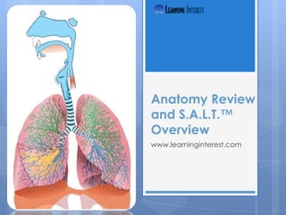
SlideShare a Scribd company logo
1 of 38
Anatomy Review
and S.A.L.T.™
Overview
www.learninginterest.com
Respiratory Review and S.A.L.T.
In-Service
Objectives
 Name   the major components of the upper
  and lower airways
 Describe the functions of the upper and
  lower airways
 Describe the process of ventilation
 Describe the process of respiration
 Identify the S.A.L.T.™ device
 Demonstrate use of the S.A.L.T.™ device
 Explain the SMO for the S.A.L.T.™ device
Terminology
   Air                        Nasal Turbinates
   Alveoli                    Nasopharynx
   Bronchial Tree             Oropharynx
   Bronchiole                 Paranasal sinuses
   Bronchus                   Pharynx
   Capillaries                Respiration
   Carina                     Rima glottidis
   Conchae                    S.A.L.T.™
   Cribriform plate           Sphenoid
   Corniculate cartilage      Surface tension
   Diffusion                  Surfactant
   Epiglottis                 Tidal Volume
   Ethmoid                    Trachea
   Glottis                    Ventilation
   Minute Volume              Vibrissae
   Mucosa                     Vocal folds
Upper and Lower Airways
Upper Airway Anatomy
   Anterior Nares
   Vestibule
   Mucosal Lining
   Septum
   Nasal Turbinates
   Posterior Nares
   Nasopharynx
   Oropharynx
   Pharynx
   Larynx
   Epiglottis
   Vocal Folds
   Glottis
Upper Airway
   Anterior Nares - These are the holes in the nose on the front of the
    face through which air is entrained for the purpose of ventilation.
   Vestibule – This space is the area just inside the nose that serves as a
    collection area as air enters the respiratory system through the nose.
    The Vibrissae (nose hair) are located here and this is where large
    particulate matter is trapped in the first step of filtering and purifying
    the air we breathe.
   Mucosal lining – The lining of the entire respiratory tract all the way
    down to the smallest of the bronchioles is made of mucosal
    epithelium which is rich with goblet cells that produce mucus. Also
    noteworthy is that these cells have cilia. This important structure is very
    vascular as well. It serves multiple functions including
    warming, adding moisture, further filtering the air. The rich blood
    supply warms the air. The mucus it produces serves to moisten the air
    and to trap the smaller particulate matter to further purify the air. The
    cilia move the mucus.
   Septum – This structure forms the middle ridge line of the nose dividing
    it into left and right halves. The majority of the septum is made of
    cartilage which is attached to the septal bone of the nose.
Upper Airway
   Nasal turbinates / Conchae – There are three sets of turbinates
    (a.k.a., Conchae). The inferior are the largest and have the most
    surface area in which to warm and moisten the air. Then there are
    the middle and the superior turbinates. The mucosal lining becomes
    thinner and less vascular as it goes up and in the superior turbinates it
    is more yellow than red. It is here that the olfactory nerve endings
    extend through the Ethmoid bone and air is gathered for the sense of
    smell. The Conchae are located on the lateral walls of the interior of
    the nose.
   Posterior nares – The posterior nares are the openings of the posterior
    nose into the Nasopharynx.
   Nasopharynx – The superior portion of the throat that is directly behind
    the nose and extends down to the opening of the oral cavity at the
    posterior soft palate.
   Oropharynx – The portion of the throat directly behind the oral cavity
    from the soft palate down to the superior rim of the hyoid bone.
   Pharynx - Is about 12.5 cm long extending from the base of the skull to
    the esophagus encompassing the Nasopharynx and the oropharynx.
Upper Airway
   Larynx – (a.k.a., voice box) serves as the entry point into the
    trachea and extends from the base of the tongue to the top
    of the trachea using the third – the sixth vertebrae as a
    reference in adult men. It is typically higher in women and
    children.
   Epiglottis – Is a cartilaginous structure that is the superior
    portion of the larynx. It serves as a flap to cover the glottis
    during swallowing since the oropharynx is a passage way for
    both food and air.
   Vocal folds – There are two sets of vocal folds. The false vocal
    folds, so called because they do not make actual
    vocalization, are the two white appearing lines of the vocal
    cords that vibrate as air passes through them to make sound.
   Rima glottidis – The space between the two true vocal folds
    (cords).
   Glottis - The entirety of the false and true vocal folds and the
    rima glottidis.
Flow of Air in the Upper Airway
                   Flow of Air
                       Anterior nares
                       Vestibule
                       Vibrissae
                       Nasal turbinates(conchae)
                           Inferior
                           Middle
                           Superior
                       Posterior nares
                       Nasopharynx
                       Oropharynx
                       Epiglottis
                       Glottis
                           Vocal folds
                           Rima glottidis
Upper and Lower Airways
Lower Airway Anatomy
   Trachea
   Mucous membrane
   C-shaped cartilaginous
    rings
   Carina
   Left main bronchus
   Right main bronchus
   Bronchial Tree
   Bronchioles
   Alveolar ducts
   Alveolar sacs
   Alveoli
       ̴300,000,000
   Capillaries
Lower Airway
   Trachea – Sometimes referred to as the “wind pipe”, the trachea the
    first structure of the lower airway. It is, as noted earlier, lined with the
    same type of mucus membrane as the rest of the airways. It extends
    from the inferior edge of the larynx to the carina.
   C-Shaped cartilaginous rings – As the name suggests, these structures
    are „C‟ shaped with the opening in the posterior and they are made
    of cartilage. The serve to provide shape, support and protection to
    the trachea. Without these rings, the trachea could collapse shutting
    off the vital supply of air. They are not complete rings so as to allow
    expansion and contraction of the trachea as its smooth muscles
    respond to the ever-changing conditions of the human body.
   Carina – This is the inferior wall of the trachea that sits at the
    bifurcation of the trachea into two main bronchi.
   Right main bronchus – This is the right branch of the trachea and is
    often larger and has a less acute angle to the trachea. Its size and
    position make it the more common path of endotracheal tubes that
    are placed too deep. It serves as the primary branch for all other
    branches of the bronchial tree in the right lung.
Lower Airway
   Left main bronchus – The primary branch of the bronchial tree
    that supplies the left lung. Its angle to the trachea is typically
    more acute and it is often smaller in diameter than its right
    counterpart.
   Bronchial Tree – This term is used to describe the branching of
    the bronchi down to the innumerable bronchioles that serve
    to transmit air into the alveoli. As the branches get
    smaller, the cartilaginous rings become more irregularly
    shaped and complete so as to support the small airways and
    prevent collapse.
   Bronchioles – These are the smallest airways of the lower
    airway anatomy. They terminate into the alveolar ducts
    which are the connection to the alveoli. At this level, the rings
    of cartilage have disappeared.
   Alveolar ducts – These are the small, acartilaginous airways
    that transmit air into the alveolar sacs.
Lower Airway
   Alveolar sacs – The terminal of gas exchange is the
    alveolar sac. These are often said to look like a
    bunch of grapes. These tiny (microscopic) sacs are
    enveloped in capillaries that are in contact with
    their surface. The „grape-like‟ structure provides a
    large surface area for efficient gas exchange.
   Alveoli – This is the term applied to the „bunches of
    grapes‟ and it has been estimated that there are
    around 300,000,000 alveoli in the human lungs.
   Capillaries – The capillaries are the smallest of blood
    vessels and are typically only one cell thick. This
    allows for efficient exchange of gases, glucose, and
    waste products between individual cells and the
    blood.
Ventilation vs. Respiration
       Ventilation  is the                         Respiration is said to
          mechanical                                 be the “processes
          movement of air                            that result in the
          into and out of the                        absorption, transport,
                                                      and utilization or
          body.
                                                     exchange of
                                                     respiratory gases
                                                     between an
                                                     organism and its
                                                     environment.”₁


We can produce artificial ventilation but we cannot produce artificial respiration!
Respiration
   Respiration begins at the anterior nares and ends at
    the anterior nares and it includes everything that
    happens in between.
   Ventilation moves the oxygen rich air in and displaces
    the carbon dioxide rich air out.
   It is in the alveoli that gas exchange takes place and
    the airways are the transmission pathways for the
    gases.
   As mentioned before, the walls of the capillaries are in
    contact with the walls of the alveoli and this contact is
    the point at which CO2 trades places with O2 making it
    possible for the body to remain homeostatic.
   Keeping in mind that all of this occurs at the cellular
    level, remember that our job is keeping as many cells
    alive as we can for as long as we can.
“the lungs serve the alveoli” just
as “the circulatory system serves
the capillaries.”
                ₁
Alveolar Function
   Gas exchange easily
    takes place in the
    alveoli
       Alveoli are thin-
        walled
       Each alveolus is in
        contact with the
        capillaries that
        surround it
       Capillary walls at this
        level are only 1 cell
        thick
Supraglottic Airway
Laryngopharangeal Tube
                 “The S.A.L.T.™ is a
                  unique single patient
                  use oropharyngeal
                  airway which can be
                  utilized to facilitate
                  blind, endotracheal
                  intubation. The
                  S.A.L.T.™ can also be
                  utilized to reduce
                  accidental
                  endotracheal tube
                  extubation.”
S.A.L.T.™
   Supraglottic Airway Laryngopharangeal Tube –“ The S.A.L.T.™ is a unique single
    patient use oropharyngeal airway which can be utilized to facilitate
    blind, endotracheal intubation. The S.A.L.T.™ can also be utilized to reduce
    accidental endotracheal tube extubation.”(Though this is not grammatically
    correct, it is a direct quotation and so was not changed.)
   This device will be used as an adjunct to open the airway and to facilitate
    endotracheal intubation in the difficult airway patients.
   We, here at National EMS, will use it after two (2) attempts at intubation via direct
    laryngoscopy or when patient access is compromised making direct
    laryngoscopy unreasonable.
   This device can be inserted using the tongue blade method the same way
    standard OPA‟s are inserted or by direct laryngoscopy.
   Remember that good BLS is a prerequisite to any ALS procedure. Always begin
    with simple airway maneuvers such as the head-tilt/chin-lift or jaw-thrust then
    bag-valve-mask ventilation if required.
   Once the decision has been made to move on to advanced airway
    management, we will start with endotracheal intubation via direct laryngoscopy.
    This is a skill we need to maintain and practice is the only way to do that.
   If intubation by direct laryngoscopy is not successful after the second
    attempt, insertion of the S.A.L.T.™ should be done as follows:
S.A.L.T.™
Indications
1. The S.A.L.T.™ is designed and intended for
   airway management in emergency or difficult
   airway cases.
2. Absence of a gag reflex.
Contraindications
1. Responsive patients with an intact gag reflex.
2. Patients with known esophageal disease.
3. Patients who have ingested caustic
   substances.
Suggested Instructions for Use
1.   Confirm anatomical compatibility by measuring the
     S.A.L.T.™ from the level of the patient's lips to the superior
     edge of the larynx.
2.   Open-patient's airway utilizing appropriate manual
     maneuvers.
3.   Ventilate/oxygenate patient via bag-valve mask or pocket
     face mask and observe for adequate chest rise/expansion
     and ventilatory compliance. If patient's airway is
     obstructed, remove obstruction prior to insertion of the
     S.A.L.T.™ If the patient exhibits no signs of foreign body
     airway obstruction and has no gag reflex, prepare for
     insertion of the SALT
4.   Lubricate the distal end of the S.A.L.T.™ with a water soluble
     lubricant.
5.   Grasp the distal end of the S.A.L.T.™ by placing the thumb
     against one lateral wall and the index finger against the
     opposite lateral wall.
Suggested Instructions for Use
6.         Align the patient's airway in a neutral position.
7.         Insert tongue depressor into patient's posterior oropharynx
           and push anteriorly, retracting the tongue. This action will
           aid in displacing the epiglottis superiorly allowing the
           S.A.L.T.™ to be advanced beneath the epiglottis.
8.         Insert the S.A.L.T.™ into the patient's oropharynx, grasp the
           proximal end of the S.A.L.T.™ and advance following the
           anatomical contour of the airway until:
      a)     resistance is encountered (indicating the SAL.T. has abutted
             against the corniculate cartilage and/or
      b)     the depth indicator ring rests against the lips or gumline.
9.         Remove tongue depressor after the S.A.L.T.™ has been
           inserted to the proper depth.
10.        Ventilate/oxygenate the patient via bag-valve mask or
           pocket face mask and observe for adequate chest
           rise/expansion and ventilatory compliance.
Alternate Instructions for Use
      1.        Utilizing a laryngoscope, advance a laryngoscope blade
                (straight #3, curved #3, or curved #4) into the patient's
                posterior pharynx and gently lift upward. This action will
                displace the epiglottis superiorly allowing the S.A.L.T.™ to be
                advanced beneath the epiglottis. Note: Visualization of the
                epiglottis is not required.
      2.        Insert the S.A.L.T.™ into the patient's oropharynx, grasp the
                proximal end of the S.A.L.T.™ and advance the airway until:
           a)     resistance is encountered (indicating the S.A.L.T. has abutted
                  against the corniculate cartilage and/or
           b)     the depth indicator ring rests against the lips or gumline.
      3.        Remove laryngoscope blade after the S.A.L.T.™ has been
                inserted to the proper depth.
      4.        Ventilate/oxygenate the patient via bag-valve mask or
                pocket face mask and observe for adequate chest
                rise/expansion and ventilatory compliance.


Note: If rescuer encounters poor ventilatory compliance; slightly withdraw
S.A.L.T.™, re-advance S.A.L.T., and reassess ventilatory status.
Insertion of Endotracheal Tube
          1.     Ensure patient is being adequately
                 ventilated/oxygenated.
          2.     Lubricate distal end of endotracheal tube with a
                 water soluble lubricant.
          3.     Insert the endotracheal tube and advance to proper
                 depth.

          Note: If resistance is encountered during endotracheal
          tube insertion:
               a)    withdraw endotrachealtube and slightly withdraw
                     S.A.L.T.™. Readvance S.A.L.T.™ and reattempt
                     endotracheal tube insertion or
               b)    withdraw endotracheal tube, apply cricoid pressure and
                     reattempt endotracheal tube insertion


Note: If esophageal placement is either detected or suspected, the endotracheal tube should be immediately removed and the
patient should be ventilated via bag-valve mask and supplemental oxygen prior to repeated endotracheal intubation attempts or
alternative airway management.
Insertion of Endotracheal Tube
          4.        Ventilate patient via bag-valve
                    resuscitator/supplemental oxygen and confirm
                    endotracheal tube placement by:
               a)     observing chest for adequate chest rise/expansion.
               b)     auscultating over epigastric region for absence of
                      gurgling sounds.
               c)     auscultating over lung fields (left and right anterior
                      chest/left and right lateral chest) for presence of bilateral
                      breath sounds.
               d)     observing moisture in the endotracheal tube.
               e)     utilizing an end-tidal C02 detector and observing color
                      change indicative of adequate concentration of C02.
               f)     utilizing an esophageal intubation detector.
               g)     utilizing capnometry/capnography and observing
                      readings/waveforms indicative of adequate
                      concentration of C02.
Note: If esophageal placement is either detected or suspected, the endotracheal tube should be immediately removed and the
patient should be ventilated via bag-valve mask and supplemental oxygen prior to repeated endotracheal intubation attempts or
alternative airway management.
Instructions for ET Tube
Securing/Clamping Device
1.   Place ET Tube securing clamp around ET Tube
     at the superior edge of the SAL.T. Note tube
     depth.
2.   Secure clamp firmly around ET Tube. DO NOT
     CRUSH or COLLAPSE ET TUBE.
3.   Place strap under/behind patients
     head/neck.
4.   Pull strap tight and connect to securing post
     on opposite side of ET Tube Clamp securing
     post.
5.   Verify ET Tube placement after securing of
     tube.
Instructions for Removal of
      S.A.L.T. with ET Tube in Place
       1.    Ensure the patient is well oxygenated.
       2.    Remove tape or tube securing device.
       3.    Disconnect the 15 mm adapter from the
             endotracheal tube.
       4.    Gently retract the S.A.L.T.™, allowing it to slide over
             the endotracheal tube.
       5.    As soon as practical, grip the endotracheal tube
             while removing the S.A.L.T.™ from the oropharynx
             to ensure minimal movement of the endotracheal
             tube.
       6.    Confirm endotracheal tube placement in
             accordance with local protocol.
       7.    Secure endotracheal tube in accordance with
             local protocol.
Note: Because the S.A.L.T.™ serves to protect against accidental endotracheal tube extubation, it is
recommended that the S.A.L.T.™ not be removed during the prehospital phase of care, unless absolutely
necessary.³
Standing Medical Order*
      A.        Open Airway
           1.      Manual maneuvers
           2.      Clear obstructions using the appropriate
                   techniques/suction
           3.      If necessary, insert appropriate airway
                   device to maintain the airway (i.e.
                   oropharyngeal, nasopharyngeal, endotrach
                   eal tube, S.A.L.T. ™, Combi-tube/King
                   Airway, cricothyrotomy)


*The following SMO is provided as an example only. Check with your Medical Director for the current Airway Management SMO
at your service.
Standing Medical Order
 4.        Intubate any unconscious patient without a gag
           reflex
      a.     monitor patient‟s pulse oximetry and cardiac rhythm
             at all times to prevent unrecognized hypoxia
      b.     hyper oxygenate prior to intubation attempt
      c.     if not able to place tube within 30
             sec., withdraw, hyper oxygenate, and re-attempt
      d.     verify placement using Ambu tube check
             device, observing appropriate chest rise, end tidal
             CO2 monitoring, and auscultation of breath sounds
      e.     orotracheal or nasotracheal intubation as indicated
      f.     secure tube with ET tube holder (pediatric – use tape)
      g.     in the cardiac arrest situation, initial airway
             management should be completed with manual
             maneuvers, & simple adjuncts.
Standing Medical Order
 5.   After two unsuccessful attempts at intubation by
      direct laryngoscopy, hyper oxygenate the
      patient, place S.A.L.T. ™ adjunct, hyper
      oxygenate, then intubate through the S.A.L.T. ™.
      The S.A.L.T. ™ is only indicated in patients for
      whom 6.5mm through 9.0mm ETT is appropriate.
 6.   Nasotracheal intubation and nasal airways
      should be avoided in the patient with facial
      trauma, or suspected basal skull fracture.
 7.   Extreme caution should be exercised in any
      patient experiencing significant head injury, or
      with signs of rising intracranial pressure.
Standing Medical Order
 8.    With suspected head injuries, administer
       Lidocaine 1.5 mg/kg prior to ETT intubation to
       help prevent rise in ICP.
 9.    For any patient with a GCS < 8, complete
       endotracheal intubation
 10.   Only if necessary, in the unusually difficult
       intubation, and when the patient can not
       otherwise be oxygenated by basic life
       support measures, consider giving Versed
       (or valium) 5 mg IVP + Morphine Sulfate 2 mg
       IVP to facilitate intubation per Medication
       Facilitated Intubation Standing Order.
Standing Medical Order
 11.        A Combi-tube/King Airway should be used if
            attempts at intubation with the S.A.L.T. ™ are
            unsuccessful. For EMT-I‟s, the Combi-tube/King
            Airway is the advanced airway for utilization. The
            Combi-tube/King Airway is contraindicated in
            the following:
       a.     patients under 5 feet in height or over 6‟4” in
              height
       b.     patients who are less than 16 years of age
       c.     patients who weigh less than 90 lbs
       d.     patients who have known esophageal disease
       e.     patients who have ingested caustic substances
Objectives Review
 Name   the major components of the upper
  and lower airways
 Describe the functions of the upper and
  lower airways
 Describe the process of ventilation
 Describe the process of respiration
 Identify the S.A.L.T.™ device
 Demonstrate use of the S.A.L.T.™ device
 Explain the SMO for the S.A.L.T.™ device
http://www.learning
interest.com/wp-
content/uploads/
SIS/sisc.html
Thank you for completing this
training program. As part of this
research study, please click on
the link above to complete the
related survey.

More Related Content

What's hot

Human Respiratry system
Human Respiratry systemHuman Respiratry system
Human Respiratry systemGladys Kalpana
 
Anatomy of the lower respiratory tract
Anatomy of the lower respiratory tractAnatomy of the lower respiratory tract
Anatomy of the lower respiratory tractDr.RMLIMS lucknow
 
Respiratory System
Respiratory SystemRespiratory System
Respiratory Systemguest065135
 
Respiratory system
Respiratory systemRespiratory system
Respiratory systemayesexy
 
Therespiratorysystem/dhsani/geb,sust
Therespiratorysystem/dhsani/geb,sustTherespiratorysystem/dhsani/geb,sust
Therespiratorysystem/dhsani/geb,sustDh Sani
 
ANATOMY AND PHYSIOLOGY OF RESPIRATORY SYSTEM
ANATOMY AND PHYSIOLOGY OF RESPIRATORY SYSTEM ANATOMY AND PHYSIOLOGY OF RESPIRATORY SYSTEM
ANATOMY AND PHYSIOLOGY OF RESPIRATORY SYSTEM paramesh Researcher
 
IVMS-Respiratory Anatomy and Physiology Global Overview / Review
IVMS-Respiratory  Anatomy and Physiology Global Overview / ReviewIVMS-Respiratory  Anatomy and Physiology Global Overview / Review
IVMS-Respiratory Anatomy and Physiology Global Overview / ReviewImhotep Virtual Medical School
 
Respiratory system for Msefah
Respiratory system for MsefahRespiratory system for Msefah
Respiratory system for Msefahmsefah
 
Respiratory system slide show
Respiratory system slide showRespiratory system slide show
Respiratory system slide showKevin Young
 
Respiratory anat online
Respiratory anat onlineRespiratory anat online
Respiratory anat onlinekmilaniBCC
 
Respiratory
RespiratoryRespiratory
Respiratorysamrus1
 
Respiratory system
Respiratory systemRespiratory system
Respiratory systemRupali Patil
 
Therespiratorysystem/dhsani/geb,sust
Therespiratorysystem/dhsani/geb,sustTherespiratorysystem/dhsani/geb,sust
Therespiratorysystem/dhsani/geb,sustDh Sani
 
Respiratory System
Respiratory SystemRespiratory System
Respiratory Systemwhitchur
 
Respiratory System (afreen khan)
 Respiratory System (afreen khan) Respiratory System (afreen khan)
Respiratory System (afreen khan)Shoaib Akhtar
 
Medical terminology
Medical terminologyMedical terminology
Medical terminologycclark15
 
Hft respiratory system
Hft   respiratory systemHft   respiratory system
Hft respiratory systemmrlchapman
 

What's hot (20)

Functional Anatomy of Respiratory system
Functional Anatomy of Respiratory systemFunctional Anatomy of Respiratory system
Functional Anatomy of Respiratory system
 
Human Respiratry system
Human Respiratry systemHuman Respiratry system
Human Respiratry system
 
Anatomy of the lower respiratory tract
Anatomy of the lower respiratory tractAnatomy of the lower respiratory tract
Anatomy of the lower respiratory tract
 
Respiratory System
Respiratory SystemRespiratory System
Respiratory System
 
Respiratory system
Respiratory systemRespiratory system
Respiratory system
 
Therespiratorysystem/dhsani/geb,sust
Therespiratorysystem/dhsani/geb,sustTherespiratorysystem/dhsani/geb,sust
Therespiratorysystem/dhsani/geb,sust
 
ANATOMY AND PHYSIOLOGY OF RESPIRATORY SYSTEM
ANATOMY AND PHYSIOLOGY OF RESPIRATORY SYSTEM ANATOMY AND PHYSIOLOGY OF RESPIRATORY SYSTEM
ANATOMY AND PHYSIOLOGY OF RESPIRATORY SYSTEM
 
IVMS-Respiratory Anatomy and Physiology Global Overview / Review
IVMS-Respiratory  Anatomy and Physiology Global Overview / ReviewIVMS-Respiratory  Anatomy and Physiology Global Overview / Review
IVMS-Respiratory Anatomy and Physiology Global Overview / Review
 
Respiratory System
Respiratory SystemRespiratory System
Respiratory System
 
Respiratory system for Msefah
Respiratory system for MsefahRespiratory system for Msefah
Respiratory system for Msefah
 
Respiratory system slide show
Respiratory system slide showRespiratory system slide show
Respiratory system slide show
 
Respiratory anat online
Respiratory anat onlineRespiratory anat online
Respiratory anat online
 
Respiratory
RespiratoryRespiratory
Respiratory
 
Respiratory system
Respiratory systemRespiratory system
Respiratory system
 
Therespiratorysystem/dhsani/geb,sust
Therespiratorysystem/dhsani/geb,sustTherespiratorysystem/dhsani/geb,sust
Therespiratorysystem/dhsani/geb,sust
 
Respiratory system
Respiratory systemRespiratory system
Respiratory system
 
Respiratory System
Respiratory SystemRespiratory System
Respiratory System
 
Respiratory System (afreen khan)
 Respiratory System (afreen khan) Respiratory System (afreen khan)
Respiratory System (afreen khan)
 
Medical terminology
Medical terminologyMedical terminology
Medical terminology
 
Hft respiratory system
Hft   respiratory systemHft   respiratory system
Hft respiratory system
 

Viewers also liked

Laryngeal Mask Airway & Igel - An Introduction
Laryngeal Mask Airway & Igel - An IntroductionLaryngeal Mask Airway & Igel - An Introduction
Laryngeal Mask Airway & Igel - An IntroductionHIRANGER
 
Supraglottic airway device
Supraglottic airway deviceSupraglottic airway device
Supraglottic airway deviceDebojyoti Dutta
 
Manejo de dispositivos para via aerea
Manejo de dispositivos para via aereaManejo de dispositivos para via aerea
Manejo de dispositivos para via aereacarmenzarivera
 
1 manejo de la via aerea
1 manejo de la via aerea1 manejo de la via aerea
1 manejo de la via aereaomarmedico
 
ObstruccióN Via Aerea
ObstruccióN Via AereaObstruccióN Via Aerea
ObstruccióN Via AereaGINGER
 
Manejo de vía aérea y ventilación
Manejo de vía aérea y ventilaciónManejo de vía aérea y ventilación
Manejo de vía aérea y ventilaciónErick Calero
 
CapíTulo 07 Manejo BáSico De La VíA AéRea
CapíTulo 07   Manejo BáSico De La VíA AéReaCapíTulo 07   Manejo BáSico De La VíA AéRea
CapíTulo 07 Manejo BáSico De La VíA AéReaAlan Lopez
 
Via Aerea. Manejo
Via Aerea. ManejoVia Aerea. Manejo
Via Aerea. Manejozuritamd
 
Manejo de via aerea avanzada
Manejo de via aerea avanzadaManejo de via aerea avanzada
Manejo de via aerea avanzadaEdser Lugo-Ferrer
 
Dispositivos de la vía aérea
Dispositivos de la vía aéreaDispositivos de la vía aérea
Dispositivos de la vía aéreaanestesiahsb
 

Viewers also liked (12)

Laryngeal Mask Airway & Igel - An Introduction
Laryngeal Mask Airway & Igel - An IntroductionLaryngeal Mask Airway & Igel - An Introduction
Laryngeal Mask Airway & Igel - An Introduction
 
Manejo De La Via Aerea
Manejo De La Via AereaManejo De La Via Aerea
Manejo De La Via Aerea
 
Supraglottic airway device
Supraglottic airway deviceSupraglottic airway device
Supraglottic airway device
 
Manejo de dispositivos para via aerea
Manejo de dispositivos para via aereaManejo de dispositivos para via aerea
Manejo de dispositivos para via aerea
 
1 manejo de la via aerea
1 manejo de la via aerea1 manejo de la via aerea
1 manejo de la via aerea
 
ObstruccióN Via Aerea
ObstruccióN Via AereaObstruccióN Via Aerea
ObstruccióN Via Aerea
 
Manejo de vía aérea y ventilación
Manejo de vía aérea y ventilaciónManejo de vía aérea y ventilación
Manejo de vía aérea y ventilación
 
CapíTulo 07 Manejo BáSico De La VíA AéRea
CapíTulo 07   Manejo BáSico De La VíA AéReaCapíTulo 07   Manejo BáSico De La VíA AéRea
CapíTulo 07 Manejo BáSico De La VíA AéRea
 
Via Aerea. Manejo
Via Aerea. ManejoVia Aerea. Manejo
Via Aerea. Manejo
 
Manejo de vía aérea y ventilacion (atls)
Manejo de vía aérea y ventilacion (atls)Manejo de vía aérea y ventilacion (atls)
Manejo de vía aérea y ventilacion (atls)
 
Manejo de via aerea avanzada
Manejo de via aerea avanzadaManejo de via aerea avanzada
Manejo de via aerea avanzada
 
Dispositivos de la vía aérea
Dispositivos de la vía aéreaDispositivos de la vía aérea
Dispositivos de la vía aérea
 

Similar to Anatomy Review and S.A.L.T.™ Overview

Structure of respiratory system
Structure of respiratory systemStructure of respiratory system
Structure of respiratory systemShahriar Ahmed
 
Anatomy of respiratory system.
Anatomy of respiratory system.Anatomy of respiratory system.
Anatomy of respiratory system.Joan Delgado
 
Respiratory system by A.H..pptx
Respiratory system by A.H..pptxRespiratory system by A.H..pptx
Respiratory system by A.H..pptxSadiyaAbubakar7
 
Respiratory system presentation
Respiratory system presentationRespiratory system presentation
Respiratory system presentationNoorZahra8
 
Anatomy of respiratory system
Anatomy of respiratory systemAnatomy of respiratory system
Anatomy of respiratory systemgaurav gautam
 
Respiratory Pathway.pptx
Respiratory Pathway.pptxRespiratory Pathway.pptx
Respiratory Pathway.pptxAkashAnand55
 
Johny's A&P structure and function of respiratory system
Johny's A&P structure and function of respiratory systemJohny's A&P structure and function of respiratory system
Johny's A&P structure and function of respiratory systemJohny Kutty Joseph
 
Anatomy of respiratory system
Anatomy of respiratory systemAnatomy of respiratory system
Anatomy of respiratory systemSelva Kumar
 
Lesson6 [2 Oth Oct 2008]
Lesson6 [2 Oth Oct 2008]Lesson6 [2 Oth Oct 2008]
Lesson6 [2 Oth Oct 2008]Marc Potter
 
Anatomy of respiratory system
Anatomy of respiratory systemAnatomy of respiratory system
Anatomy of respiratory systemaveekdatta7
 
RESPIRATORY FAILURE & CHEST TRAUMA.pptx
RESPIRATORY FAILURE & CHEST TRAUMA.pptxRESPIRATORY FAILURE & CHEST TRAUMA.pptx
RESPIRATORY FAILURE & CHEST TRAUMA.pptxSaraswathy Karuppasamy
 
The respiratory system
The respiratory systemThe respiratory system
The respiratory systemitutor
 
Respiratory System - Human Anatomy and Physiology bPharm
Respiratory System - Human Anatomy and Physiology bPharmRespiratory System - Human Anatomy and Physiology bPharm
Respiratory System - Human Anatomy and Physiology bPharmPankaj Saha
 
therespiratorysystem-130524022043-phpapp01.pptx
therespiratorysystem-130524022043-phpapp01.pptxtherespiratorysystem-130524022043-phpapp01.pptx
therespiratorysystem-130524022043-phpapp01.pptxPawanKanherkar1
 

Similar to Anatomy Review and S.A.L.T.™ Overview (20)

Structure of respiratory system
Structure of respiratory systemStructure of respiratory system
Structure of respiratory system
 
Anatomy of respiratory system.
Anatomy of respiratory system.Anatomy of respiratory system.
Anatomy of respiratory system.
 
Respiratory system by A.H..pptx
Respiratory system by A.H..pptxRespiratory system by A.H..pptx
Respiratory system by A.H..pptx
 
anatomy of Respsystem
anatomy of Respsystem anatomy of Respsystem
anatomy of Respsystem
 
Respiratory system presentation
Respiratory system presentationRespiratory system presentation
Respiratory system presentation
 
Anatomy of respiratory system
Anatomy of respiratory systemAnatomy of respiratory system
Anatomy of respiratory system
 
Respiratory Pathway.pptx
Respiratory Pathway.pptxRespiratory Pathway.pptx
Respiratory Pathway.pptx
 
Johny's A&P structure and function of respiratory system
Johny's A&P structure and function of respiratory systemJohny's A&P structure and function of respiratory system
Johny's A&P structure and function of respiratory system
 
Anatomy of respiratory system
Anatomy of respiratory systemAnatomy of respiratory system
Anatomy of respiratory system
 
Lesson6 [2 Oth Oct 2008]
Lesson6 [2 Oth Oct 2008]Lesson6 [2 Oth Oct 2008]
Lesson6 [2 Oth Oct 2008]
 
Anatomy of respiratory system
Anatomy of respiratory systemAnatomy of respiratory system
Anatomy of respiratory system
 
Respiration
RespirationRespiration
Respiration
 
Respiratory system
Respiratory systemRespiratory system
Respiratory system
 
RESPIRATORY SYSTEM.pptx
RESPIRATORY SYSTEM.pptxRESPIRATORY SYSTEM.pptx
RESPIRATORY SYSTEM.pptx
 
Unit VI
Unit VIUnit VI
Unit VI
 
RESPIRATORY FAILURE & CHEST TRAUMA.pptx
RESPIRATORY FAILURE & CHEST TRAUMA.pptxRESPIRATORY FAILURE & CHEST TRAUMA.pptx
RESPIRATORY FAILURE & CHEST TRAUMA.pptx
 
Respiratory system
Respiratory systemRespiratory system
Respiratory system
 
The respiratory system
The respiratory systemThe respiratory system
The respiratory system
 
Respiratory System - Human Anatomy and Physiology bPharm
Respiratory System - Human Anatomy and Physiology bPharmRespiratory System - Human Anatomy and Physiology bPharm
Respiratory System - Human Anatomy and Physiology bPharm
 
therespiratorysystem-130524022043-phpapp01.pptx
therespiratorysystem-130524022043-phpapp01.pptxtherespiratorysystem-130524022043-phpapp01.pptx
therespiratorysystem-130524022043-phpapp01.pptx
 

More from Tonia A. Dousay

Students’ Use of Digital Storytelling to Enhance Media Literacy Skills
Students’ Use of Digital Storytelling to Enhance Media Literacy SkillsStudents’ Use of Digital Storytelling to Enhance Media Literacy Skills
Students’ Use of Digital Storytelling to Enhance Media Literacy SkillsTonia A. Dousay
 
Measuring the Impact of Multimedia Design on Situational Interest of Learners
Measuring the Impact of Multimedia Design on Situational Interest of LearnersMeasuring the Impact of Multimedia Design on Situational Interest of Learners
Measuring the Impact of Multimedia Design on Situational Interest of LearnersTonia A. Dousay
 
From death by PowerPoint to engaging Captivate: Lessons learned when implemen...
From death by PowerPoint to engaging Captivate: Lessons learned when implemen...From death by PowerPoint to engaging Captivate: Lessons learned when implemen...
From death by PowerPoint to engaging Captivate: Lessons learned when implemen...Tonia A. Dousay
 
Final Dissertation Defense
Final Dissertation DefenseFinal Dissertation Defense
Final Dissertation DefenseTonia A. Dousay
 
Making Design Interesting
Making Design InterestingMaking Design Interesting
Making Design InterestingTonia A. Dousay
 
Effect of Multimedia Design Principles on Situational Interest of Adult Learners
Effect of Multimedia Design Principles on Situational Interest of Adult LearnersEffect of Multimedia Design Principles on Situational Interest of Adult Learners
Effect of Multimedia Design Principles on Situational Interest of Adult LearnersTonia A. Dousay
 
Presenting Research Findings with Visuals
Presenting Research Findings with VisualsPresenting Research Findings with Visuals
Presenting Research Findings with VisualsTonia A. Dousay
 
Graphical Presentation & Deception
Graphical Presentation & DeceptionGraphical Presentation & Deception
Graphical Presentation & DeceptionTonia A. Dousay
 
Research & Scholars in Information Design
Research & Scholars in Information DesignResearch & Scholars in Information Design
Research & Scholars in Information DesignTonia A. Dousay
 
History of Information Design
History of Information DesignHistory of Information Design
History of Information DesignTonia A. Dousay
 
EDIT 9990 Course Introduction & Overview
EDIT 9990 Course Introduction & OverviewEDIT 9990 Course Introduction & Overview
EDIT 9990 Course Introduction & OverviewTonia A. Dousay
 
Americans with Disabilities Act (ADA): Implications for Information Design
Americans with Disabilities Act (ADA): Implications for Information DesignAmericans with Disabilities Act (ADA): Implications for Information Design
Americans with Disabilities Act (ADA): Implications for Information DesignTonia A. Dousay
 
Anatomy Review and S.A.L.T.™ OveRview
Anatomy Review and S.A.L.T.™ OveRviewAnatomy Review and S.A.L.T.™ OveRview
Anatomy Review and S.A.L.T.™ OveRviewTonia A. Dousay
 
Anatomy Review and S.A.L.T.™ OvervieW
Anatomy Review and S.A.L.T.™ OvervieWAnatomy Review and S.A.L.T.™ OvervieW
Anatomy Review and S.A.L.T.™ OvervieWTonia A. Dousay
 
Anatomy Review and S.A.L.T.™ OveRvieW
Anatomy Review and S.A.L.T.™ OveRvieWAnatomy Review and S.A.L.T.™ OveRvieW
Anatomy Review and S.A.L.T.™ OveRvieWTonia A. Dousay
 
Evaluating & Analyzing the Phases of ADDIE
Evaluating & Analyzing the Phases of ADDIEEvaluating & Analyzing the Phases of ADDIE
Evaluating & Analyzing the Phases of ADDIETonia A. Dousay
 

More from Tonia A. Dousay (20)

Students’ Use of Digital Storytelling to Enhance Media Literacy Skills
Students’ Use of Digital Storytelling to Enhance Media Literacy SkillsStudents’ Use of Digital Storytelling to Enhance Media Literacy Skills
Students’ Use of Digital Storytelling to Enhance Media Literacy Skills
 
Measuring the Impact of Multimedia Design on Situational Interest of Learners
Measuring the Impact of Multimedia Design on Situational Interest of LearnersMeasuring the Impact of Multimedia Design on Situational Interest of Learners
Measuring the Impact of Multimedia Design on Situational Interest of Learners
 
From death by PowerPoint to engaging Captivate: Lessons learned when implemen...
From death by PowerPoint to engaging Captivate: Lessons learned when implemen...From death by PowerPoint to engaging Captivate: Lessons learned when implemen...
From death by PowerPoint to engaging Captivate: Lessons learned when implemen...
 
Final Dissertation Defense
Final Dissertation DefenseFinal Dissertation Defense
Final Dissertation Defense
 
Making Design Interesting
Making Design InterestingMaking Design Interesting
Making Design Interesting
 
Effect of Multimedia Design Principles on Situational Interest of Adult Learners
Effect of Multimedia Design Principles on Situational Interest of Adult LearnersEffect of Multimedia Design Principles on Situational Interest of Adult Learners
Effect of Multimedia Design Principles on Situational Interest of Adult Learners
 
Presenting Research Findings with Visuals
Presenting Research Findings with VisualsPresenting Research Findings with Visuals
Presenting Research Findings with Visuals
 
Graphical Presentation & Deception
Graphical Presentation & DeceptionGraphical Presentation & Deception
Graphical Presentation & Deception
 
Research & Scholars in Information Design
Research & Scholars in Information DesignResearch & Scholars in Information Design
Research & Scholars in Information Design
 
Types of Visuals
Types of VisualsTypes of Visuals
Types of Visuals
 
History of Information Design
History of Information DesignHistory of Information Design
History of Information Design
 
Information Design
Information DesignInformation Design
Information Design
 
EDIT 9990 Course Introduction & Overview
EDIT 9990 Course Introduction & OverviewEDIT 9990 Course Introduction & Overview
EDIT 9990 Course Introduction & Overview
 
Americans with Disabilities Act (ADA): Implications for Information Design
Americans with Disabilities Act (ADA): Implications for Information DesignAmericans with Disabilities Act (ADA): Implications for Information Design
Americans with Disabilities Act (ADA): Implications for Information Design
 
Anatomy Review and S.A.L.T.™ OveRview
Anatomy Review and S.A.L.T.™ OveRviewAnatomy Review and S.A.L.T.™ OveRview
Anatomy Review and S.A.L.T.™ OveRview
 
Anatomy Review and S.A.L.T.™ OvervieW
Anatomy Review and S.A.L.T.™ OvervieWAnatomy Review and S.A.L.T.™ OvervieW
Anatomy Review and S.A.L.T.™ OvervieW
 
Anatomy Review and S.A.L.T.™ OveRvieW
Anatomy Review and S.A.L.T.™ OveRvieWAnatomy Review and S.A.L.T.™ OveRvieW
Anatomy Review and S.A.L.T.™ OveRvieW
 
Mendeley Seminar
Mendeley SeminarMendeley Seminar
Mendeley Seminar
 
Evaluating & Analyzing the Phases of ADDIE
Evaluating & Analyzing the Phases of ADDIEEvaluating & Analyzing the Phases of ADDIE
Evaluating & Analyzing the Phases of ADDIE
 
2010 GREPIT Orientation
2010 GREPIT Orientation2010 GREPIT Orientation
2010 GREPIT Orientation
 

Recently uploaded

Pharmacognosy Flower 3. Compositae 2023.pdf
Pharmacognosy Flower 3. Compositae 2023.pdfPharmacognosy Flower 3. Compositae 2023.pdf
Pharmacognosy Flower 3. Compositae 2023.pdfMahmoud M. Sallam
 
EPANDING THE CONTENT OF AN OUTLINE using notes.pptx
EPANDING THE CONTENT OF AN OUTLINE using notes.pptxEPANDING THE CONTENT OF AN OUTLINE using notes.pptx
EPANDING THE CONTENT OF AN OUTLINE using notes.pptxRaymartEstabillo3
 
Incoming and Outgoing Shipments in 1 STEP Using Odoo 17
Incoming and Outgoing Shipments in 1 STEP Using Odoo 17Incoming and Outgoing Shipments in 1 STEP Using Odoo 17
Incoming and Outgoing Shipments in 1 STEP Using Odoo 17Celine George
 
Framing an Appropriate Research Question 6b9b26d93da94caf993c038d9efcdedb.pdf
Framing an Appropriate Research Question 6b9b26d93da94caf993c038d9efcdedb.pdfFraming an Appropriate Research Question 6b9b26d93da94caf993c038d9efcdedb.pdf
Framing an Appropriate Research Question 6b9b26d93da94caf993c038d9efcdedb.pdfUjwalaBharambe
 
Final demo Grade 9 for demo Plan dessert.pptx
Final demo Grade 9 for demo Plan dessert.pptxFinal demo Grade 9 for demo Plan dessert.pptx
Final demo Grade 9 for demo Plan dessert.pptxAvyJaneVismanos
 
Meghan Sutherland In Media Res Media Component
Meghan Sutherland In Media Res Media ComponentMeghan Sutherland In Media Res Media Component
Meghan Sutherland In Media Res Media ComponentInMediaRes1
 
DATA STRUCTURE AND ALGORITHM for beginners
DATA STRUCTURE AND ALGORITHM for beginnersDATA STRUCTURE AND ALGORITHM for beginners
DATA STRUCTURE AND ALGORITHM for beginnersSabitha Banu
 
भारत-रोम व्यापार.pptx, Indo-Roman Trade,
भारत-रोम व्यापार.pptx, Indo-Roman Trade,भारत-रोम व्यापार.pptx, Indo-Roman Trade,
भारत-रोम व्यापार.pptx, Indo-Roman Trade,Virag Sontakke
 
Full Stack Web Development Course for Beginners
Full Stack Web Development Course  for BeginnersFull Stack Web Development Course  for Beginners
Full Stack Web Development Course for BeginnersSabitha Banu
 
ECONOMIC CONTEXT - PAPER 1 Q3: NEWSPAPERS.pptx
ECONOMIC CONTEXT - PAPER 1 Q3: NEWSPAPERS.pptxECONOMIC CONTEXT - PAPER 1 Q3: NEWSPAPERS.pptx
ECONOMIC CONTEXT - PAPER 1 Q3: NEWSPAPERS.pptxiammrhaywood
 
History Class XII Ch. 3 Kinship, Caste and Class (1).pptx
History Class XII Ch. 3 Kinship, Caste and Class (1).pptxHistory Class XII Ch. 3 Kinship, Caste and Class (1).pptx
History Class XII Ch. 3 Kinship, Caste and Class (1).pptxsocialsciencegdgrohi
 
Crayon Activity Handout For the Crayon A
Crayon Activity Handout For the Crayon ACrayon Activity Handout For the Crayon A
Crayon Activity Handout For the Crayon AUnboundStockton
 
CELL CYCLE Division Science 8 quarter IV.pptx
CELL CYCLE Division Science 8 quarter IV.pptxCELL CYCLE Division Science 8 quarter IV.pptx
CELL CYCLE Division Science 8 quarter IV.pptxJiesonDelaCerna
 
Presiding Officer Training module 2024 lok sabha elections
Presiding Officer Training module 2024 lok sabha electionsPresiding Officer Training module 2024 lok sabha elections
Presiding Officer Training module 2024 lok sabha electionsanshu789521
 
internship ppt on smartinternz platform as salesforce developer
internship ppt on smartinternz platform as salesforce developerinternship ppt on smartinternz platform as salesforce developer
internship ppt on smartinternz platform as salesforce developerunnathinaik
 

Recently uploaded (20)

9953330565 Low Rate Call Girls In Rohini Delhi NCR
9953330565 Low Rate Call Girls In Rohini  Delhi NCR9953330565 Low Rate Call Girls In Rohini  Delhi NCR
9953330565 Low Rate Call Girls In Rohini Delhi NCR
 
Pharmacognosy Flower 3. Compositae 2023.pdf
Pharmacognosy Flower 3. Compositae 2023.pdfPharmacognosy Flower 3. Compositae 2023.pdf
Pharmacognosy Flower 3. Compositae 2023.pdf
 
EPANDING THE CONTENT OF AN OUTLINE using notes.pptx
EPANDING THE CONTENT OF AN OUTLINE using notes.pptxEPANDING THE CONTENT OF AN OUTLINE using notes.pptx
EPANDING THE CONTENT OF AN OUTLINE using notes.pptx
 
Incoming and Outgoing Shipments in 1 STEP Using Odoo 17
Incoming and Outgoing Shipments in 1 STEP Using Odoo 17Incoming and Outgoing Shipments in 1 STEP Using Odoo 17
Incoming and Outgoing Shipments in 1 STEP Using Odoo 17
 
Framing an Appropriate Research Question 6b9b26d93da94caf993c038d9efcdedb.pdf
Framing an Appropriate Research Question 6b9b26d93da94caf993c038d9efcdedb.pdfFraming an Appropriate Research Question 6b9b26d93da94caf993c038d9efcdedb.pdf
Framing an Appropriate Research Question 6b9b26d93da94caf993c038d9efcdedb.pdf
 
Final demo Grade 9 for demo Plan dessert.pptx
Final demo Grade 9 for demo Plan dessert.pptxFinal demo Grade 9 for demo Plan dessert.pptx
Final demo Grade 9 for demo Plan dessert.pptx
 
Meghan Sutherland In Media Res Media Component
Meghan Sutherland In Media Res Media ComponentMeghan Sutherland In Media Res Media Component
Meghan Sutherland In Media Res Media Component
 
DATA STRUCTURE AND ALGORITHM for beginners
DATA STRUCTURE AND ALGORITHM for beginnersDATA STRUCTURE AND ALGORITHM for beginners
DATA STRUCTURE AND ALGORITHM for beginners
 
भारत-रोम व्यापार.pptx, Indo-Roman Trade,
भारत-रोम व्यापार.pptx, Indo-Roman Trade,भारत-रोम व्यापार.pptx, Indo-Roman Trade,
भारत-रोम व्यापार.pptx, Indo-Roman Trade,
 
TataKelola dan KamSiber Kecerdasan Buatan v022.pdf
TataKelola dan KamSiber Kecerdasan Buatan v022.pdfTataKelola dan KamSiber Kecerdasan Buatan v022.pdf
TataKelola dan KamSiber Kecerdasan Buatan v022.pdf
 
ESSENTIAL of (CS/IT/IS) class 06 (database)
ESSENTIAL of (CS/IT/IS) class 06 (database)ESSENTIAL of (CS/IT/IS) class 06 (database)
ESSENTIAL of (CS/IT/IS) class 06 (database)
 
Full Stack Web Development Course for Beginners
Full Stack Web Development Course  for BeginnersFull Stack Web Development Course  for Beginners
Full Stack Web Development Course for Beginners
 
ECONOMIC CONTEXT - PAPER 1 Q3: NEWSPAPERS.pptx
ECONOMIC CONTEXT - PAPER 1 Q3: NEWSPAPERS.pptxECONOMIC CONTEXT - PAPER 1 Q3: NEWSPAPERS.pptx
ECONOMIC CONTEXT - PAPER 1 Q3: NEWSPAPERS.pptx
 
History Class XII Ch. 3 Kinship, Caste and Class (1).pptx
History Class XII Ch. 3 Kinship, Caste and Class (1).pptxHistory Class XII Ch. 3 Kinship, Caste and Class (1).pptx
History Class XII Ch. 3 Kinship, Caste and Class (1).pptx
 
Crayon Activity Handout For the Crayon A
Crayon Activity Handout For the Crayon ACrayon Activity Handout For the Crayon A
Crayon Activity Handout For the Crayon A
 
CELL CYCLE Division Science 8 quarter IV.pptx
CELL CYCLE Division Science 8 quarter IV.pptxCELL CYCLE Division Science 8 quarter IV.pptx
CELL CYCLE Division Science 8 quarter IV.pptx
 
Presiding Officer Training module 2024 lok sabha elections
Presiding Officer Training module 2024 lok sabha electionsPresiding Officer Training module 2024 lok sabha elections
Presiding Officer Training module 2024 lok sabha elections
 
internship ppt on smartinternz platform as salesforce developer
internship ppt on smartinternz platform as salesforce developerinternship ppt on smartinternz platform as salesforce developer
internship ppt on smartinternz platform as salesforce developer
 
Model Call Girl in Bikash Puri Delhi reach out to us at 🔝9953056974🔝
Model Call Girl in Bikash Puri  Delhi reach out to us at 🔝9953056974🔝Model Call Girl in Bikash Puri  Delhi reach out to us at 🔝9953056974🔝
Model Call Girl in Bikash Puri Delhi reach out to us at 🔝9953056974🔝
 
OS-operating systems- ch04 (Threads) ...
OS-operating systems- ch04 (Threads) ...OS-operating systems- ch04 (Threads) ...
OS-operating systems- ch04 (Threads) ...
 

Anatomy Review and S.A.L.T.™ Overview

  • 2. Respiratory Review and S.A.L.T. In-Service
  • 3. Objectives  Name the major components of the upper and lower airways  Describe the functions of the upper and lower airways  Describe the process of ventilation  Describe the process of respiration  Identify the S.A.L.T.™ device  Demonstrate use of the S.A.L.T.™ device  Explain the SMO for the S.A.L.T.™ device
  • 4. Terminology  Air  Nasal Turbinates  Alveoli  Nasopharynx  Bronchial Tree  Oropharynx  Bronchiole  Paranasal sinuses  Bronchus  Pharynx  Capillaries  Respiration  Carina  Rima glottidis  Conchae  S.A.L.T.™  Cribriform plate  Sphenoid  Corniculate cartilage  Surface tension  Diffusion  Surfactant  Epiglottis  Tidal Volume  Ethmoid  Trachea  Glottis  Ventilation  Minute Volume  Vibrissae  Mucosa  Vocal folds
  • 5. Upper and Lower Airways
  • 7. Upper Airway Anatomy  Anterior Nares  Vestibule  Mucosal Lining  Septum  Nasal Turbinates  Posterior Nares  Nasopharynx  Oropharynx  Pharynx  Larynx  Epiglottis  Vocal Folds  Glottis
  • 8. Upper Airway  Anterior Nares - These are the holes in the nose on the front of the face through which air is entrained for the purpose of ventilation.  Vestibule – This space is the area just inside the nose that serves as a collection area as air enters the respiratory system through the nose. The Vibrissae (nose hair) are located here and this is where large particulate matter is trapped in the first step of filtering and purifying the air we breathe.  Mucosal lining – The lining of the entire respiratory tract all the way down to the smallest of the bronchioles is made of mucosal epithelium which is rich with goblet cells that produce mucus. Also noteworthy is that these cells have cilia. This important structure is very vascular as well. It serves multiple functions including warming, adding moisture, further filtering the air. The rich blood supply warms the air. The mucus it produces serves to moisten the air and to trap the smaller particulate matter to further purify the air. The cilia move the mucus.  Septum – This structure forms the middle ridge line of the nose dividing it into left and right halves. The majority of the septum is made of cartilage which is attached to the septal bone of the nose.
  • 9. Upper Airway  Nasal turbinates / Conchae – There are three sets of turbinates (a.k.a., Conchae). The inferior are the largest and have the most surface area in which to warm and moisten the air. Then there are the middle and the superior turbinates. The mucosal lining becomes thinner and less vascular as it goes up and in the superior turbinates it is more yellow than red. It is here that the olfactory nerve endings extend through the Ethmoid bone and air is gathered for the sense of smell. The Conchae are located on the lateral walls of the interior of the nose.  Posterior nares – The posterior nares are the openings of the posterior nose into the Nasopharynx.  Nasopharynx – The superior portion of the throat that is directly behind the nose and extends down to the opening of the oral cavity at the posterior soft palate.  Oropharynx – The portion of the throat directly behind the oral cavity from the soft palate down to the superior rim of the hyoid bone.  Pharynx - Is about 12.5 cm long extending from the base of the skull to the esophagus encompassing the Nasopharynx and the oropharynx.
  • 10. Upper Airway  Larynx – (a.k.a., voice box) serves as the entry point into the trachea and extends from the base of the tongue to the top of the trachea using the third – the sixth vertebrae as a reference in adult men. It is typically higher in women and children.  Epiglottis – Is a cartilaginous structure that is the superior portion of the larynx. It serves as a flap to cover the glottis during swallowing since the oropharynx is a passage way for both food and air.  Vocal folds – There are two sets of vocal folds. The false vocal folds, so called because they do not make actual vocalization, are the two white appearing lines of the vocal cords that vibrate as air passes through them to make sound.  Rima glottidis – The space between the two true vocal folds (cords).  Glottis - The entirety of the false and true vocal folds and the rima glottidis.
  • 11. Flow of Air in the Upper Airway  Flow of Air  Anterior nares  Vestibule  Vibrissae  Nasal turbinates(conchae)  Inferior  Middle  Superior  Posterior nares  Nasopharynx  Oropharynx  Epiglottis  Glottis  Vocal folds  Rima glottidis
  • 12. Upper and Lower Airways
  • 13. Lower Airway Anatomy  Trachea  Mucous membrane  C-shaped cartilaginous rings  Carina  Left main bronchus  Right main bronchus  Bronchial Tree  Bronchioles  Alveolar ducts  Alveolar sacs  Alveoli  ̴300,000,000  Capillaries
  • 14. Lower Airway  Trachea – Sometimes referred to as the “wind pipe”, the trachea the first structure of the lower airway. It is, as noted earlier, lined with the same type of mucus membrane as the rest of the airways. It extends from the inferior edge of the larynx to the carina.  C-Shaped cartilaginous rings – As the name suggests, these structures are „C‟ shaped with the opening in the posterior and they are made of cartilage. The serve to provide shape, support and protection to the trachea. Without these rings, the trachea could collapse shutting off the vital supply of air. They are not complete rings so as to allow expansion and contraction of the trachea as its smooth muscles respond to the ever-changing conditions of the human body.  Carina – This is the inferior wall of the trachea that sits at the bifurcation of the trachea into two main bronchi.  Right main bronchus – This is the right branch of the trachea and is often larger and has a less acute angle to the trachea. Its size and position make it the more common path of endotracheal tubes that are placed too deep. It serves as the primary branch for all other branches of the bronchial tree in the right lung.
  • 15. Lower Airway  Left main bronchus – The primary branch of the bronchial tree that supplies the left lung. Its angle to the trachea is typically more acute and it is often smaller in diameter than its right counterpart.  Bronchial Tree – This term is used to describe the branching of the bronchi down to the innumerable bronchioles that serve to transmit air into the alveoli. As the branches get smaller, the cartilaginous rings become more irregularly shaped and complete so as to support the small airways and prevent collapse.  Bronchioles – These are the smallest airways of the lower airway anatomy. They terminate into the alveolar ducts which are the connection to the alveoli. At this level, the rings of cartilage have disappeared.  Alveolar ducts – These are the small, acartilaginous airways that transmit air into the alveolar sacs.
  • 16. Lower Airway  Alveolar sacs – The terminal of gas exchange is the alveolar sac. These are often said to look like a bunch of grapes. These tiny (microscopic) sacs are enveloped in capillaries that are in contact with their surface. The „grape-like‟ structure provides a large surface area for efficient gas exchange.  Alveoli – This is the term applied to the „bunches of grapes‟ and it has been estimated that there are around 300,000,000 alveoli in the human lungs.  Capillaries – The capillaries are the smallest of blood vessels and are typically only one cell thick. This allows for efficient exchange of gases, glucose, and waste products between individual cells and the blood.
  • 17. Ventilation vs. Respiration  Ventilation is the  Respiration is said to mechanical be the “processes movement of air that result in the into and out of the absorption, transport, and utilization or body. exchange of respiratory gases between an organism and its environment.”₁ We can produce artificial ventilation but we cannot produce artificial respiration!
  • 18. Respiration  Respiration begins at the anterior nares and ends at the anterior nares and it includes everything that happens in between.  Ventilation moves the oxygen rich air in and displaces the carbon dioxide rich air out.  It is in the alveoli that gas exchange takes place and the airways are the transmission pathways for the gases.  As mentioned before, the walls of the capillaries are in contact with the walls of the alveoli and this contact is the point at which CO2 trades places with O2 making it possible for the body to remain homeostatic.  Keeping in mind that all of this occurs at the cellular level, remember that our job is keeping as many cells alive as we can for as long as we can.
  • 19. “the lungs serve the alveoli” just as “the circulatory system serves the capillaries.” ₁
  • 20. Alveolar Function  Gas exchange easily takes place in the alveoli  Alveoli are thin- walled  Each alveolus is in contact with the capillaries that surround it  Capillary walls at this level are only 1 cell thick
  • 21.
  • 22. Supraglottic Airway Laryngopharangeal Tube  “The S.A.L.T.™ is a unique single patient use oropharyngeal airway which can be utilized to facilitate blind, endotracheal intubation. The S.A.L.T.™ can also be utilized to reduce accidental endotracheal tube extubation.”
  • 23. S.A.L.T.™  Supraglottic Airway Laryngopharangeal Tube –“ The S.A.L.T.™ is a unique single patient use oropharyngeal airway which can be utilized to facilitate blind, endotracheal intubation. The S.A.L.T.™ can also be utilized to reduce accidental endotracheal tube extubation.”(Though this is not grammatically correct, it is a direct quotation and so was not changed.)  This device will be used as an adjunct to open the airway and to facilitate endotracheal intubation in the difficult airway patients.  We, here at National EMS, will use it after two (2) attempts at intubation via direct laryngoscopy or when patient access is compromised making direct laryngoscopy unreasonable.  This device can be inserted using the tongue blade method the same way standard OPA‟s are inserted or by direct laryngoscopy.  Remember that good BLS is a prerequisite to any ALS procedure. Always begin with simple airway maneuvers such as the head-tilt/chin-lift or jaw-thrust then bag-valve-mask ventilation if required.  Once the decision has been made to move on to advanced airway management, we will start with endotracheal intubation via direct laryngoscopy. This is a skill we need to maintain and practice is the only way to do that.  If intubation by direct laryngoscopy is not successful after the second attempt, insertion of the S.A.L.T.™ should be done as follows:
  • 24. S.A.L.T.™ Indications 1. The S.A.L.T.™ is designed and intended for airway management in emergency or difficult airway cases. 2. Absence of a gag reflex. Contraindications 1. Responsive patients with an intact gag reflex. 2. Patients with known esophageal disease. 3. Patients who have ingested caustic substances.
  • 25. Suggested Instructions for Use 1. Confirm anatomical compatibility by measuring the S.A.L.T.™ from the level of the patient's lips to the superior edge of the larynx. 2. Open-patient's airway utilizing appropriate manual maneuvers. 3. Ventilate/oxygenate patient via bag-valve mask or pocket face mask and observe for adequate chest rise/expansion and ventilatory compliance. If patient's airway is obstructed, remove obstruction prior to insertion of the S.A.L.T.™ If the patient exhibits no signs of foreign body airway obstruction and has no gag reflex, prepare for insertion of the SALT 4. Lubricate the distal end of the S.A.L.T.™ with a water soluble lubricant. 5. Grasp the distal end of the S.A.L.T.™ by placing the thumb against one lateral wall and the index finger against the opposite lateral wall.
  • 26. Suggested Instructions for Use 6. Align the patient's airway in a neutral position. 7. Insert tongue depressor into patient's posterior oropharynx and push anteriorly, retracting the tongue. This action will aid in displacing the epiglottis superiorly allowing the S.A.L.T.™ to be advanced beneath the epiglottis. 8. Insert the S.A.L.T.™ into the patient's oropharynx, grasp the proximal end of the S.A.L.T.™ and advance following the anatomical contour of the airway until: a) resistance is encountered (indicating the SAL.T. has abutted against the corniculate cartilage and/or b) the depth indicator ring rests against the lips or gumline. 9. Remove tongue depressor after the S.A.L.T.™ has been inserted to the proper depth. 10. Ventilate/oxygenate the patient via bag-valve mask or pocket face mask and observe for adequate chest rise/expansion and ventilatory compliance.
  • 27. Alternate Instructions for Use 1. Utilizing a laryngoscope, advance a laryngoscope blade (straight #3, curved #3, or curved #4) into the patient's posterior pharynx and gently lift upward. This action will displace the epiglottis superiorly allowing the S.A.L.T.™ to be advanced beneath the epiglottis. Note: Visualization of the epiglottis is not required. 2. Insert the S.A.L.T.™ into the patient's oropharynx, grasp the proximal end of the S.A.L.T.™ and advance the airway until: a) resistance is encountered (indicating the S.A.L.T. has abutted against the corniculate cartilage and/or b) the depth indicator ring rests against the lips or gumline. 3. Remove laryngoscope blade after the S.A.L.T.™ has been inserted to the proper depth. 4. Ventilate/oxygenate the patient via bag-valve mask or pocket face mask and observe for adequate chest rise/expansion and ventilatory compliance. Note: If rescuer encounters poor ventilatory compliance; slightly withdraw S.A.L.T.™, re-advance S.A.L.T., and reassess ventilatory status.
  • 28. Insertion of Endotracheal Tube 1. Ensure patient is being adequately ventilated/oxygenated. 2. Lubricate distal end of endotracheal tube with a water soluble lubricant. 3. Insert the endotracheal tube and advance to proper depth. Note: If resistance is encountered during endotracheal tube insertion: a) withdraw endotrachealtube and slightly withdraw S.A.L.T.™. Readvance S.A.L.T.™ and reattempt endotracheal tube insertion or b) withdraw endotracheal tube, apply cricoid pressure and reattempt endotracheal tube insertion Note: If esophageal placement is either detected or suspected, the endotracheal tube should be immediately removed and the patient should be ventilated via bag-valve mask and supplemental oxygen prior to repeated endotracheal intubation attempts or alternative airway management.
  • 29. Insertion of Endotracheal Tube 4. Ventilate patient via bag-valve resuscitator/supplemental oxygen and confirm endotracheal tube placement by: a) observing chest for adequate chest rise/expansion. b) auscultating over epigastric region for absence of gurgling sounds. c) auscultating over lung fields (left and right anterior chest/left and right lateral chest) for presence of bilateral breath sounds. d) observing moisture in the endotracheal tube. e) utilizing an end-tidal C02 detector and observing color change indicative of adequate concentration of C02. f) utilizing an esophageal intubation detector. g) utilizing capnometry/capnography and observing readings/waveforms indicative of adequate concentration of C02. Note: If esophageal placement is either detected or suspected, the endotracheal tube should be immediately removed and the patient should be ventilated via bag-valve mask and supplemental oxygen prior to repeated endotracheal intubation attempts or alternative airway management.
  • 30. Instructions for ET Tube Securing/Clamping Device 1. Place ET Tube securing clamp around ET Tube at the superior edge of the SAL.T. Note tube depth. 2. Secure clamp firmly around ET Tube. DO NOT CRUSH or COLLAPSE ET TUBE. 3. Place strap under/behind patients head/neck. 4. Pull strap tight and connect to securing post on opposite side of ET Tube Clamp securing post. 5. Verify ET Tube placement after securing of tube.
  • 31. Instructions for Removal of S.A.L.T. with ET Tube in Place 1. Ensure the patient is well oxygenated. 2. Remove tape or tube securing device. 3. Disconnect the 15 mm adapter from the endotracheal tube. 4. Gently retract the S.A.L.T.™, allowing it to slide over the endotracheal tube. 5. As soon as practical, grip the endotracheal tube while removing the S.A.L.T.™ from the oropharynx to ensure minimal movement of the endotracheal tube. 6. Confirm endotracheal tube placement in accordance with local protocol. 7. Secure endotracheal tube in accordance with local protocol. Note: Because the S.A.L.T.™ serves to protect against accidental endotracheal tube extubation, it is recommended that the S.A.L.T.™ not be removed during the prehospital phase of care, unless absolutely necessary.³
  • 32. Standing Medical Order* A. Open Airway 1. Manual maneuvers 2. Clear obstructions using the appropriate techniques/suction 3. If necessary, insert appropriate airway device to maintain the airway (i.e. oropharyngeal, nasopharyngeal, endotrach eal tube, S.A.L.T. ™, Combi-tube/King Airway, cricothyrotomy) *The following SMO is provided as an example only. Check with your Medical Director for the current Airway Management SMO at your service.
  • 33. Standing Medical Order 4. Intubate any unconscious patient without a gag reflex a. monitor patient‟s pulse oximetry and cardiac rhythm at all times to prevent unrecognized hypoxia b. hyper oxygenate prior to intubation attempt c. if not able to place tube within 30 sec., withdraw, hyper oxygenate, and re-attempt d. verify placement using Ambu tube check device, observing appropriate chest rise, end tidal CO2 monitoring, and auscultation of breath sounds e. orotracheal or nasotracheal intubation as indicated f. secure tube with ET tube holder (pediatric – use tape) g. in the cardiac arrest situation, initial airway management should be completed with manual maneuvers, & simple adjuncts.
  • 34. Standing Medical Order 5. After two unsuccessful attempts at intubation by direct laryngoscopy, hyper oxygenate the patient, place S.A.L.T. ™ adjunct, hyper oxygenate, then intubate through the S.A.L.T. ™. The S.A.L.T. ™ is only indicated in patients for whom 6.5mm through 9.0mm ETT is appropriate. 6. Nasotracheal intubation and nasal airways should be avoided in the patient with facial trauma, or suspected basal skull fracture. 7. Extreme caution should be exercised in any patient experiencing significant head injury, or with signs of rising intracranial pressure.
  • 35. Standing Medical Order 8. With suspected head injuries, administer Lidocaine 1.5 mg/kg prior to ETT intubation to help prevent rise in ICP. 9. For any patient with a GCS < 8, complete endotracheal intubation 10. Only if necessary, in the unusually difficult intubation, and when the patient can not otherwise be oxygenated by basic life support measures, consider giving Versed (or valium) 5 mg IVP + Morphine Sulfate 2 mg IVP to facilitate intubation per Medication Facilitated Intubation Standing Order.
  • 36. Standing Medical Order 11. A Combi-tube/King Airway should be used if attempts at intubation with the S.A.L.T. ™ are unsuccessful. For EMT-I‟s, the Combi-tube/King Airway is the advanced airway for utilization. The Combi-tube/King Airway is contraindicated in the following: a. patients under 5 feet in height or over 6‟4” in height b. patients who are less than 16 years of age c. patients who weigh less than 90 lbs d. patients who have known esophageal disease e. patients who have ingested caustic substances
  • 37. Objectives Review  Name the major components of the upper and lower airways  Describe the functions of the upper and lower airways  Describe the process of ventilation  Describe the process of respiration  Identify the S.A.L.T.™ device  Demonstrate use of the S.A.L.T.™ device  Explain the SMO for the S.A.L.T.™ device
  • 38. http://www.learning interest.com/wp- content/uploads/ SIS/sisc.html Thank you for completing this training program. As part of this research study, please click on the link above to complete the related survey.