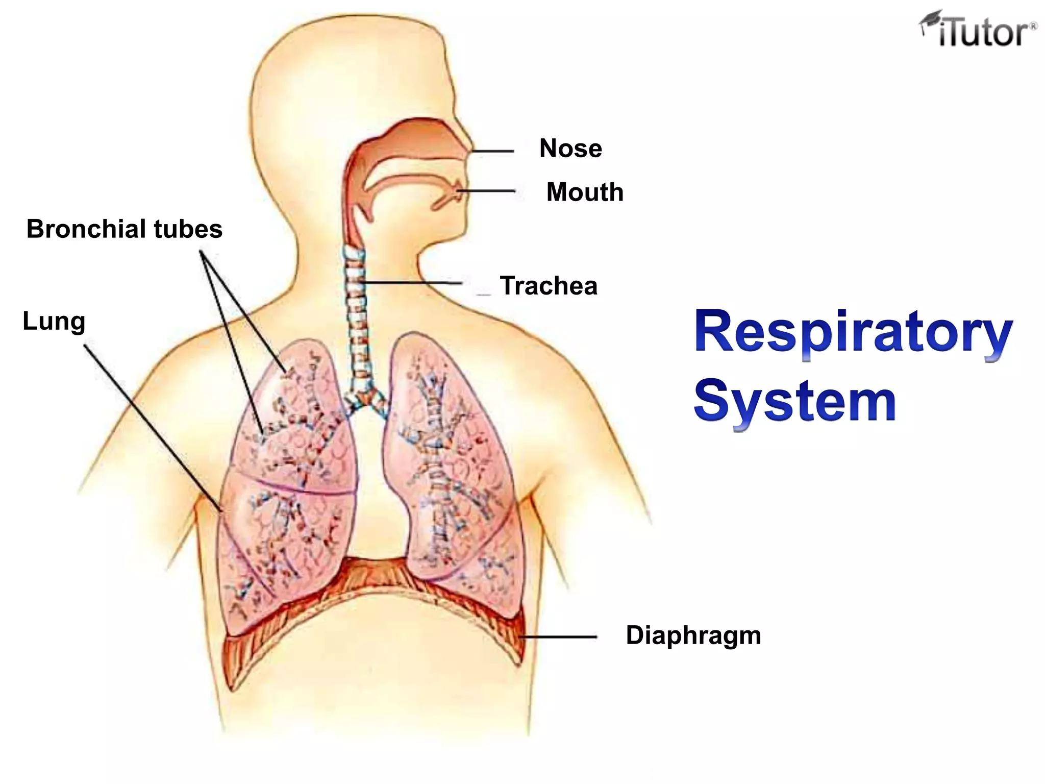
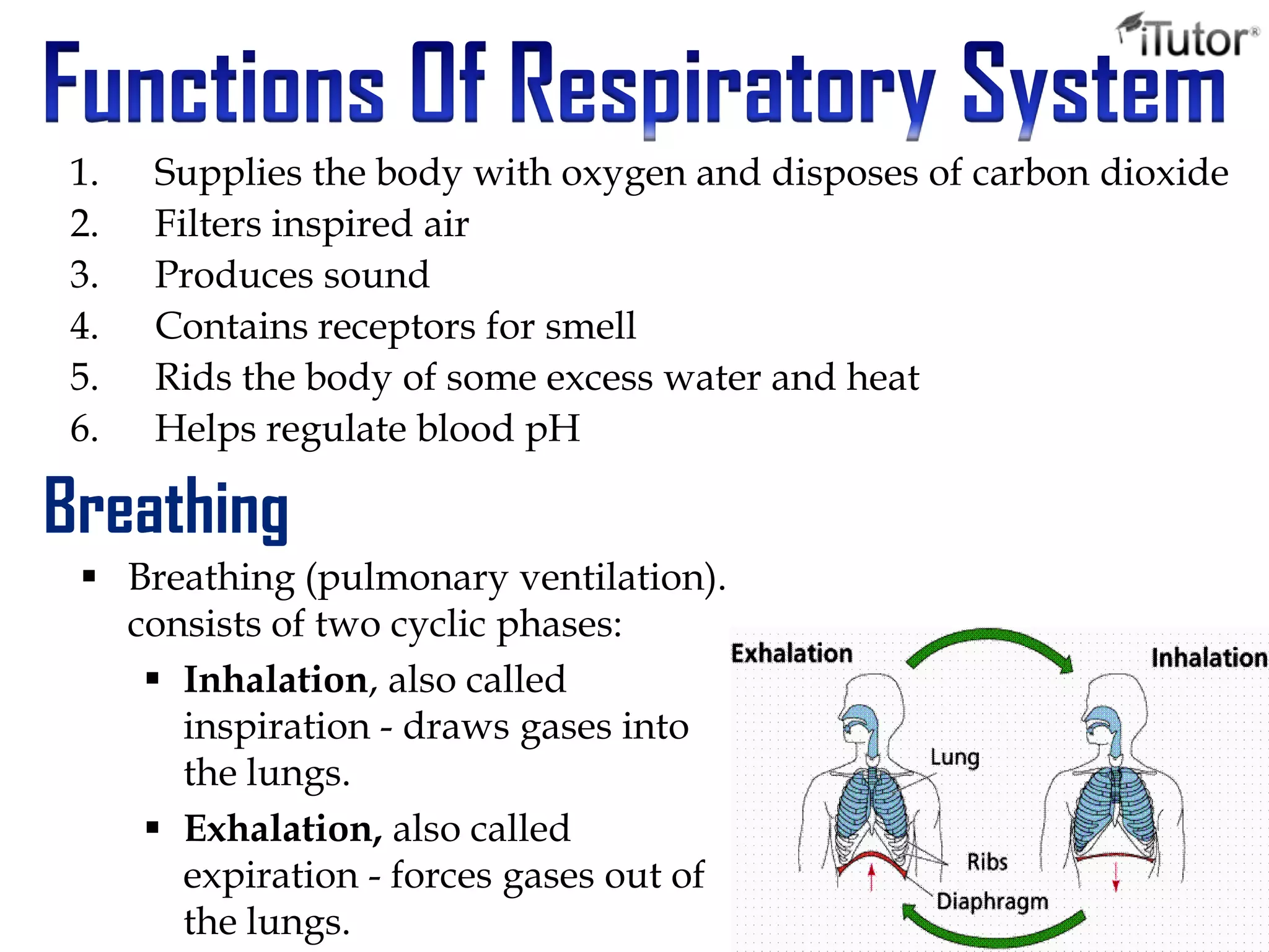
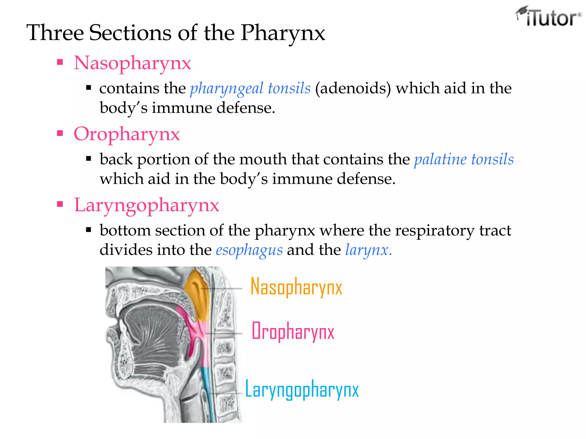
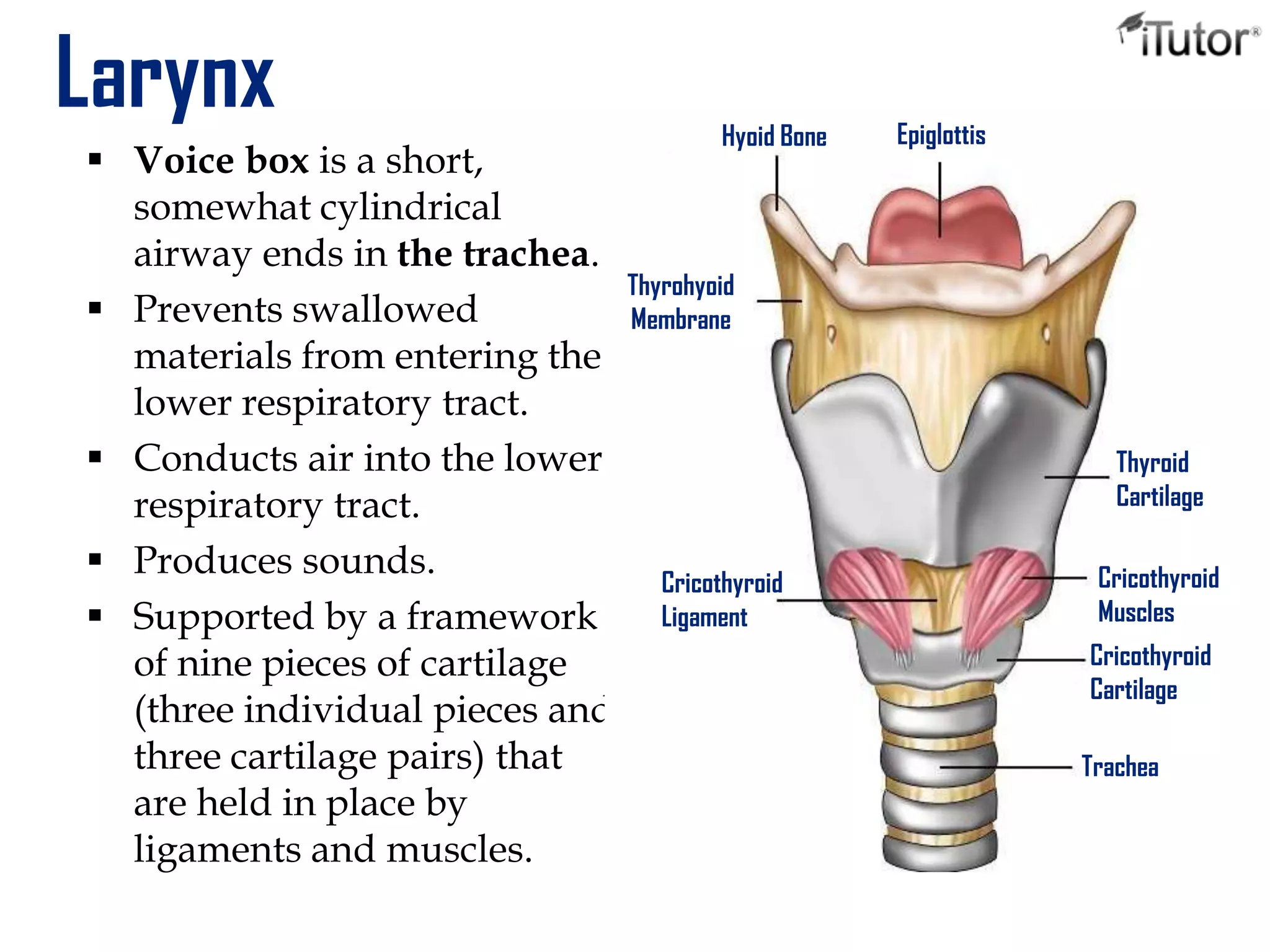
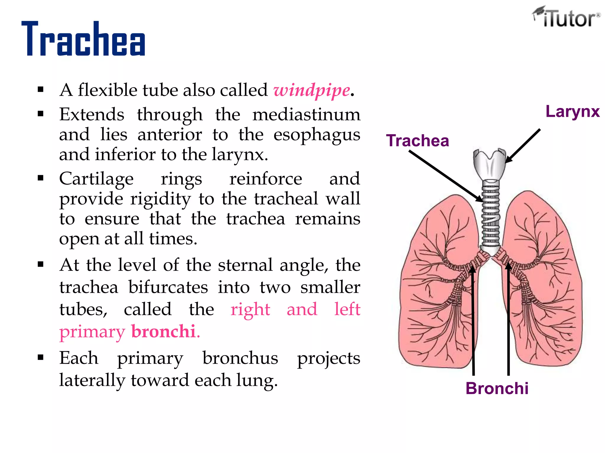
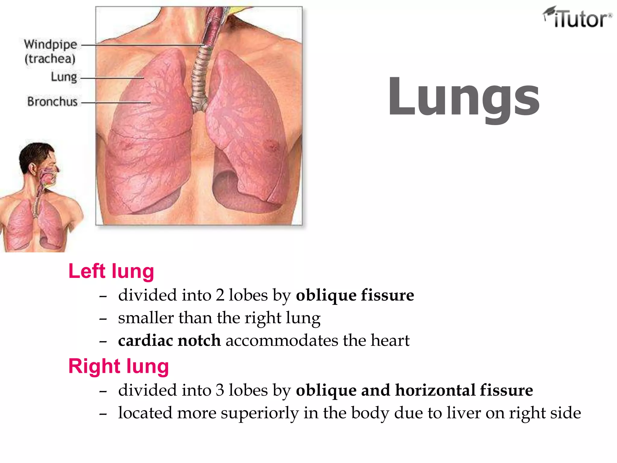
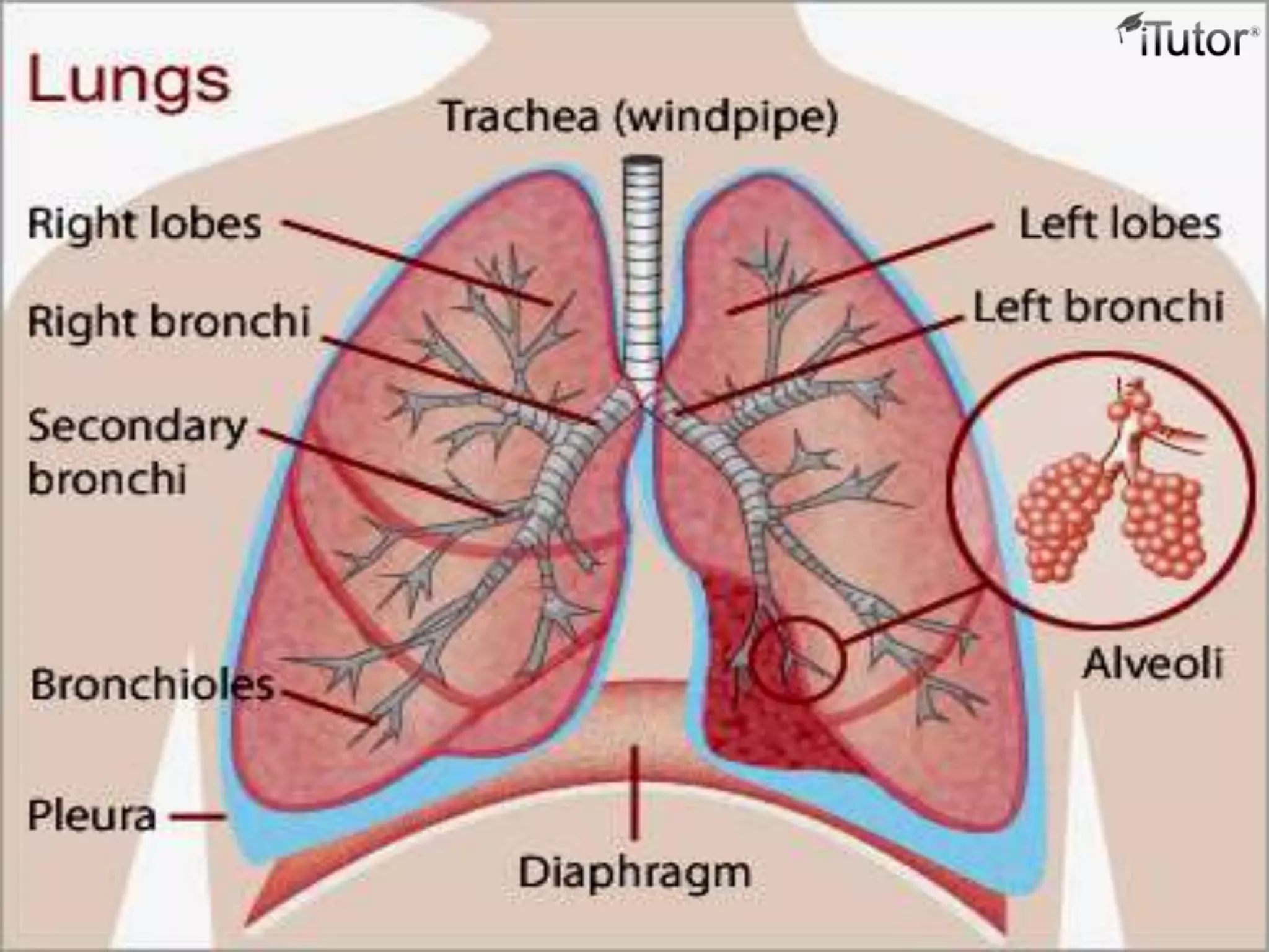
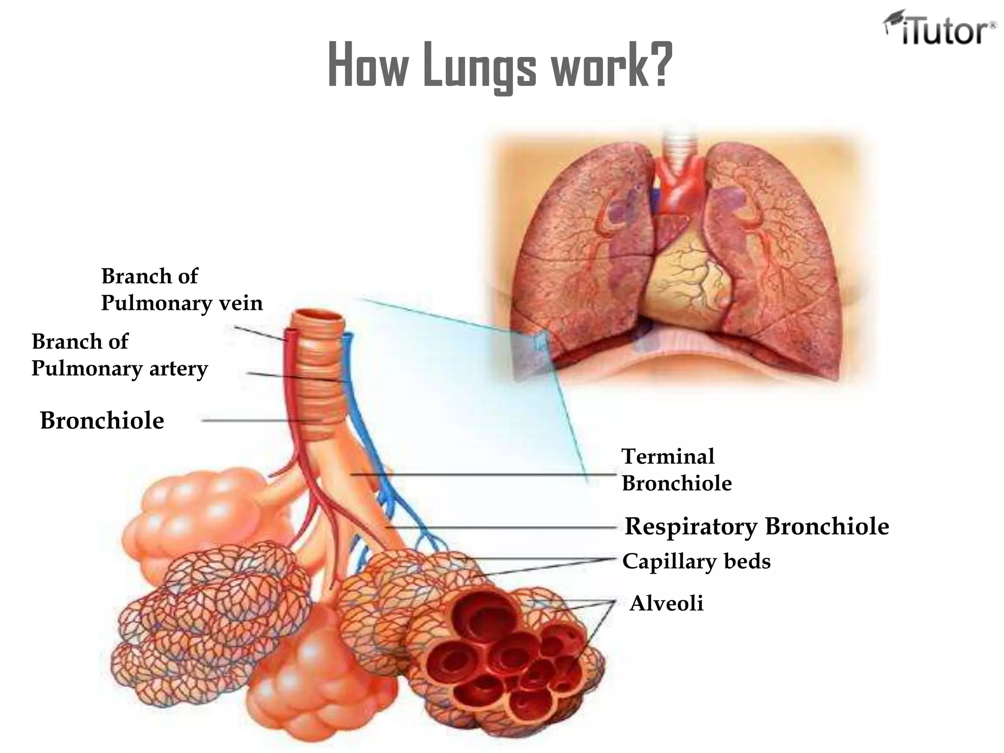
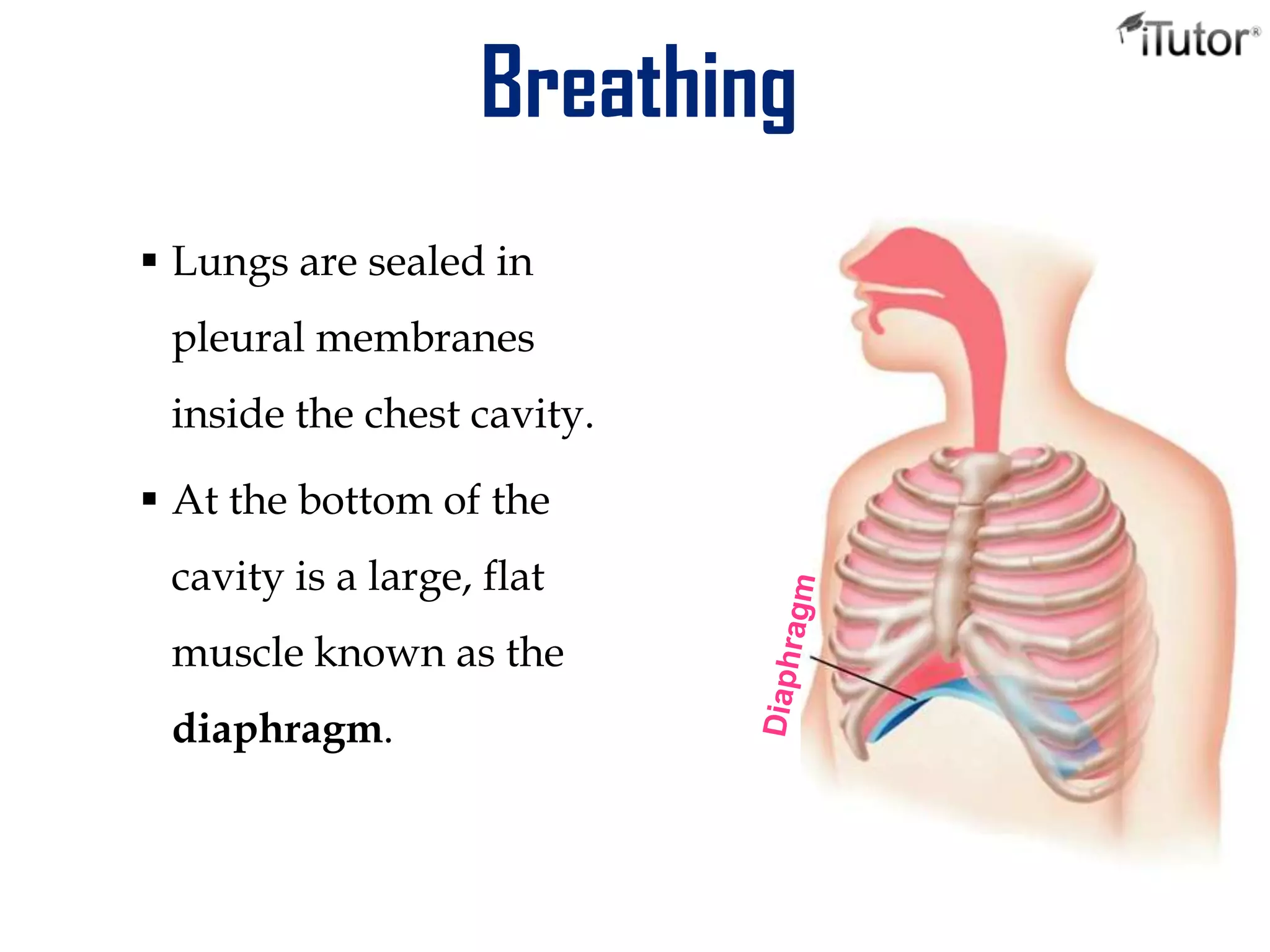
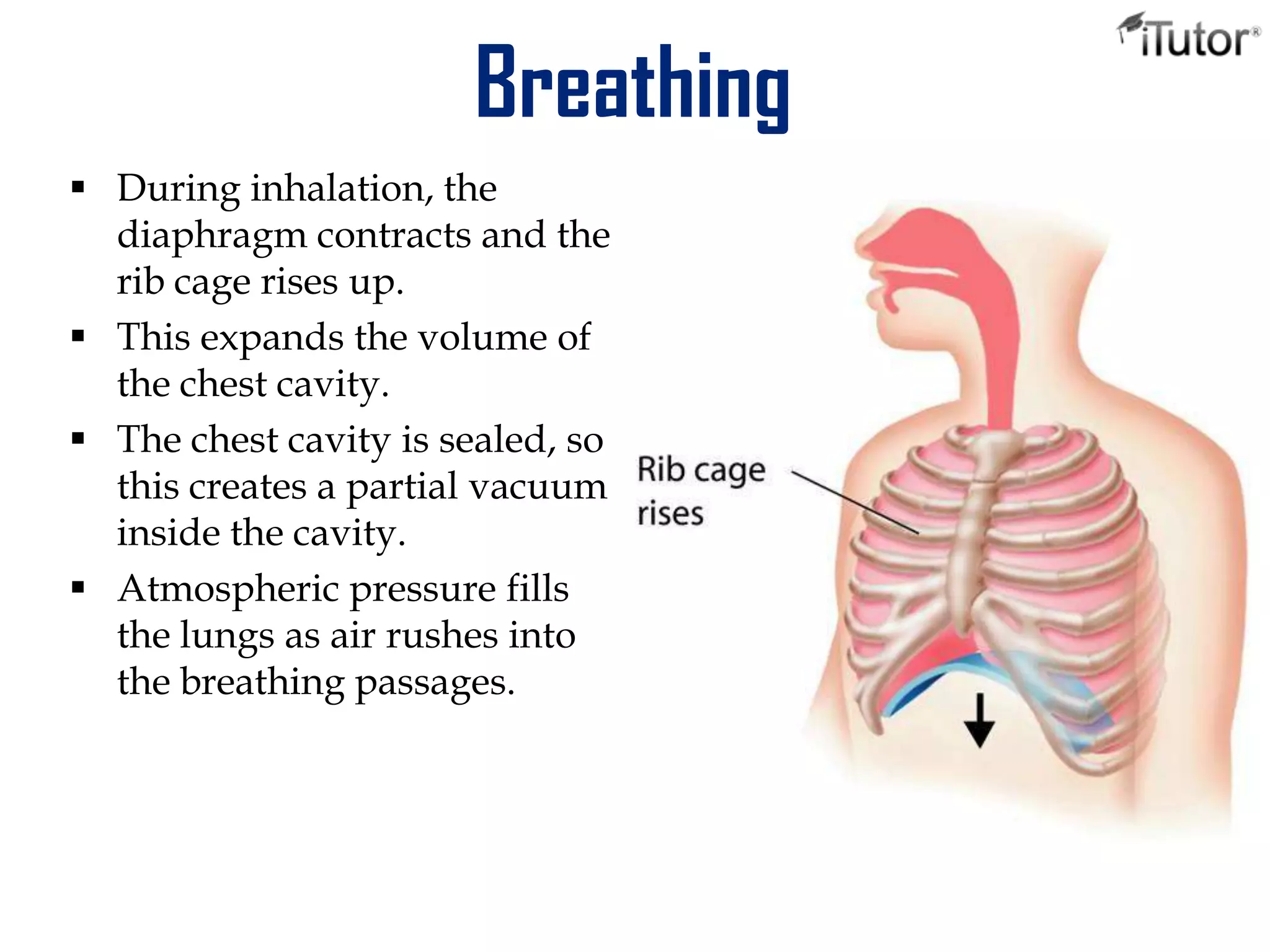
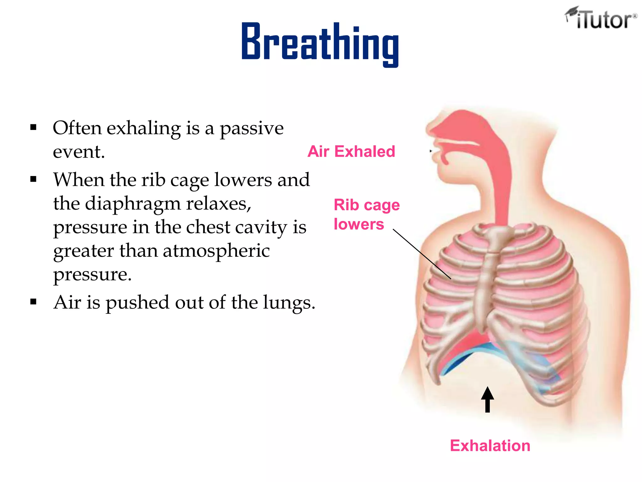
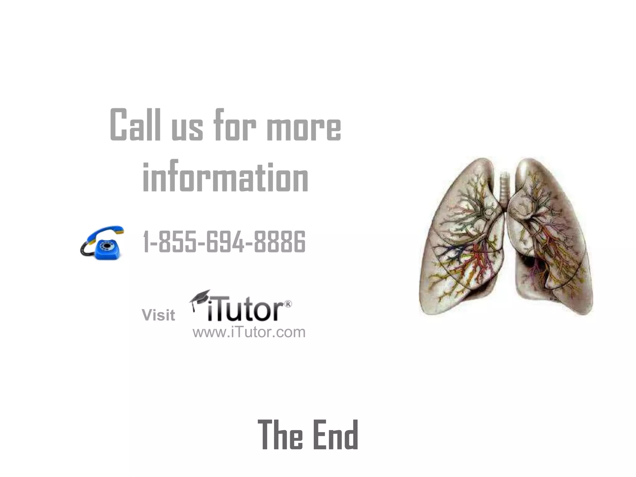
The Respiratory
system
T- 1-855-694-8886
Email- info@iTutor.com
By iTutor.com
Respiratory System: Oxygen Delivery System
 The respiratory system is the set of organs that allows a person to
breathe and exchange oxygen and carbon dioxide throughout the
body.
 The integrated system of organs involved in the intake and
exchange of oxygen and carbon dioxide between the body and
the environment and including the nasal passages, larynx,
trachea, bronchial tubes, and lungs.
The respiratory system performs two major tasks:
 Exchanging air between the body and the outside
environment known as external respiration.
 Bringing oxygen to the cells and removing carbon dioxide
from them referred to as internal respiration.
Lung
Bronchial tubes
Nose
Mouth
Trachea
Diaphragm
1. Supplies the body with oxygen and disposes of carbon dioxide
2. Filters inspired air
3. Produces sound
4. Contains receptors for smell
5. Rids the body of some excess water and heat
6. Helps regulate blood pH
Breathing
 Breathing (pulmonary ventilation).
consists of two cyclic phases:
 Inhalation, also called
inspiration - draws gases into
the lungs.
 Exhalation, also called
expiration - forces gases out of
the lungs.
 Air from the outside environment enters the nose or mouth
during inspiration (inhalation).
 Composed of the nose and nasal cavity, paranasal sinuses,
pharynx (throat), larynx.
 All part of the conducting portion of the respiratory system.
Nostril
Mouth
Nasal Cavity
Throat
(pharynx)
Voice box(Larynx)
Nose Also called external nares.
 Divided into two halves by the nasal septum.
 Contains the paranasal sinuses where air is warmed.
 Contains cilia which is responsible for filtering out foreign
bodies.
Nose and Nasal Cavities
Nasal concha Sphenoid sinus
Internal naris
Nasopharynx
External naris
Frontal sinus
Middle nasal concha
Inferior nasal
concha
 Internal nares - opening to exterior
 External nares - opening to pharynx
 Nasal conchae - folds in the mucous membrane that
increase air turbulence and ensures that most air contacts
the mucous membranes
Provides and airway for respiration
 Moistens and warms entering air
 Filters and cleans inspired air
 Resonating chamber for speech
- detects odors in the air stream
Pharynx Common space used by both the respiratory and digestive
systems.
 Commonly called the throat.
 Originates posterior to the nasal and oral cavities and extends
inferiorly near the level of the bifurcation of the larynx and
esophagus.
 Common pathway for both air and food.
 Walls are lined by a mucosa and contain skeletal muscles that
are primarily used for swallowing.
 Flexible lateral walls are distensible in order to force swallowed
food into the esophagus.
Three Sections of the Pharynx
 Nasopharynx
 contains the pharyngeal tonsils (adenoids) which aid in the
body’s immune defense.
 Oropharynx
 back portion of the mouth that contains the palatine tonsils
which aid in the body’s immune defense.
 Laryngopharynx
 bottom section of the pharynx where the respiratory tract
divides into the esophagus and the larynx.
Nasopharynx
Oropharynx
Laryngopharynx
Larynx
 Voice box is a short,
somewhat cylindrical
airway ends in the trachea.
 Prevents swallowed
materials from entering the
lower respiratory tract.
 Conducts air into the lower
respiratory tract.
 Produces sounds.
 Supported by a framework
of nine pieces of cartilage
(three individual pieces and
three cartilage pairs) that
are held in place by
ligaments and muscles.
Hyoid Bone Epiglottis
Thyrohyoid
Membrane
Cricothyroid
Ligament
Cricothyroid
Muscles
Cricothyroid
Cartilage
Trachea
Thyroid
Cartilage
Trachea
 A flexible tube also called windpipe.
 Extends through the mediastinum
and lies anterior to the esophagus
and inferior to the larynx.
 Cartilage rings reinforce and
provide rigidity to the tracheal wall
to ensure that the trachea remains
open at all times.
 At the level of the sternal angle, the
trachea bifurcates into two smaller
tubes, called the right and left
primary bronchi.
 Each primary bronchus projects
laterally toward each lung.
Trachea
Bronchi
Larynx
Lungs
 Each lung has a conical shape. Its wide, concave base rests
upon the muscular diaphragm.
 Its superior region called the apex projects superiorly to a
point that is slightly superior and posterior to the clavicle.
 Both lungs are bordered by the thoracic wall anteriorly,
laterally, and posteriorly, and supported by the rib cage.
 Toward the midline, the lungs are separated from each
other by the mediastinum.
 The relatively broad, rounded surface in contact with the
thoracic wall is called the costal surface of the lung.
Left lung
– divided into 2 lobes by oblique fissure
– smaller than the right lung
– cardiac notch accommodates the heart
Right lung
– divided into 3 lobes by oblique and horizontal fissure
– located more superiorly in the body due to liver on right side
Lungs
Pleura
 The outer surface of each lung and the adjacent internal
thoracic wall are lined by a serous membrane called pleura.
 The outer surface of each lung is tightly covered by the
visceral pleura.
 while the internal thoracic walls, the lateral surfaces of the
mediastinum, and the superior surface of the diaphragm are
lined by the parietal pleura.
 The parietal and visceral pleural layers are continuous at the
hilus of each lung
Pleural Cavities
The potential space between the serous membrane layers is a
pleural cavity.
 The pleural membranes produce a thin, serous pleural fluid that
circulates in the pleural cavity and acts as a lubricant, ensuring
minimal friction during breathing.
 Pleural effusion – pleuritis with too much fluid
 Air enters your lungs through a system of pipes called the bronchi.
 The alveoli are where the important work of gas exchange takes place
between the air and your blood. Covering each alveolus is a whole
network of little blood vessel called capillaries,
 It is important that the air in the alveoli and the blood in the capillaries
are very close together, so that oxygen and carbon dioxide can move (or
diffuse) between them.
 When you breathe in, air comes down the trachea and through the
bronchi into the alveoli.
 This fresh air has lots of oxygen in it, and some of this oxygen will travel
across the walls of the alveoli into your blood stream.
 Travelling in the opposite direction is carbon dioxide, which crosses from
the blood in the capillaries into the air in the alveoli and is then breathed
out.
 In this way, you bring in to your body the oxygen that you need to live,
and get rid of the waste product carbon dioxide.
How Lungs work?
Branch of
Pulmonary vein
Branch of
Pulmonary artery
Bronchiole
Terminal
Bronchiole
Respiratory Bronchiole
Capillary beds
Alveoli
Breathing
 Lungs are sealed in
pleural membranes
inside the chest cavity.
 At the bottom of the
cavity is a large, flat
muscle known as the
diaphragm.
Breathing
 During inhalation, the
diaphragm contracts and the
rib cage rises up.
 This expands the volume of
the chest cavity.
 The chest cavity is sealed, so
this creates a partial vacuum
inside the cavity.
 Atmospheric pressure fills
the lungs as air rushes into
the breathing passages.
Breathing
 Often exhaling is a passive
event.
 When the rib cage lowers and
the diaphragm relaxes,
pressure in the chest cavity is
greater than atmospheric
pressure.
 Air is pushed out of the lungs.
Exhalation
Rib cage
lowers
Air Exhaled
The End
Call us for more
information
www.iTutor.com
1-855-694-8886
Visit

The respiratory system

  • 1.
  • 2.
    Respiratory System: OxygenDelivery System  The respiratory system is the set of organs that allows a person to breathe and exchange oxygen and carbon dioxide throughout the body.  The integrated system of organs involved in the intake and exchange of oxygen and carbon dioxide between the body and the environment and including the nasal passages, larynx, trachea, bronchial tubes, and lungs. The respiratory system performs two major tasks:  Exchanging air between the body and the outside environment known as external respiration.  Bringing oxygen to the cells and removing carbon dioxide from them referred to as internal respiration.
  • 3.
  • 4.
    1. Supplies thebody with oxygen and disposes of carbon dioxide 2. Filters inspired air 3. Produces sound 4. Contains receptors for smell 5. Rids the body of some excess water and heat 6. Helps regulate blood pH Breathing  Breathing (pulmonary ventilation). consists of two cyclic phases:  Inhalation, also called inspiration - draws gases into the lungs.  Exhalation, also called expiration - forces gases out of the lungs.
  • 5.
     Air fromthe outside environment enters the nose or mouth during inspiration (inhalation).  Composed of the nose and nasal cavity, paranasal sinuses, pharynx (throat), larynx.  All part of the conducting portion of the respiratory system. Nostril Mouth Nasal Cavity Throat (pharynx) Voice box(Larynx)
  • 6.
    Nose Also calledexternal nares.  Divided into two halves by the nasal septum.  Contains the paranasal sinuses where air is warmed.  Contains cilia which is responsible for filtering out foreign bodies. Nose and Nasal Cavities Nasal concha Sphenoid sinus Internal naris Nasopharynx External naris Frontal sinus Middle nasal concha Inferior nasal concha
  • 7.
     Internal nares- opening to exterior  External nares - opening to pharynx  Nasal conchae - folds in the mucous membrane that increase air turbulence and ensures that most air contacts the mucous membranes Provides and airway for respiration  Moistens and warms entering air  Filters and cleans inspired air  Resonating chamber for speech - detects odors in the air stream
  • 8.
    Pharynx Common spaceused by both the respiratory and digestive systems.  Commonly called the throat.  Originates posterior to the nasal and oral cavities and extends inferiorly near the level of the bifurcation of the larynx and esophagus.  Common pathway for both air and food.  Walls are lined by a mucosa and contain skeletal muscles that are primarily used for swallowing.  Flexible lateral walls are distensible in order to force swallowed food into the esophagus.
  • 9.
    Three Sections ofthe Pharynx  Nasopharynx  contains the pharyngeal tonsils (adenoids) which aid in the body’s immune defense.  Oropharynx  back portion of the mouth that contains the palatine tonsils which aid in the body’s immune defense.  Laryngopharynx  bottom section of the pharynx where the respiratory tract divides into the esophagus and the larynx. Nasopharynx Oropharynx Laryngopharynx
  • 10.
    Larynx  Voice boxis a short, somewhat cylindrical airway ends in the trachea.  Prevents swallowed materials from entering the lower respiratory tract.  Conducts air into the lower respiratory tract.  Produces sounds.  Supported by a framework of nine pieces of cartilage (three individual pieces and three cartilage pairs) that are held in place by ligaments and muscles. Hyoid Bone Epiglottis Thyrohyoid Membrane Cricothyroid Ligament Cricothyroid Muscles Cricothyroid Cartilage Trachea Thyroid Cartilage
  • 11.
    Trachea  A flexibletube also called windpipe.  Extends through the mediastinum and lies anterior to the esophagus and inferior to the larynx.  Cartilage rings reinforce and provide rigidity to the tracheal wall to ensure that the trachea remains open at all times.  At the level of the sternal angle, the trachea bifurcates into two smaller tubes, called the right and left primary bronchi.  Each primary bronchus projects laterally toward each lung. Trachea Bronchi Larynx
  • 12.
    Lungs  Each lunghas a conical shape. Its wide, concave base rests upon the muscular diaphragm.  Its superior region called the apex projects superiorly to a point that is slightly superior and posterior to the clavicle.  Both lungs are bordered by the thoracic wall anteriorly, laterally, and posteriorly, and supported by the rib cage.  Toward the midline, the lungs are separated from each other by the mediastinum.  The relatively broad, rounded surface in contact with the thoracic wall is called the costal surface of the lung.
  • 13.
    Left lung – dividedinto 2 lobes by oblique fissure – smaller than the right lung – cardiac notch accommodates the heart Right lung – divided into 3 lobes by oblique and horizontal fissure – located more superiorly in the body due to liver on right side Lungs
  • 14.
    Pleura  The outersurface of each lung and the adjacent internal thoracic wall are lined by a serous membrane called pleura.  The outer surface of each lung is tightly covered by the visceral pleura.  while the internal thoracic walls, the lateral surfaces of the mediastinum, and the superior surface of the diaphragm are lined by the parietal pleura.  The parietal and visceral pleural layers are continuous at the hilus of each lung Pleural Cavities The potential space between the serous membrane layers is a pleural cavity.  The pleural membranes produce a thin, serous pleural fluid that circulates in the pleural cavity and acts as a lubricant, ensuring minimal friction during breathing.  Pleural effusion – pleuritis with too much fluid
  • 16.
     Air entersyour lungs through a system of pipes called the bronchi.  The alveoli are where the important work of gas exchange takes place between the air and your blood. Covering each alveolus is a whole network of little blood vessel called capillaries,  It is important that the air in the alveoli and the blood in the capillaries are very close together, so that oxygen and carbon dioxide can move (or diffuse) between them.  When you breathe in, air comes down the trachea and through the bronchi into the alveoli.  This fresh air has lots of oxygen in it, and some of this oxygen will travel across the walls of the alveoli into your blood stream.  Travelling in the opposite direction is carbon dioxide, which crosses from the blood in the capillaries into the air in the alveoli and is then breathed out.  In this way, you bring in to your body the oxygen that you need to live, and get rid of the waste product carbon dioxide.
  • 17.
    How Lungs work? Branchof Pulmonary vein Branch of Pulmonary artery Bronchiole Terminal Bronchiole Respiratory Bronchiole Capillary beds Alveoli
  • 18.
    Breathing  Lungs aresealed in pleural membranes inside the chest cavity.  At the bottom of the cavity is a large, flat muscle known as the diaphragm.
  • 19.
    Breathing  During inhalation,the diaphragm contracts and the rib cage rises up.  This expands the volume of the chest cavity.  The chest cavity is sealed, so this creates a partial vacuum inside the cavity.  Atmospheric pressure fills the lungs as air rushes into the breathing passages.
  • 20.
    Breathing  Often exhalingis a passive event.  When the rib cage lowers and the diaphragm relaxes, pressure in the chest cavity is greater than atmospheric pressure.  Air is pushed out of the lungs. Exhalation Rib cage lowers Air Exhaled
  • 21.
    The End Call usfor more information www.iTutor.com 1-855-694-8886 Visit