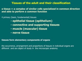
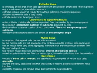
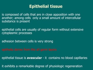
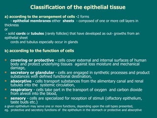
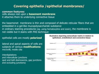
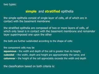
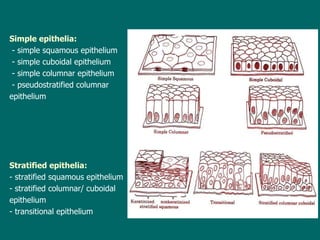
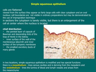
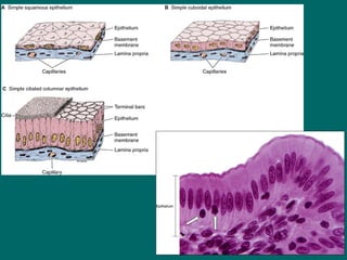
Lecture 7     ESS_2nd semester Epithelial tissue – definition ,  classification   and  histogenesis Overview of covering and glandular   epithelia .  Characteristics of glandular cells Absorptive, respiratory, and sensory epithelia
T issues of the adult and their classification a tissue = a  complex of similar cells specialized in common direction and able to perform a common function  4  primary (basic, fundamental) tissues   - epithelial tissue (epithelium)   - connective and supporting tissues   - muscle (muscular) tissue   - nerve tissue tissues  form elementary components of organs t he occurrence, arrangement and proportions of tissues in individual organs are different  and  are  object of study  in   the microscopic anatomy
Epithelial   tissue   is composed of cells  that  are in close apposition with one another; among cells  there is present only a small amount of intercellular substance e pithelial cells are usually of regular form without extensive cytoplasmic processes a dhesion between the cells is very strong e pithelia derive from  the  all germ layers Connective and supporting tissues unlike epithelia, contain  cells  that are separated  from one another  by  intervening spaces , these   contain   intercellular material   or  substance  produced by cells t he intercellular substance consists of two components:  fibers  and  amorphous ground substance  c onnective and supporting tissues are always of  mesenchymal origin Muscle tissue   is composed of elongated  cells that  are able to  contract f or this function cells are well adapted as they contain contractile proteins: actin and myosin cells or muscle fibers tend to be aggregated in bundles  that  are conspicuously different from the surroundings tissues t wo types of muscle tissue are distinguished:  smooth , skeletal and cardiac   s mooth muscle tissue  derives  from the mesenchyme, striated muscle tissue from the mesoderm Nerve tissue consists of  nerve cells  -  neurons , and associated supporting cells of various type called  neuroglia n eurons are highly specialized cells that  show  ability to receive, generate and transmit nerve impulses e xcept the microglia, the nervous tissue derives from the neuroectoderm
Epithelial   tissue   is composed of cells  that  are in close apposition with one another; among cells  only   a small amount of intercellular substance   is present  e pithelial cells are usually of regular form without extensive cytoplasmic processes a dhesion between cells is very strong e pithelia derive from  the  all germ layers e pithelial tissue is  avascular  -  it  contains no blood capillaries it  exhibit s  a remarkable degree of physiologic regeneration
Classification of the epithelial tissue a) according to the arrangement of cells - 2 forms -  epithelial membranes   either  sheets   -  composed of one or more  cell  layers in  thickness  or - solid  cords  or  tubules  (rarely follicles) that have developed as out- growths from an  epithelial sheet cords and tubulus especially occur in  glands b)  according to the function of cells covering or  protective  - cells cover external and internal surfaces of human body and protect underlying tissues  against loss moisture and mechanical damage, secretory  or glandular  - cells are engaged in synthetic processes and product substances with defined functional destination, absorptive  - cells transport substances from the alimentary canal and renal tubules into the  systemic circulation, respiratory  - cells  take part  in  the  transport of oxygen  and carbon dioxide from alveoli into the blood, sensory  - cells are specialized for reception of stimuli (olfactory epithelium, taste buds etc.) a  given epithelium may serve one or more functions, depending upon the cell types presented, eg.  protective and secretory functions of  the epithelium in the stomach or protective and absorptive
Covering   epithelia /e pithelial membranes / common features: cells  always rest upon  a  basement membrane   it  attach es  the m  to underlying connective tissue t he basemenet  membrane is thin and composed of delicate reticular fibers that are embedded in a gel-like mucopolysaccharide substance i n ordinary staining procedures  ( eg. using hematoxylin e  and eosin ),  the membrane is  not visible but it stains with PAS technique  epithelial cells  are mostly   polarized lateral and apical aspects  of cells  are  subjects of various  modifications :  microvilli, motile cilia interdigitations and intercellular junctions spot and belt desmosome, gap junctions  and occluding junctions)
two  types: simple  and  stratified  epithelia t he simple epithelia consist of single layer of cells, all of which are in contact with the basement membrane t he stratified epithelia are composed of two or more layers of cells, of which only  basal  is in contact with the basement membrane and remainder  layer  superimposed one upon the other the both   are  further subdivided according to the shape of cells : t he component cells may be  squamous  -  the width and depth of the cell is greater   than its height;  cuboidal  -  the width, depth and height are approximately the   same;  and   columnar  -  the height of the cell appreciably exceeds the width and   depth t he classification  based on  both criteria is :
Simple epithelia:   - simple squamous epithelium - simple cuboidal epithelium - simple columnar epithelium - pseudostratified columnar  epithelium Stratified epithelia:   - stratified squamous epithelium  - stratified columnar/ cuboidal epithelium - transitional epithelium
Simple squamous epithelium cells are flattened v iewed from the surface they appear as fairly large cells with clear cytoplasm and a n  oval nucleus ; c ell boundaries are  not visible in ordinary preparations   but may be   demonstrated by  the use of impregnation technique i n sections the cytoplasm is barely visible, but there is an enlargement of the cell at center where the nucleus is  locate d c hief distribution :  the parietal layer of capsule of  Bowman and descending limb of the  loop of Henle in the kidney inner surface of the wall of the  membranous labyrinth and inner  surface of the tympanic membrane the smallest excretory ducts of  many glands i n two locations, simple squamous epithelium  is modified and has special functions there is a  mesothelium   -  lines serous cavities   and  is deriving  from the mesoderm  a nd  the   endothelium   -  lines the lumina of blood and lymph vessels and arises from the   mesenchyme
 
Simple cuboidal epithelium cells of cuboidal epithelium are smaller in surface view than those of the   simple  squamous type  and are roughly hexagonal c ell boundaries are often clearly visible i n vertical sections the cells are square with a  spherical  nucleus in the center of   each th e square shape is modified to that of truncated prism when cells line  s mall  ducts c hief distribution :  free surface of the ovary, the choroid plexus surface,  the inner surface of the lens, the excretory ducts of many glands Simple columnar epithelium the  surface view of columnar epithelium is like that of the cuboidal epithelium  i n  sections,  the cells are seen to be taller than they are broad;  t he ir  nucleus  is  near  the  base of the cell  a s in the case  of the  cuboidal epithelium ,  the rectangular shape of columnar cells is  changed to pyramidal when they are grouped around small lumen  in order to right diagnosis of  this  epithelium it is important to   select  a region ,  in  which the section passes through the tissue in a plane   perpendicular to  the  surface c hief distribution:  the stomach, larger excretory ducts, and uterus; in the case of oviduct  and small bronchi, apices of some epithelial cells are ciliated
a simple cuboidal epithelium
simple columnar epithelium  with striated border (microvilli)
a simple columnar ciliated epithelium  (with kinocilia)
Pseudostratified columnar epithelium all cells are in contact with the basement membrane but only a part of them extends   to the free surface i n pseudostratified epithelium cells are variable in   shape  and their  nuclei  lie  at different levels ;  a ccording to cell height, three different cell types  are  distinguished: small basal or supporting cells spindle - shape d  cells  tall superficial cells  that  are connected with the basement membrane via slender processes , a pices of tall superficial cells are often ciliated goblet cells producing mucous  s ecretion are usually present in this epithelium chief distribution:  the  respiratory passages
Stratified epithelia small cuboidal or columnar cells lie next to the basement membrane above them  one or more layers of polygonal cells are the  free surface is covered by a layer of cells  that are important for diag n osis Stratified squamous epithelium the thickness  of the epithelium  and number of cells vary in different parts of the body the shape and arrangement of component cells, however, follow the same general plan : consists of small cuboidal or even columnar cells  are  are covered  by a varying number of  layers of more or less polygonal (polyhedral) cells  they  gradually begin to flatten out toward to the epithelium surface they become scale-like form, their nuclei are pyknotic but they may be yet considered as viable such  epithelium is found in  the mouth, the esophagus, the vagina, and on the surface of the cornea on the exposed outer surfaces , t he stratified squamous epithelium  is  modified as  cornified  or  keratinized t he surface cells loose their nuclei and undergo a transformation into dry and dead scales t he keratinized stratified squamous epithelium covers the skin as a  epidermis
stratified squamous epithelium stratified squamous epithelium  (cornified)  = epidermis
 
Stratified columnar epithelium it  differs f rom  the pseudostratified  epithelium  in having a continuous layer of small,   rounded  cells next to the basement membrane ,   a varying number (3-4) of more or   less polyhedral cells and by presence of  superficial cells  that are  tall and prismatic  the epithelium is truly stratified chief distribution :  the fornix of the conjunctiva ,  the cavernous part of the male urethra ,   large excretory ducts of some glands Transitional epithelium it  consists of several layers of cells t he basal cells are like those of stratified columnar epithelium a bove them there is a varying number of layers of polygonal  or pear-shaped  cells , t he cell layer at the free   surface is composed of large,  rather  flattened cells,  usually  described as dome shaped o ne of  dome shaped cell  often covers two or three pear-shaped cells  that  underl ie it cells   show  unusual degree the ability to change their position, sliding over each other w hen an organ is distended,  the epithelium is reduced to three or four layers and vice versa  (by organ   contraction the cells heap up  forming several layers between the basal and surface cells) chief distribution:  it covers  the mucosa of the excretory passages from the renal calyces  to the urethra
transitional epithelium
Secretory /glandular/epithelium the epithelium  whose  cells are able to synthesize special products -  secretions substances that are for organism and right function of many organs very important  in many cases they cannot be substituted (GLANDULAR CORDS AND NOT SHEATHS) Secretory products are released either  upon free surfaces or into blood  (rarely lymphatic) vessels for distribution  / hormones / Histophysiology of secretion s ecretory process in volves  3 phases as follows: ingestion of single substances or precursors from the surrounding  or blood synthesis of secretory products including their segregation and accumulation release of secretory products Ingestion of precursors  is usually realized by 3 ways: - passive diffusion down a concentration gradient, - active transport against a concentration gradient using ATP as an energy source, - pinocytosis - the way providing of transport of macromolecules across the plasma  membrane
synthesis of secretory products   under cooperation of  many cell organelles  -  free ribosomes or unbounded polyribosomes and the rough endoplasmic reticulum s ynthesized products are then transferred from this compartment to the  Golgi  apparatus, where  they  become final form and are  directed to their final destination in each cell s ynthetic processes need an energy  -  is produced by mitochondria a ll the mentioned cell organelles are typical of glandular cells  release or extrusion of secretory products three  modes   have been distinguished: - merocrine  secretion  - apocrine   secretion  -  holocrine   secretion
Release of secretion products merocrine secretion   products are released through the cell membrane  without the loss any cytoplasm  (during the whole secretory cycle ) the cell does not change  its shape goblet cells
apocrine  secretion   by a loss of the apical  parts  that  contain s   an accumulated  secret ory  material   glandular cells of follicles in thyroid apocrine sweet glands
holocrine secretion the discharge of product is a ssociated with  a destruction of  the whole  glandular cells cells of  sebaceous glands
secretory  cells  constitute special organs  called  glands Classification of glands With respect to the mode of secretion With respect to the number of glandular cells With respect to the chemical properties of secreted product a) with respect to the mode of secretion: exocrine glands  - discharge their product via ducts at an external or internal surface, endocrine glands  release their product into the blood or lymph vessels have  no ducts  (ductless glands) secretions are called  hormones  and regulate and coordinate the activities  of all the cells in the body in cooperation with the nervous system b) with respect to the number of glandular cells: unicellular  glands   - the glandular components consist of a single cell among  other cells  t hat are not  glandular multicellular glands  - the glandular components consist of more than one cell
With respect to the mode of secretion With respect to the number of glandular cells With respect to the chemical properties of secreted product With respect to the  ducts Exocrine Endocrine Unicellular Multicellular Serous Mucous Seromucous Simple Compound mucous cells or goblet cells  and - enteroendocrine  (GEP) cells I ntraepithelial glands  E xtraepithelial glands   secretory units and ducts
Exocrine glands or  glands of external secretion (eg. Directly into the GIT)   retain connections with surfaces exocrine glands occur as  uni - and multicellular  ones u nicellular glands   are -  mucous cells or goblet cells  and - enteroendocrine  (GEP) cells goblet  cells   are scattered among the columnar cells of the simple columnar or pseudostratified columnar  e pithelia c ells secrete mucin  Mucous e ach cell has an expanded, oval apex filled with  pale droplets of mucigen and a slender basal  end containing  a compressed nucleus and  a small amount  of deeply staining basophilic  cytoplasm enteroendocrine  (GEP) cells  occur  in the wall of  the stomach, small and large intestine, bile ducts (hepatic, cystic or common bile) and  islets of Langerhans produce  catecholamines or peptidehormones ; for visualization of endocrine cells were formerly used  impregnation techniques, recently is used TEM or immunohistochemistry  goblet cells
M ulticellular glands   I ntraepithelial glands  are  relative rare  and are composed of small accumulations of glandular cells that lie wholly within the thickness of an epithelial membrane chief distribution:  mucous glands of lacunae urethrales Morgagni in the male urethra E xtraepithelial glands   are always in the connective tissue of respective organ or often more deeper glnds  develop as invaginations of the epithelial membrane into the underlying tissues t he extraepithelial glands consist of  secretory units  and  ducts secretory  units are of three type s: tubules  (unit is shaped like a tube) alveoli (acini)  (unit is shaped like a flask) tuboalveolar  portions (combination of  the tube and alveolus which  is located at  one end of the tube)  ducts  may be short and unbranched -  simple glands or  ducts  are long a branched -  compound glands t he shape of secretory units and the length of ducts are used  in the anatomical  classification of glands as follows
Multicellular glands


b esides the anatomical classification of glands it may be taken in account the chemical properties of secreted product the secretory units are divided into   m ucous  units -  the shape of straight tubules with a small lum ina cells are usually of  cuboidal  form or of truncated pyramids ,  nuclei are usually small, dark, and are flattened against the plasma membrane of the cells the  cytoplasm stains light in HE preparation and may have a foamy appearance (alcian blue, mucicarmine, and the PAS  reaction are used to  demonstrate mucous tubules ) serous units   -  are the form of alveoli and are composed  of pyramidal  cells whose nuclei are located centrally secretion granules are more or less acidophilic and are primarily protein in character t he cytoplasm always stains  in pink or pinkish purple, more  darkly  than that of mucous cells t he lumen of alveoli (acini) is definite and and smaller in diameter than that of mucous tubules seromucous  units -  mixed  units are composed  of both mucous and serous cells a  mixed unit is composed of mucous cells lining  a tubular part (short tubule) to  which one end is added a collection of serous cells arranged in a half-moon fashion t hese  serous cells  collections  are called as  demilune  complexes  or  lunulae  of  Giannuzzi i n a great number they are occurred in the human  subma ndibular  gland
acinar / alveoli /
tubular /tubules /
tubuloacinar / tubules  with serous demilunes /
tubule with serous demilune   = demilune of Gianuzzi
Absorptive epithelium cells whose apices are provided with microvilli striated border  (enterocytes) brush border  (cells of proximal tubule of the nephron)
Respiratory   epithelium is extremely thin and lines alveoli   membranous pneumocytes  (type I) granular pneumocytes  (type II)  secrete pulmonary surfactant
Respira tory  epit h el ium the type I alveolar cell s   or  membranous pneumocyte s   -  flattened, with micropino - cytotic vesicles near basal and apical surfaces  and thin processes ( 20–25 nm ), cells cover cca   97 %  of the alveolar surface the type II  alveolar cell s or  granular pneumocyte s  -  cuboidal cell s  with secretory  granules in  the  cytoplasm ; they  contain phospholipid- protein and are secreted on the surface of  epithelium as monomolecular film  "lining  complex" (surfactant)   that  is primarily  composed of dipalmitoyl lecithin t he layer of surfactant (about 30 nm thick) prevents from atelectasis (alveolar collapse) lamina basalis
Sensory epithelium sensory organs   sensory areas (fields) supporting cells – basal cells sensory cells  -  receipt stimuli from external environment ,  stimuli induce a special  status  in their bodies  -   irritation s ensory cells are  of  two  type s: a)  primary sensory cells   ( neuroepithelial cells or unipolar neurons ) they consist  of  receptor area (can be photosensitive or registrate odoriferous substances), cell body and axon or neurite  that  conducts impulses to the next neuron i n the human  -  olfactory cells  in the olfactory epithelium and by  rod cells and cone cells  in the retina b)  secondary sensory cells   are only  modified cells of covering epithelia cells  have spindle-like or flask-like form and consist of only receptor area and body the process corresponding to the axon is always missing  (s ignals from cells are conducted via dendritic processes of sensory nerves  that  endon the surface of these cells ) cells of taste buds, and hair cells  in the membranouslabyrinth
cone  cell  rod cell
taste bud with taste cells hair cells (utricle)

Lecture7

  • 1.
    Lecture 7 ESS_2nd semester Epithelial tissue – definition , classification and histogenesis Overview of covering and glandular epithelia . Characteristics of glandular cells Absorptive, respiratory, and sensory epithelia
  • 2.
    T issues ofthe adult and their classification a tissue = a complex of similar cells specialized in common direction and able to perform a common function 4 primary (basic, fundamental) tissues - epithelial tissue (epithelium) - connective and supporting tissues - muscle (muscular) tissue - nerve tissue tissues form elementary components of organs t he occurrence, arrangement and proportions of tissues in individual organs are different and are object of study in the microscopic anatomy
  • 3.
    Epithelial tissue is composed of cells that are in close apposition with one another; among cells there is present only a small amount of intercellular substance e pithelial cells are usually of regular form without extensive cytoplasmic processes a dhesion between the cells is very strong e pithelia derive from the all germ layers Connective and supporting tissues unlike epithelia, contain cells that are separated from one another by intervening spaces , these contain intercellular material or substance produced by cells t he intercellular substance consists of two components: fibers and amorphous ground substance c onnective and supporting tissues are always of mesenchymal origin Muscle tissue is composed of elongated cells that are able to contract f or this function cells are well adapted as they contain contractile proteins: actin and myosin cells or muscle fibers tend to be aggregated in bundles that are conspicuously different from the surroundings tissues t wo types of muscle tissue are distinguished: smooth , skeletal and cardiac s mooth muscle tissue derives from the mesenchyme, striated muscle tissue from the mesoderm Nerve tissue consists of nerve cells - neurons , and associated supporting cells of various type called neuroglia n eurons are highly specialized cells that show ability to receive, generate and transmit nerve impulses e xcept the microglia, the nervous tissue derives from the neuroectoderm
  • 4.
    Epithelial tissue is composed of cells that are in close apposition with one another; among cells only a small amount of intercellular substance is present e pithelial cells are usually of regular form without extensive cytoplasmic processes a dhesion between cells is very strong e pithelia derive from the all germ layers e pithelial tissue is avascular - it contains no blood capillaries it exhibit s a remarkable degree of physiologic regeneration
  • 5.
    Classification of theepithelial tissue a) according to the arrangement of cells - 2 forms - epithelial membranes either sheets - composed of one or more cell layers in thickness or - solid cords or tubules (rarely follicles) that have developed as out- growths from an epithelial sheet cords and tubulus especially occur in glands b) according to the function of cells covering or protective - cells cover external and internal surfaces of human body and protect underlying tissues against loss moisture and mechanical damage, secretory or glandular - cells are engaged in synthetic processes and product substances with defined functional destination, absorptive - cells transport substances from the alimentary canal and renal tubules into the systemic circulation, respiratory - cells take part in the transport of oxygen and carbon dioxide from alveoli into the blood, sensory - cells are specialized for reception of stimuli (olfactory epithelium, taste buds etc.) a given epithelium may serve one or more functions, depending upon the cell types presented, eg. protective and secretory functions of the epithelium in the stomach or protective and absorptive
  • 6.
    Covering epithelia /e pithelial membranes / common features: cells always rest upon a basement membrane it attach es the m to underlying connective tissue t he basemenet membrane is thin and composed of delicate reticular fibers that are embedded in a gel-like mucopolysaccharide substance i n ordinary staining procedures ( eg. using hematoxylin e and eosin ), the membrane is not visible but it stains with PAS technique epithelial cells are mostly polarized lateral and apical aspects of cells are subjects of various modifications : microvilli, motile cilia interdigitations and intercellular junctions spot and belt desmosome, gap junctions and occluding junctions)
  • 7.
    two types:simple and stratified epithelia t he simple epithelia consist of single layer of cells, all of which are in contact with the basement membrane t he stratified epithelia are composed of two or more layers of cells, of which only basal is in contact with the basement membrane and remainder layer superimposed one upon the other the both are further subdivided according to the shape of cells : t he component cells may be squamous - the width and depth of the cell is greater than its height; cuboidal - the width, depth and height are approximately the same; and columnar - the height of the cell appreciably exceeds the width and depth t he classification based on both criteria is :
  • 8.
    Simple epithelia: - simple squamous epithelium - simple cuboidal epithelium - simple columnar epithelium - pseudostratified columnar epithelium Stratified epithelia: - stratified squamous epithelium - stratified columnar/ cuboidal epithelium - transitional epithelium
  • 9.
    Simple squamous epitheliumcells are flattened v iewed from the surface they appear as fairly large cells with clear cytoplasm and a n oval nucleus ; c ell boundaries are not visible in ordinary preparations but may be demonstrated by the use of impregnation technique i n sections the cytoplasm is barely visible, but there is an enlargement of the cell at center where the nucleus is locate d c hief distribution : the parietal layer of capsule of Bowman and descending limb of the loop of Henle in the kidney inner surface of the wall of the membranous labyrinth and inner surface of the tympanic membrane the smallest excretory ducts of many glands i n two locations, simple squamous epithelium is modified and has special functions there is a mesothelium - lines serous cavities and is deriving from the mesoderm a nd the endothelium - lines the lumina of blood and lymph vessels and arises from the mesenchyme
  • 10.
  • 11.
    Simple cuboidal epitheliumcells of cuboidal epithelium are smaller in surface view than those of the simple squamous type and are roughly hexagonal c ell boundaries are often clearly visible i n vertical sections the cells are square with a spherical nucleus in the center of each th e square shape is modified to that of truncated prism when cells line s mall ducts c hief distribution : free surface of the ovary, the choroid plexus surface, the inner surface of the lens, the excretory ducts of many glands Simple columnar epithelium the surface view of columnar epithelium is like that of the cuboidal epithelium i n sections, the cells are seen to be taller than they are broad; t he ir nucleus is near the base of the cell a s in the case of the cuboidal epithelium , the rectangular shape of columnar cells is changed to pyramidal when they are grouped around small lumen in order to right diagnosis of this epithelium it is important to select a region , in which the section passes through the tissue in a plane perpendicular to the surface c hief distribution: the stomach, larger excretory ducts, and uterus; in the case of oviduct and small bronchi, apices of some epithelial cells are ciliated
  • 12.
  • 13.
    simple columnar epithelium with striated border (microvilli)
  • 14.
    a simple columnarciliated epithelium (with kinocilia)
  • 15.
    Pseudostratified columnar epitheliumall cells are in contact with the basement membrane but only a part of them extends to the free surface i n pseudostratified epithelium cells are variable in shape and their nuclei lie at different levels ; a ccording to cell height, three different cell types are distinguished: small basal or supporting cells spindle - shape d cells tall superficial cells that are connected with the basement membrane via slender processes , a pices of tall superficial cells are often ciliated goblet cells producing mucous s ecretion are usually present in this epithelium chief distribution: the respiratory passages
  • 16.
    Stratified epithelia smallcuboidal or columnar cells lie next to the basement membrane above them one or more layers of polygonal cells are the free surface is covered by a layer of cells that are important for diag n osis Stratified squamous epithelium the thickness of the epithelium and number of cells vary in different parts of the body the shape and arrangement of component cells, however, follow the same general plan : consists of small cuboidal or even columnar cells are are covered by a varying number of layers of more or less polygonal (polyhedral) cells they gradually begin to flatten out toward to the epithelium surface they become scale-like form, their nuclei are pyknotic but they may be yet considered as viable such epithelium is found in the mouth, the esophagus, the vagina, and on the surface of the cornea on the exposed outer surfaces , t he stratified squamous epithelium is modified as cornified or keratinized t he surface cells loose their nuclei and undergo a transformation into dry and dead scales t he keratinized stratified squamous epithelium covers the skin as a epidermis
  • 17.
    stratified squamous epitheliumstratified squamous epithelium (cornified) = epidermis
  • 18.
  • 19.
    Stratified columnar epitheliumit differs f rom the pseudostratified epithelium in having a continuous layer of small, rounded cells next to the basement membrane , a varying number (3-4) of more or less polyhedral cells and by presence of superficial cells that are tall and prismatic the epithelium is truly stratified chief distribution : the fornix of the conjunctiva , the cavernous part of the male urethra , large excretory ducts of some glands Transitional epithelium it consists of several layers of cells t he basal cells are like those of stratified columnar epithelium a bove them there is a varying number of layers of polygonal or pear-shaped cells , t he cell layer at the free surface is composed of large, rather flattened cells, usually described as dome shaped o ne of dome shaped cell often covers two or three pear-shaped cells that underl ie it cells show unusual degree the ability to change their position, sliding over each other w hen an organ is distended, the epithelium is reduced to three or four layers and vice versa (by organ contraction the cells heap up forming several layers between the basal and surface cells) chief distribution: it covers the mucosa of the excretory passages from the renal calyces to the urethra
  • 20.
  • 21.
    Secretory /glandular/epithelium theepithelium whose cells are able to synthesize special products - secretions substances that are for organism and right function of many organs very important in many cases they cannot be substituted (GLANDULAR CORDS AND NOT SHEATHS) Secretory products are released either upon free surfaces or into blood (rarely lymphatic) vessels for distribution / hormones / Histophysiology of secretion s ecretory process in volves 3 phases as follows: ingestion of single substances or precursors from the surrounding or blood synthesis of secretory products including their segregation and accumulation release of secretory products Ingestion of precursors is usually realized by 3 ways: - passive diffusion down a concentration gradient, - active transport against a concentration gradient using ATP as an energy source, - pinocytosis - the way providing of transport of macromolecules across the plasma membrane
  • 22.
    synthesis of secretoryproducts under cooperation of many cell organelles - free ribosomes or unbounded polyribosomes and the rough endoplasmic reticulum s ynthesized products are then transferred from this compartment to the Golgi apparatus, where they become final form and are directed to their final destination in each cell s ynthetic processes need an energy - is produced by mitochondria a ll the mentioned cell organelles are typical of glandular cells release or extrusion of secretory products three modes have been distinguished: - merocrine secretion - apocrine secretion - holocrine secretion
  • 23.
    Release of secretionproducts merocrine secretion products are released through the cell membrane without the loss any cytoplasm (during the whole secretory cycle ) the cell does not change its shape goblet cells
  • 24.
    apocrine secretion by a loss of the apical parts that contain s an accumulated secret ory material glandular cells of follicles in thyroid apocrine sweet glands
  • 25.
    holocrine secretion thedischarge of product is a ssociated with a destruction of the whole glandular cells cells of sebaceous glands
  • 26.
    secretory cells constitute special organs called glands Classification of glands With respect to the mode of secretion With respect to the number of glandular cells With respect to the chemical properties of secreted product a) with respect to the mode of secretion: exocrine glands - discharge their product via ducts at an external or internal surface, endocrine glands release their product into the blood or lymph vessels have no ducts (ductless glands) secretions are called hormones and regulate and coordinate the activities of all the cells in the body in cooperation with the nervous system b) with respect to the number of glandular cells: unicellular glands - the glandular components consist of a single cell among other cells t hat are not glandular multicellular glands - the glandular components consist of more than one cell
  • 27.
    With respect tothe mode of secretion With respect to the number of glandular cells With respect to the chemical properties of secreted product With respect to the ducts Exocrine Endocrine Unicellular Multicellular Serous Mucous Seromucous Simple Compound mucous cells or goblet cells and - enteroendocrine (GEP) cells I ntraepithelial glands E xtraepithelial glands secretory units and ducts
  • 28.
    Exocrine glands or glands of external secretion (eg. Directly into the GIT) retain connections with surfaces exocrine glands occur as uni - and multicellular ones u nicellular glands are - mucous cells or goblet cells and - enteroendocrine (GEP) cells goblet cells are scattered among the columnar cells of the simple columnar or pseudostratified columnar e pithelia c ells secrete mucin Mucous e ach cell has an expanded, oval apex filled with pale droplets of mucigen and a slender basal end containing a compressed nucleus and a small amount of deeply staining basophilic cytoplasm enteroendocrine (GEP) cells occur in the wall of the stomach, small and large intestine, bile ducts (hepatic, cystic or common bile) and islets of Langerhans produce catecholamines or peptidehormones ; for visualization of endocrine cells were formerly used impregnation techniques, recently is used TEM or immunohistochemistry goblet cells
  • 29.
    M ulticellular glands I ntraepithelial glands are relative rare and are composed of small accumulations of glandular cells that lie wholly within the thickness of an epithelial membrane chief distribution: mucous glands of lacunae urethrales Morgagni in the male urethra E xtraepithelial glands are always in the connective tissue of respective organ or often more deeper glnds develop as invaginations of the epithelial membrane into the underlying tissues t he extraepithelial glands consist of secretory units and ducts secretory units are of three type s: tubules (unit is shaped like a tube) alveoli (acini) (unit is shaped like a flask) tuboalveolar portions (combination of the tube and alveolus which is located at one end of the tube) ducts may be short and unbranched - simple glands or ducts are long a branched - compound glands t he shape of secretory units and the length of ducts are used in the anatomical classification of glands as follows
  • 30.
  • 31.
  • 32.
  • 33.
  • 34.
    b esides theanatomical classification of glands it may be taken in account the chemical properties of secreted product the secretory units are divided into m ucous units - the shape of straight tubules with a small lum ina cells are usually of cuboidal form or of truncated pyramids , nuclei are usually small, dark, and are flattened against the plasma membrane of the cells the cytoplasm stains light in HE preparation and may have a foamy appearance (alcian blue, mucicarmine, and the PAS reaction are used to demonstrate mucous tubules ) serous units - are the form of alveoli and are composed of pyramidal cells whose nuclei are located centrally secretion granules are more or less acidophilic and are primarily protein in character t he cytoplasm always stains in pink or pinkish purple, more darkly than that of mucous cells t he lumen of alveoli (acini) is definite and and smaller in diameter than that of mucous tubules seromucous units - mixed units are composed of both mucous and serous cells a mixed unit is composed of mucous cells lining a tubular part (short tubule) to which one end is added a collection of serous cells arranged in a half-moon fashion t hese serous cells collections are called as demilune complexes or lunulae of Giannuzzi i n a great number they are occurred in the human subma ndibular gland
  • 35.
  • 36.
  • 37.
    tubuloacinar / tubules with serous demilunes /
  • 38.
    tubule with serousdemilune = demilune of Gianuzzi
  • 39.
    Absorptive epithelium cellswhose apices are provided with microvilli striated border (enterocytes) brush border (cells of proximal tubule of the nephron)
  • 40.
    Respiratory epithelium is extremely thin and lines alveoli membranous pneumocytes (type I) granular pneumocytes (type II) secrete pulmonary surfactant
  • 41.
    Respira tory epit h el ium the type I alveolar cell s or membranous pneumocyte s - flattened, with micropino - cytotic vesicles near basal and apical surfaces and thin processes ( 20–25 nm ), cells cover cca 97 % of the alveolar surface the type II alveolar cell s or granular pneumocyte s - cuboidal cell s with secretory granules in the cytoplasm ; they contain phospholipid- protein and are secreted on the surface of epithelium as monomolecular film "lining complex" (surfactant) that is primarily composed of dipalmitoyl lecithin t he layer of surfactant (about 30 nm thick) prevents from atelectasis (alveolar collapse) lamina basalis
  • 42.
    Sensory epithelium sensoryorgans sensory areas (fields) supporting cells – basal cells sensory cells - receipt stimuli from external environment , stimuli induce a special status in their bodies - irritation s ensory cells are of two type s: a) primary sensory cells ( neuroepithelial cells or unipolar neurons ) they consist of receptor area (can be photosensitive or registrate odoriferous substances), cell body and axon or neurite that conducts impulses to the next neuron i n the human - olfactory cells in the olfactory epithelium and by rod cells and cone cells in the retina b) secondary sensory cells are only modified cells of covering epithelia cells have spindle-like or flask-like form and consist of only receptor area and body the process corresponding to the axon is always missing (s ignals from cells are conducted via dendritic processes of sensory nerves that endon the surface of these cells ) cells of taste buds, and hair cells in the membranouslabyrinth
  • 43.
    cone cell rod cell
  • 44.
    taste bud withtaste cells hair cells (utricle)

Editor's Notes

  • #7 covering or protective - cells cover external and internal surfaces of human body and protect underlying tissues against loss moisture and mechanical damage,
  • #24 Goblet cells are glandular simple columnar epithelial cells whose sole function is to secrete mucin , which dissolves in water to form mucus .