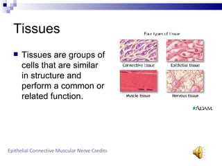
Tissues Tissues are groups of cells that are similar in structure and perform a common or related function. Epithelial   Connective   Muscular   Nerve   Credits
Epithelial Tissue Epithelial tissues are composed entirely of cells. They contain no blood vessels, but are supplied by nerve fibers. They are classified by level and shape. Simple ,  Stratified ,  Transitional ,  Home
Stratified Epithelial Consists of two or more cells layers stacked on top of one another. Common in high abrasion areas where protection is important, such as the skin. Squamous ,  Cuboidal ,  Columnar ,  Home
Stratified Squamous Composed of several layers, it is thick and well suited for its protective purposes. Found in areas subjected to wear and tear. Columnar ,  Cuboidal ,  Home
Stratified Cuboidal Very rare in the human body. Found in some mammary glands. Typically two cell layers thick. Squamous ,  Columnar ,  Home
Stratified Columnar Small amounts are found in the pharynx, the male urethra, and lining some glandular ducts. Occurs at transition areas or junctions between two other types of epithelia. Squamous ,  Cuboidal ,  Home
Simple Epithelial Composed of a single layer of cells. Found where absorption and filtration occur and a thin layer of epithelial tissue is desired. Squamous ,  Cuboidal ,  Columnar ,  Home
Simple Squamous Contains a single layer of flattened cells with disc shaped nuclei and sparse cytoplasm. Main functions are diffusion and filtration. Cuboidal ,  Columnar ,  Home
Simple Cuboidal Single layer of cube like cells Function in secretion and absorption. Columnar ,  Squamous ,  Home
Simple Columnar Composed of simple layers of tall cells with oval nuclei. Contain cilia. The non-ciliated type line the digestive tract and gallbladder. Cuboidal ,  Squamous ,  Home
Connective Tissue Connective tissues are found throughout the body. Main functions are binding, support, protection, insulation, and transportation. C.T. proper ,  cartilage ,  bone ,  blood ,  Home
Blood Blood is the fluid within blood vessels. Most atypical connective tissue. Does not connect things or give mechanical support. Home
Bone Also known as osseous tissue. Great ability to support and protect body. Provide cavities for fat storage and synthesis of blood cells. Home
Cartilage Stands up to both tension and compression.  Tough and flexible. Home
Hyaline Cartilage Also known as gristle Most abundant cartilage in the body. Home
Elastic Cartilage Much more elastic fiber then hyaline cartilage. Able to maintain its shape and structure while still allowing flexibility. Hyaline ,  Fibrocartilage ,  Home
Fibrocartilage Matrix has thick collagen fibers. Provides tensile strength and absorbs compression shock Elastic Cartilage ,  Hyaline Cartilage ,  Home
Connective Tissue Proper connective tissue proper is divided into two subclasses. Loose ,  Dense
Loose Connective Tissue Loose arrangement of fibers.  Matrix is occupied by ground substance, and appears to be empty space when viewed through a microscope. Adipose ,  Reticular ,  Areolar
Dense Connective Tissue Subclass of connective tissue proper. Regular ,  Irregular
Reticular Has loose ground substance with reticular fibers Reticular  cells lie in a fiber network. Adipose ,  Areolar
Areoloar Most widely distributed connective tissue in the body. Serves as a universal packing material. Reticular ,  Adipose
Dense Regular connective Tissue Proper Parallel collagen fibers with a few elastic fibers. Major cell type is fibroblasts. Found in tendons, ligaments, and aponeuroses which are flat sheet like tendons. Irregular Dense Connective tissue Proper ,  Home
Dense Irregular Connective Tissue Proper Irregularly arranged  collagen fibers with some elastic fibers. Dense Regular Connective Tissue Proper ,  Home
Adipose Adipose tissue is similar to areoloar tissue in structure and function. Great nutrient storing ability. Richly vascularized. Reticular ,  Areolar
Transitional Forms lining of hollow urinary organs.  Have the ability to change shape. Home
Muscular Tissue Muscle tissues are highly cellular. They are well vascularized tissues. They are responsible for most types of body movements. Skeletal ,  Smooth ,  Cardiac
Cardiac Found only in the wall of the heart. Contractions help propel blood through the blood vessels to all parts of the body. Smooth ,  Skeletal
Smooth Cells have no visible striations Cells are spindle shaped and contain one centrally located nucleus. Cardiac ,  Skeletal
Skeletal Long, centrically, multinucleate cells with obvious striations. Initiate electrical signals from sensory receptors to effectors. Cardiac ,  Smooth
Nerve Nerve tissues are the main component of the nervous system. Regulate and control body functions. Neurons
Neurons Highly specialized nerve cells that generate and conduct nerve impulses.  Typically branching cells with cytoplasmic extensions or processes.
Credits Stratified Cuboidal Voice: Calida Morris Picture:http://www3.umdnj.edu/histsweb/lab2/images/fstratcuboidal.jpg  Startified Columnar Voice: Calida Morris Picture:  http://cytochemistry.net/Cell-biology/Medical/25092.JPG Blood Voice: Calida Morris Picture: Calida, Justin, Amanda Bone Voice: Calida Morris Picture: Calida, Justin, Amanda Fibrocartilage Voice: calida Morris Picture: Calida, Justin, Amanda Dense Regular connective Tissue Proper Voice: Calida Morris Picture: Calida, Justin, Amanda Transitional Voice: Calida Morris Picture:  Calida, Justin, Amanda Cardiac Voice: Calida Morris Picture: Calida, Justin, Amanda
Credits Simple Columnar Voice: Calida Morris Picture:http://www.kumc.edu/instruction/medicine/anatomy/histoweb/epithel/small/Epth005s.JPG  Loose Connective Tissue Voice: Calida Morris Picture:  http://www.drbio.cornell.edu/Infrastructure/MPM_WWW/MPM_hist/loosehis.jpg Smooth Voice: Calida Morris Picture: Calida, Justin, Amanda
Credits Connective Tissue Proper Voice: Calida Morris Picture:http://kentsimmons.uwinnipeg.ca/cm1504/15lab42006/lb4pg6_files/image002.jpg Dense Connective Tissue Voice: Calida Morris Picture:http://www.drbio.cornell.edu/Infrastructure/MPM_WWW/MPM_hist/loosehis.jpg
Credits Dense Connective Tissue Voice: Calida Morris Picture:http://w3.ouhsc.edu/histology/Glass%20slides/62_02.jpg Reticular Voice: Calida Morris Picture:http://www.gwc.maricopa.edu/class/bio201/histoprc/retic2.jpg
Credits Areoloar Voice: Calida Morris Picture:http://www.gwc.maricopa.edu/class/bio201/histoprc/areol4_s.jpg Dense Irregular Connective Tissue Proper Voice: Calida Morris Picture:http://www.pc.ctc.edu/hart/ctprop/ctprimag/dirr.jpg
Credits Adipose Voice: Calida Morris Picture:http://www.unm.edu/~vscience/images/31-2728%20Human%20Adipose%20Tissue%20sec.%20(100x).jpg Skeletal Voice: Calida Morris Picture:http://www.siumed.edu/~dking2/ssb/images/NM016c.jpg
Credits Muscular Tissue Voice: Calida Morris Picture:http://www.up.ac.za/academic/medicine/telemed/Hist/018/img/018-001.jpg Neurons Voice: Calida Morris Picture:http://www.mirrorservice.org/sites/home.ubalt.edu/ntsbarsh/Business-stat/opre/neurons.gif
Credits Cartilage Voice: Calida Morris Picture:http://www.botany.uwc.ac.za/sci_ed/grade10/mammal/images/cartilage1.gif Stratified Epithelium Voice: Calida Morris Picture:http://www.lab.anhb.uwa.edu.au/mb140/CorePages/Epithelia/Images/oes041he1.jpg
Credits Tissues Voice: Calida Morris Picture:  http://z.abut.com/f/p/440/graphics/images/en/8682.jpg Epithelial tissue Voice: Calida Morris Picture:  http://web.mit.edu/spectroscopy/research/rsch_figs/LSS_fig02.jpg Simple Epithelia Voice: Calida Morris Picture:http://nte-serveur.univ-lyon1.fr/nte/EMBRYON/www.uoguelph.ca/zoology/devobio/miller/01362fig6-1.gif Stratified Squamous Voice: Calida Morris Picture: Calida, Justin, Amanda Simple Squamous Voice: Calida Morris Picture: Calida, Justin, Amanda Nerve Voice: Calida Morris Picture: Calida, Justin, Amanda Simple Cuboidal Voice: calida Morris Picture:  calida, Justin, Amanda Connective Tissue Voice:Calida Morris Picture: http://www.steve.gb.com/images/science/connective_tissue.png

Tissues

  • 1.
    Tissues Tissues aregroups of cells that are similar in structure and perform a common or related function. Epithelial Connective Muscular Nerve Credits
  • 2.
    Epithelial Tissue Epithelialtissues are composed entirely of cells. They contain no blood vessels, but are supplied by nerve fibers. They are classified by level and shape. Simple , Stratified , Transitional , Home
  • 3.
    Stratified Epithelial Consistsof two or more cells layers stacked on top of one another. Common in high abrasion areas where protection is important, such as the skin. Squamous , Cuboidal , Columnar , Home
  • 4.
    Stratified Squamous Composedof several layers, it is thick and well suited for its protective purposes. Found in areas subjected to wear and tear. Columnar , Cuboidal , Home
  • 5.
    Stratified Cuboidal Veryrare in the human body. Found in some mammary glands. Typically two cell layers thick. Squamous , Columnar , Home
  • 6.
    Stratified Columnar Smallamounts are found in the pharynx, the male urethra, and lining some glandular ducts. Occurs at transition areas or junctions between two other types of epithelia. Squamous , Cuboidal , Home
  • 7.
    Simple Epithelial Composedof a single layer of cells. Found where absorption and filtration occur and a thin layer of epithelial tissue is desired. Squamous , Cuboidal , Columnar , Home
  • 8.
    Simple Squamous Containsa single layer of flattened cells with disc shaped nuclei and sparse cytoplasm. Main functions are diffusion and filtration. Cuboidal , Columnar , Home
  • 9.
    Simple Cuboidal Singlelayer of cube like cells Function in secretion and absorption. Columnar , Squamous , Home
  • 10.
    Simple Columnar Composedof simple layers of tall cells with oval nuclei. Contain cilia. The non-ciliated type line the digestive tract and gallbladder. Cuboidal , Squamous , Home
  • 11.
    Connective Tissue Connectivetissues are found throughout the body. Main functions are binding, support, protection, insulation, and transportation. C.T. proper , cartilage , bone , blood , Home
  • 12.
    Blood Blood isthe fluid within blood vessels. Most atypical connective tissue. Does not connect things or give mechanical support. Home
  • 13.
    Bone Also knownas osseous tissue. Great ability to support and protect body. Provide cavities for fat storage and synthesis of blood cells. Home
  • 14.
    Cartilage Stands upto both tension and compression. Tough and flexible. Home
  • 15.
    Hyaline Cartilage Alsoknown as gristle Most abundant cartilage in the body. Home
  • 16.
    Elastic Cartilage Muchmore elastic fiber then hyaline cartilage. Able to maintain its shape and structure while still allowing flexibility. Hyaline , Fibrocartilage , Home
  • 17.
    Fibrocartilage Matrix hasthick collagen fibers. Provides tensile strength and absorbs compression shock Elastic Cartilage , Hyaline Cartilage , Home
  • 18.
    Connective Tissue Properconnective tissue proper is divided into two subclasses. Loose , Dense
  • 19.
    Loose Connective TissueLoose arrangement of fibers. Matrix is occupied by ground substance, and appears to be empty space when viewed through a microscope. Adipose , Reticular , Areolar
  • 20.
    Dense Connective TissueSubclass of connective tissue proper. Regular , Irregular
  • 21.
    Reticular Has looseground substance with reticular fibers Reticular cells lie in a fiber network. Adipose , Areolar
  • 22.
    Areoloar Most widelydistributed connective tissue in the body. Serves as a universal packing material. Reticular , Adipose
  • 23.
    Dense Regular connectiveTissue Proper Parallel collagen fibers with a few elastic fibers. Major cell type is fibroblasts. Found in tendons, ligaments, and aponeuroses which are flat sheet like tendons. Irregular Dense Connective tissue Proper , Home
  • 24.
    Dense Irregular ConnectiveTissue Proper Irregularly arranged collagen fibers with some elastic fibers. Dense Regular Connective Tissue Proper , Home
  • 25.
    Adipose Adipose tissueis similar to areoloar tissue in structure and function. Great nutrient storing ability. Richly vascularized. Reticular , Areolar
  • 26.
    Transitional Forms liningof hollow urinary organs. Have the ability to change shape. Home
  • 27.
    Muscular Tissue Muscletissues are highly cellular. They are well vascularized tissues. They are responsible for most types of body movements. Skeletal , Smooth , Cardiac
  • 28.
    Cardiac Found onlyin the wall of the heart. Contractions help propel blood through the blood vessels to all parts of the body. Smooth , Skeletal
  • 29.
    Smooth Cells haveno visible striations Cells are spindle shaped and contain one centrally located nucleus. Cardiac , Skeletal
  • 30.
    Skeletal Long, centrically,multinucleate cells with obvious striations. Initiate electrical signals from sensory receptors to effectors. Cardiac , Smooth
  • 31.
    Nerve Nerve tissuesare the main component of the nervous system. Regulate and control body functions. Neurons
  • 32.
    Neurons Highly specializednerve cells that generate and conduct nerve impulses. Typically branching cells with cytoplasmic extensions or processes.
  • 33.
    Credits Stratified CuboidalVoice: Calida Morris Picture:http://www3.umdnj.edu/histsweb/lab2/images/fstratcuboidal.jpg Startified Columnar Voice: Calida Morris Picture: http://cytochemistry.net/Cell-biology/Medical/25092.JPG Blood Voice: Calida Morris Picture: Calida, Justin, Amanda Bone Voice: Calida Morris Picture: Calida, Justin, Amanda Fibrocartilage Voice: calida Morris Picture: Calida, Justin, Amanda Dense Regular connective Tissue Proper Voice: Calida Morris Picture: Calida, Justin, Amanda Transitional Voice: Calida Morris Picture: Calida, Justin, Amanda Cardiac Voice: Calida Morris Picture: Calida, Justin, Amanda
  • 34.
    Credits Simple ColumnarVoice: Calida Morris Picture:http://www.kumc.edu/instruction/medicine/anatomy/histoweb/epithel/small/Epth005s.JPG Loose Connective Tissue Voice: Calida Morris Picture: http://www.drbio.cornell.edu/Infrastructure/MPM_WWW/MPM_hist/loosehis.jpg Smooth Voice: Calida Morris Picture: Calida, Justin, Amanda
  • 35.
    Credits Connective TissueProper Voice: Calida Morris Picture:http://kentsimmons.uwinnipeg.ca/cm1504/15lab42006/lb4pg6_files/image002.jpg Dense Connective Tissue Voice: Calida Morris Picture:http://www.drbio.cornell.edu/Infrastructure/MPM_WWW/MPM_hist/loosehis.jpg
  • 36.
    Credits Dense ConnectiveTissue Voice: Calida Morris Picture:http://w3.ouhsc.edu/histology/Glass%20slides/62_02.jpg Reticular Voice: Calida Morris Picture:http://www.gwc.maricopa.edu/class/bio201/histoprc/retic2.jpg
  • 37.
    Credits Areoloar Voice:Calida Morris Picture:http://www.gwc.maricopa.edu/class/bio201/histoprc/areol4_s.jpg Dense Irregular Connective Tissue Proper Voice: Calida Morris Picture:http://www.pc.ctc.edu/hart/ctprop/ctprimag/dirr.jpg
  • 38.
    Credits Adipose Voice:Calida Morris Picture:http://www.unm.edu/~vscience/images/31-2728%20Human%20Adipose%20Tissue%20sec.%20(100x).jpg Skeletal Voice: Calida Morris Picture:http://www.siumed.edu/~dking2/ssb/images/NM016c.jpg
  • 39.
    Credits Muscular TissueVoice: Calida Morris Picture:http://www.up.ac.za/academic/medicine/telemed/Hist/018/img/018-001.jpg Neurons Voice: Calida Morris Picture:http://www.mirrorservice.org/sites/home.ubalt.edu/ntsbarsh/Business-stat/opre/neurons.gif
  • 40.
    Credits Cartilage Voice:Calida Morris Picture:http://www.botany.uwc.ac.za/sci_ed/grade10/mammal/images/cartilage1.gif Stratified Epithelium Voice: Calida Morris Picture:http://www.lab.anhb.uwa.edu.au/mb140/CorePages/Epithelia/Images/oes041he1.jpg
  • 41.
    Credits Tissues Voice:Calida Morris Picture: http://z.abut.com/f/p/440/graphics/images/en/8682.jpg Epithelial tissue Voice: Calida Morris Picture: http://web.mit.edu/spectroscopy/research/rsch_figs/LSS_fig02.jpg Simple Epithelia Voice: Calida Morris Picture:http://nte-serveur.univ-lyon1.fr/nte/EMBRYON/www.uoguelph.ca/zoology/devobio/miller/01362fig6-1.gif Stratified Squamous Voice: Calida Morris Picture: Calida, Justin, Amanda Simple Squamous Voice: Calida Morris Picture: Calida, Justin, Amanda Nerve Voice: Calida Morris Picture: Calida, Justin, Amanda Simple Cuboidal Voice: calida Morris Picture: calida, Justin, Amanda Connective Tissue Voice:Calida Morris Picture: http://www.steve.gb.com/images/science/connective_tissue.png