THE UPPER LIMB
CLAVICLE
Lateral – convex
Medial – concave
Lateral aspect:
• Conoid tubercle
• Trapezoid line
Conoid ligament
Trapezoid ligament
…make up the coracoclavicular ligament complex.
AXILLA (ARMPIT)
• Anterior wall: pectorial region
o Superficial:
 Pec major – forms anteroir fold
 Associated deep fasica
o Deep:
 Subclavius
 Pec minor
 Clavipectorial fascia
• Posterior wall: scapular region (front of scapula)
o Subscapularis
o Teres major
o Latissimus tendon – winds round lower border to form posterior fold
Anterior fold = pec major
Posterior fold = lat dorsi
• Medial wall: serratus anterior (overlying the ribs of the chest wall)
• Base: skin of armpit
• Deltoid drapes over whole shoulder joint – deltoid region
• Boney inlet:
o Medial: rib 1
o Anterior: clavicle
o Posterior: superior scapula to coronoid process
PECTORAL REGION
PECTORIAL REGION:
• In front of shoulder joint
• 3 bones make up the pectorial girdle:
o Proximal humerus
o Scapula
o Clavicle
HUMERUS:
• Spherical head:
o covered with articular catilage
o fits into glenoid cavity of scapula
• Stout anatomical neck
• Greater and lesser tubercles:
o Distal to head and neck
o Insertion of many muscle
o Track down the humerus as the crests of
greater and lesser tubercles.
o Intertubercular groove between the crests.
• Surgical neck of humerus:
o Distal to all of the above
o Point where humerus normally breaks
• Deltoid tuberosity:
o Distal to surgical neck
o Rough area
o Where deltoid inserts
SCAPULA:
• Triangular
• Glenoid cavity:
o faces outward at lateral side
o Covered in articular cartilage
o Associates with head of humerus
• Caracoid process:
o Projects forwards
• Acromion:
o Projects backwards
o Articulates with lateral end of clavicle
• Scapular notch:
o Allows nerovascular bundle to reach muscles on back of scapular
• Subscapular fossa:
o Large flat surface on front of scapular
CLAVICLE:
• S-shaped
• Subcutaneous – easy to palpate
• 2 synovial articulation:
 Laterally with the acromion – acromioclavicular joint
 Medially with manubrium (of sternum) – sternoclavicular joint
o Only true articulation between trunk and pectorial girdle
 Medially with costocartilage of 1st
rib – costoclavicular ligament
Supraglenoid tubercle
Infraglenoid tubercle
Suprascapular notch
Greater scapular notch (spinoglenoid notch)
MUSCLES OF THE PECTORIAL REGION:
 2 layers
 Bigger superficial layer:
o Pectoralis major
 Deep layer:
o Pectoralis minus
o Subclavius
Pectoralis minor:
o Triangular
o Action:
 Depresses the shoulder
 BUT if pectorial girlde is fixed, origin and insertion switch – accessory muscle of
respiration, raising rib-cage.
o Origin: middle 3 true ribs – III, IV & V (base of triangle)
o Insertion: coracoid process (apex of triangle)
Subclavius:
o Action: steadying action by joining 1st
rib which has promary cartilagionous joint to manubrium.
o Origin: upper surface of 1st
rib
o Insertion: upwards and laterally to under surface of clavicle
Clavipectorial fascia:
o Extension of deep fascia and periosteum of clavicle which encloses pectoralis minor.
Pectoralis major:
o Action:
• Adductor of upper limb
• Medial rotation of arm
o Superficial to subclavius, pec minor and clavipectoral fascia
o Origin:
1) Upper fibres: Front of medial half of clavicle
2) Lower fibers:
Front of sternum
Upper costal cartilages
External oblique aponeurosis
o Insertion: lateral lip of bicipital groove of humerus
o Cleft between lower and upper fibres
o Lower fibres twist upwards and insert above the upper (clavicular) fibres.
o Creates rounded anterior axillary fold.
o Pec major covered in deep fascia
o Deep fasica forms base of female breast.
o To test if breast cancer has invaded deep fascia / muscle:
o Hands on hips – fixes the muscle
o If lump not mobile, it has infiltrated deep fascia / muscle.
SCAPULAR REGION
Scapular region:
o Attached to trunk by muscle
o Attached to clavicle by ligaments
o Scapular is mobile
MUSCLES ARISING FROM SCAPULAR:
• Subscapularis:
o Anterior of scapular
o Multipennate muscles – long sustained contraction
 Action:
 adductor
 medial rotation
 Keeps head of humerus in glenoid cavity
 Origin: subscapular fossa (anterior surface of scapula)
 Insertion: tendon which insertes into lesser tubercle of humerus (fibres fuse with
capsule of joint as they pass over it)
o Subscapular bursa is balooning of synovial membrane of joint through a hole in the
capsule of shoulder joint – reduces friction between subscapularis and joint capsule.
• Teres major:
o Posterior of scapular
o Thick and round
 Action:
 adductor
 medial rotation
 Keeps head of humerus in glenoid cavity
 Origin: lateral border of posterior scapular blade
 Insertion: Medial lip of intertubecular sulcus
Latissimus dorsi:
• Technically muscle of posterior body wall
 Action:
 Adducts
 Medially rotates
(climbing)
 Origin:
 Lower T and L spinous processes & interspinous ligaments
 Thoracolumbar fascia
 Iliac crest
 Insertion: fuse into tendon which inserts into intertubercular groove of humerus
(lady between 2 majors)
 Tendon winds around lower border of teres major – gives rounded posterior fold of
axilla
Medial side
Lesser tubercle: subscapularis
Medial lip: teres major
Medial shaft: coracobrachialis
Lateral side:
Greater tubercle: supraspinatus
Lateral lip: pec major
Lateral shaft: deltoid
Serratus anterior:
• Forms medial wall of axilla along with the ribs
• Muscle of side of chest wall
• Aranged as slips/digitations
 Action:
o protraction – pulls scapular forwards around chest wall
o Aids rotation of scapular on chest wall, so glenoid faces upwards (esp lowermost fibres)
o Holds medial margin of scapula against the chest wall
 Operation for breat cancer can damage long thoracic nerve supply to serratus
anterior, paralysing it and causing ‘winged scapula’ – splaying of medial margin
of scapula from chest wall.
o Aids in respiration (if pectorial girdle is fixed)
 Origin: first 8 ribs
 Insertion: curve back around chest wall to insert into medial margin of scapula
DELTOID REGION
DELTOID REGION:
• Triangular cape which covers shoulder
• Multipennate – long sustained contraction
 Action:
o Anterior fibres: flex arm
o Posterior fibres: extend arm
o Middle fibres (overlying shoulder joint): continue abduction of arm
 Origin: U shaped
o Lateral clavicle  acromion  spine of scapula
 Insertion: deltoid tuberoisity of humerus
 Subacromial bursa lies between deltoid and shoulder joint to stop friction.
AXILLA AND ITS CONTENTS:
 Pyramidal
 Apex: medial to caracoid process
 Structures enter and leave at the apex – the inlet
 Boundaries of inlet of the axilla:
o Anterior: clavicle
o Medial: 1st
rib
o Lateral: coracoid process
o Posterior: upper margin of scapula
CONTENTS:
BRACHIAL PLEXUS:
• Plexus of nerves arising in the neck to supply the arm.
Rugby Teams Drink Cold Beers
Roots, Trunks, Divisions, Cords, Branches
Additionals:
C5-C6 root: nerve to subclavius
Medial cord – medial cutaneous nerve arm + forearm
Trunk: superior, middle, inferior
Cords: lateral, medial, posterior (LMP, can you feel the cords?)
Roots
• Ventral rami of C5-C8 + T1
• Emerge between scalenus anterior and scalenus medius in the neck
• Enter posterior triangle of the neck:
o Sternocleidomastiod
o Clavicle
o Trapesius
Trunks
• In posterior triangle, forms 3 trunks:
o Upper trunk: C5 + C6
o Middle trunk: C7
o Lower trunk: C8 + T1
Branches:
• 4 branches off the trunks at level of posterior triangle:
o 2 to scapular muscles on back
 Dorsal scapular nerve:
• To levator scapulae and rhomboids
 Suprascapular nerve:
• Scapular notch  back of scapula: supraspinatus and infraspinatus
muscles.
o Nerve to subclavius
o Long thoracic nerve: descends over surface of serratus anterior, supplying it along its
course (vulnerable in operations)
Anterior and posterior division of the trunks:
 Each of the upper, middle and lower trunks divides into anterior and posterior divisions behind
the clavicle.
 These divisions form 3 new cords:
o Lateral cord
o Medial cord
o Posterior cord
 Arranged around axiallary artery
 Lateral & medial cords: flexor aspects
 Posterior cord: extensor aspects
Roots: posterior triangle
Trunks
Divisions: behind clavicle
Cords: around axillary artery (LMP, cords, blood)
TERMINAL NERVES OF THE BRACHIAL PLEXUS:
Pectoral nerves:
 Lateral pectoral nerve
o From lateral cord
o Pierces clavipectorial fascia  pectoralis major
 Medial pectoral nerve
o From medial cord
o Deep surface of pec minor
o Also pierce pec minor to help supply pec major
LATERAL CORD
Musculocutaneous nerve:
 From lateral cord
 Pierces biceps and coracobrachialis
 Lateral cord continues as median nerve (+ also recieves contribution from medial cord)
 Medial cord supplies flexor aspect below elbow.
MEDIAL CORD
Ulnar nerve
 Continuation of medial cord
 Supplies flexor below elbow
Medial cutaneous nerve of arm
Medial cutaneous nerve of forearm
 Arise from medial cord
Contributes to median nerve
POSTERIOR CORD
 Extensor aspects of limb
 Continues as the radial nerve
Subscapular nerve:
 Supply subscapularis and teres major
Thoracodorsal nerve:
 Latissiumus dorsi
Axillary nerve:
 Between subscapularis and teres major
 Curls round back of humerus
 Lies deep to deltoid
 Supplies deltoid (damage due to dislocation of shoulder joint can paralyse deltoid and
desensitise skin above deltoid tuberosity)
AXILLARY ARTERY AND VEIN:
 Supply upper limb
 Continuation of subclavian artery beyond 1st
rib
 Enters the apex of axilla
 Leaves at lower border of teres major – renamed ‘brachial artery’
Subclavian – 1st
RIB – axillary – TERES MAJOR - brachial
Axillary artery:
 Deep to clavipectoral fascia  deep to pectoralis minor
 Brachial plexus aranged around it (cords)
 Below level of pec minor, the axillary artery is surrounded by terminal branches of cords of
brachial plexus.
Axillary vein:
 Lies on meidal side of neurovascular complex
 Becomes subclavian vein when it crosses over the front of the first rib.
Artery – behind anterior scalene
Vein – in front of anterior scalene
Both – behind pec minor
ARRANGEMENT OF PLEXUS AROUND AXILLARY ARTERY:
 Contribution of median artery from medial cord passes in front of artery
 Ulnar nerve (medial cord) and median nerve (lateral cord) lie either side of the artery.
 The posterior cord (radial nerve) lies behind the artery.
M-shape occurs over axillary artery
BRANCHES OF AXILLARY ARTERY:
 Supply:
o Scapular muscles
o Chest wall
o Female breast
Send The Lifegaurd he is a SAP
 Superior thoracic
 Thoracoacromial
 Lateral thoracic (esp. large in women to supply breast)
o All supply muslce, fat and breast on front of chest
 Subscapular artery
o Supplies:
 Subscapularis
 Teres major
 Latissimus dorsi
o Accompanies thoracodorsal nerve
o Circumflex scapular branch arises from it to supply scapular muscles.
 Anterior + Posterior circumflex humeral
o Around surgical neck of humerus
o Posterior accompanies axillary nerve behind humerus
Blood supply to scapular muscles:
 Branches of axillary artery
 ALSO branches from subclavian in back of neck, which cross the posterior triangle with the 2
branches of the brachial plexus which supply the posterior scapular muscles (dorsal scapular +
suprascapular)
 Allows a collateral blood flow to upper limb if axillary artery is blocked.
AXILLARY LYMPH:
 3 groups of lymph nodes:
o Lateral
 lateral to axillary vessels
 Lymph from upper limb lymphatics
o Pectoral / anterior
 Between anterior and medial walls of axilla
 Upper nodes in this group - mainly lymph from breast
 Also lymph from side wall of thorax
o Subscapular / posterior
 On posterior wall of axila, along subscapular vessels.
 Receive lymph from subscapular region and back.
 Join with central nodes in the fat of the axilla
 Central nodes then drain into apical nodes – at apex of axilla
 Apical nodes  subclavian lymph trunk  subclavian vein.
 In the pectoral region:
o Infraclavicular nodes lie on clavipectoral fascia in groove between deltoid and pec
major.
o  drain into apical nodes.
 Axillary nodes are important spread of cancer in breast carcinoma.
BREAST
LOCATION:
• Lies in the fat of the pectoral region, on the deep fascia of the pectoralis major
• Extends from the 2nd
to the 6th
rib
• At lower extremity (6th
rib), it lies on the origin of the external oblique muscle.
• Extends from side of sternum to edge of pectoralis major
• Some glandular tissue extends beyond edge of muscle to lie on the medial wall of the axilla – an
extension reaches high into the axilla and is called the axillary tail.
STRUCTURE:
• Consists of glandular tissue embedded in fat, separated into lobes by connective tissue.
• During lactation, glandular part enlarges much more than fatty part.
• Gland has no fibrous capsule  cancer can easily invade underlying fascia and pectoral muscle
• Nipple is surrounded by dark coloured skin – the areolar (darkens after first pregnancy)
• Surface of the areolar is irregular due to the presence of Montgomery’s glands (specialised
sebaceous glands).
• Glandular tissues is divided by fibrous septa (suspensory ligaments) which pass through the fat
of the breast from skin to pectoral fascia.
• Divisions of the gland are called lobes – 15+ lobes in each breast.
• Lobes are further divided into lobules.
• Milk produced in each lobe are passed towards nipple by the lactiferous duct. One lactiferous
duct arises from each lobe.
• Before opening on to the nipple, the lactiferous duct dilates to form the lactiferous sinus.
BLOOD SUPPLY TO THE BREAST:
1. Perforating branches of the internal thoracic artery – especially at 2nd
, 3rd
and 4th
intercostals
spaces.
2. Thoracic branches of the axillary artery:
• Superior thoracic artery
• Thoracoacromial artery
• Lateral thoracic artery.
3. Lateral mammary branches of cutaneous branches of the posterior intercostals arteries.
• These vessels all branch and anastosome with one another.
• Venous drainage corresponds to arterial supply.
LYMPHATIC DRAINAGE OF BREAST:
• Most of the lymph drains to the axillary lymph nodes:
o Lateral group (lies on axillary vessels)
o Pectoral / anterior group
o Subcapsular / posterior group
o  lymph from the above groups drain into the 
o Central nodes (in the fat of the axilla)
o  lymph drains from the central nodes into the 
o Apical nodes (in apex of axilla).
o  lymph drains from the apical nodes into the subclavian lymph trunk
o  lymph then drains into subclavian vein.
• From deep parts of the gland, lymphatics pierce the intercostals space and follows the
perforating branches of the internal thoracic artery to nodes which lie alongside the internal
thoracic artery – the internal thoracic nodes.
•  lymph from internal thoracic nodes follow intercostals arteries back to region of aorta where
they drain into para-aortic nodes.
• Lymphatic drainage is major route of spread of breast carcinoma.
• Often leads to metastasis in the axillary lymph nodes – hard lump in the axilla.
• By draining to the para-aortic nodes, cancer can spread to the thorax.
• Some lymph from lower breast spreads downwards through abdominal wall to spread cancer to
the abdominal cavity.
• Skin lymphatics link the two breasts across the midline, and thus allow spread of cancer from
one breast to the other.
• Lymph from the nipple and areola drains into the sub-areolar plexus, which eventually drains
into the axillary nodes.
POSTERIOR ASPECT OF SCAPULAR
STRUCTURE OF POSTERIOR SCAPULA:
 Scapula spine divides suprasinous fossa and infraspinous fossa
 Scapula spine becomes acromion laterally
 Scapular notch in upper border of scapula
 Scapular notch becomes scapular foramen due to suprascapular ligament.
 Spinoglenoid notch allows the 2 fossa to communicate with one another.
MUSCLES OF THE POSTERIOR SCAPULAR:
 Supraspinatus
 Action: initiation of abduction of arm & stability.
 Origin: supraspinous fossa
 Insertion: Passes beneath overhanging acromion, with tendon inserting into greater
tubercle of humerus.
 Some fibres fuse with capsule of shoulder joint.
 Tendon is prone to:
 Rupture in injury
 Calcific degeneration.
 Suprascapular nerve passes through the scapular notch from brachial plexus to supply the
supraspinatus.
 Branch of subclavian artery also passes through
 Infraspinatus
 Action: lateral rotation of the arm (opposite to subscapularis) & stability.
 Origin: infraspinatus fossa
 Insertion: tendon inserting into greater tubercle of humerus.
 Supplied by same vascular bundle as supplies the supraspinatus, via the scapular notch
and then the spinoglenoid notch.
 Its position corresponds with subscapularis muscle on anterior of scapular.
 Teres minor
 Small, at lower edge of infraspinatus
 Action: initiation of abduction of arm & stability.
 Origin: infraspinatus fossa
 Insertion: tendon inserting into greater tubercle of humerus.
 Supplied by axillary nerve (which also supplies deltoid) – a branch of the posterior cord
of the brachial plexus.
Posterior muscles which suspend scapular from vertebral column:
 Allow rotation of scapula on chest wall
 Levator scapulae
 Rhomboid
 Trapezius
Levator scapulae & rhomboid:
 Action: draw scapula towards midline and rotate glenoid downwards.
 Origin: vertebral column
 Insertion: medial edge of scapula
 Supplied by dorsal scapular nerve (branch of brachial plexus in neck)
Trapezius:
 Action:
o Upper and lower fibres together: rotate so glenoid faces upwards
o Upper fibres only: elevate scapula
o Middle fibres only: brace shoulders backwards
 Origin: from long linear origin:
o Back of scull
o Ligamentum nuchae
o C & T spines and supraspinous ligaments.
 Insertion (similar to origin of deltoid):
o Upper fibres: lateral 3rd
of clavicle
o Lower fibres: acromion and spine of scapula
 Trapezius supplied by spinal accessory nerve (cranial XI).
 Muscles which rotate glenoid cavity downwards:
o Levator scapulae
o Rhomboids
 Muscles which rotate glenoid cavity upwards:
o Upper and lower fibres of trapezius acting together.
SHOULDER JOINTS
• 3 main joints:
o Sternoclavicular
o Acromioclavicular
o Shoulder joint
STERNOCLAVICULAR:
• Only articulation between pectoral girdle and trunk
• Synovial
• Ball and socket:
o Ball: medial end of clavicle
o Socket: manubrium and upper border of 1st
rib (joined by 1o
cartilaginous)
• Strong capsule
• Synovial cavity divided in two by intra-articular disc; attached to clavicle above and 1st
rib below
– prevents extreme upwards displacement of medial clavicle.
• Clavicle is on a see-saw:
o Medial end swings forwards when shoulders braced backwards, and vice versa.
• Displacement of joint prevented by:
o Several strong ligaments
o Capsule
o Intra-articular disk
ACROMIOCLAVICULAR JOINT:
• Between scapcular and lateral clavicle.
• Synovial
• Limited gliding movement
 The weight of the upper limb is supported mainly by:
o Coracoclavicular ligaments – between coracoid process and lateral clavicle
o Levator scapulae & trapezius muscles – suspend from head, neck and trunk.
SHOULDER JOINT:
• Synovial
• Ball and socket:
o Ball: head of humerus (spherical except for thick anatomical neck) covered with articular
cartilage.
o Socket: glenoid cavity – pear shape and slightly concave.
• Articular surface of glenoid cavity is deepened by rim of fibrous tissue – labrum glenoidale
CAPSULE OF SHOULDER JOINT:
• Capsule is attached to both humerus and glenoid cavity at their margins: anatomical neck of
humerus and labrum glenoidale.
• Capsule passes over intertubercular groove, where it is thickened to form transverse ligament.
• NOTE that inferiorly, capsule attaches further down, below anatomical neck.
HOLES IN THE CAPSULE:
• Subscapular bursa:
o On the front of joint
o Balooning of synovial membrane through hole in capsule
• Corresponding bursa often found on the back of the joint, below the infraspinatus muscle.
• Synovial membrane also emerges beneath bridge of transverse ligament.
• This synovial membrane covers the tendon of long head of the biceps brachii muscle, which
travels from above the glenoid in the scapula, into the intertubercular groove of the humerus
within the capsule.
LIGAMENTS WHICH STENGTHEN CAPSULE:
• Glenohumeral ligaments
• Coracohumeral ligament:
o Fibrous tissue: coracoid process  greater tubercle
o Prevents excessive external rotation
• Coracoacromial ligament:
o Arch between coracoid process and acromion
o Prevents upwards displacement of the joint
• Tendon of long head of the biceps brachii muscle:
o Intracapsular
o Stabilises
NATURE OF SYNOVIAL MEMBRANE:
• SM lines all intracapsular surfaces except those covered with articular cartilage.
• ∆ in shoulder joint, SM covers:
o Inside of capsule
o Tendon of long head of biceps brachii
o Forms subscapular bursa
• Subacromial bursa:
o NOT in communication with joint SM
o Separate bag of SM
o Covers:
 Upper part of joint capsule
 Supraspinatus tendon (on its way to greater tubercle)
o Seperates these 2 from the coracoacromial arch and deltoid, so they can move freely.
• Joint capsule is surrounded by muscle on all sides except below:
o Above: supraspinatus
o Behind: infraspinatus & teres minor
o Front: subscapularis
• Tendons of these muscles fuse with the capsule – forms a sleeve called rotator cuff
• Below, because no supporting muscle, capsule falls below between subscapularis and
infraspinatus.
• Humerus usually dislocates through here in shoulder dislocation – can damage the axillary nerve
in doing so.
MOBILITY OF THE SHOULDER JOINT:
• Most mobile joint in the body:
o Large head of humerus, shallow glenoid cavity
o Loose capsule
• What stops dislocation then?
o Glenoid labrum – deepens socket
o Rotator cuff tendons – hold head in glenoid
o Long head of biceps which runs intracapuslarly
o Ligaments:
 Glenohumeral
 Coracohumeral
 Coracoaromial
o Atmospheric pressure (but dislocation is sliding, so this is not a big factor)
Movements:
• Flexion (forward in saggital plane)
o Anterior fibres of deltoid
o Pec major
• Extension (backwards in saggital plane)
o Posterior fibres of deltoid
o Latissimus dorsi
• Abduction (arm lifted away from body)
o Initiated: supraspinatus
o Continued: deltoid
 If supraspinatus is torn, abduction cannot be initiated – patients must
swing arms passively to get initial momentum.
 At 90o
abduction, articular surface of humerus head is used up
 Head must then be rotated laterally to give more articular surface.
• Adduction (arm brought inwards to body)
o Pec major
o Latissimus dorsi
• Circumduction
• Rotation (humerus rotates in long axis)
o Subscapularis
o Teres major
o Infraspinatus
NOTE all 3 joints move in any movement of the shoulder
E.g. abduction:
o Scapular rotates at the same time as the shoulder joint moves
o Trapezius & serratus anterior rotate glenoid upwards
o  passive depression of sternoclavicular joint
APPLIED ANATOMY
Shoulder joint is prone to fracture / dislocation
Breast cancer is a common site of carcinoma – must palpate both breast and axilla.
Bones and joint injuries:
• Make comparison between 2 shoulders in examination
Clavicular fracture
• Fractured clavicle is common – esp. medial to coracoclavicular ligament (2/3 way along
clavicle)
• Pectorial girdle and lateral 1/3 of clavicle fall
• Bandage used to brace shoulders back
• Clavicular fracture unites readily.
Humerus fracture
• Common at surgical neck, but heals quickly
Dislocation:
• Humerus head leaves glenoid inferiorly: pulled medially, below and infront of glenoid fossa.
• May damage axillary nerve
• Most lateral boney landmark becomes to acromion.
• Subscapularis spasms – medially rotates the humerus; elbow lies away from body
• Reducing the shoulder:
o Flex elbow
o Rotate forearm outwards
o Bring elbow medially across trunk
• Should then test function of deltoid, and sensitivity of skin overlying deltoid tuberosity – to
check for axillary nerve damage.
Supraspinatus tendon:
• Injured during falls, or torn at insertion
• Must be repaired surgically.
• Also prone to degenerative processes – calcification:
o Pain on abduction
o Inflamation of the subacromial bursa.
Injuries to brachial plexus:
• Cervical rib / birth / road traffic accident
• RTA: Force hitting shoulder pulls nerve roots from neck
• Cervical rib: pressure on T1 contribution, and blood vessels to upper limb.
• Birth: traction to head/neck – damage to brachial plexus:
o Erb-Duchenne syndrome - C5-C6 & suprascapular branch normally injured  upper limb
deformity
o C7 – paralysis of muscles supplied by radial nerve
o C8 & T1 – paralysis of small muscles of hand (Klumpke syndrome).

THE SHOULDER.............................

  • 1.
  • 2.
    Lateral – convex Medial– concave Lateral aspect: • Conoid tubercle • Trapezoid line Conoid ligament Trapezoid ligament …make up the coracoclavicular ligament complex. AXILLA (ARMPIT) • Anterior wall: pectorial region o Superficial:  Pec major – forms anteroir fold  Associated deep fasica o Deep:  Subclavius  Pec minor  Clavipectorial fascia • Posterior wall: scapular region (front of scapula) o Subscapularis o Teres major o Latissimus tendon – winds round lower border to form posterior fold
  • 3.
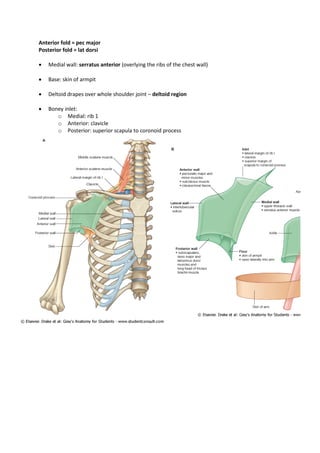
    Anterior fold =pec major Posterior fold = lat dorsi • Medial wall: serratus anterior (overlying the ribs of the chest wall) • Base: skin of armpit • Deltoid drapes over whole shoulder joint – deltoid region • Boney inlet: o Medial: rib 1 o Anterior: clavicle o Posterior: superior scapula to coronoid process
  • 4.
  • 5.
    PECTORIAL REGION: • Infront of shoulder joint • 3 bones make up the pectorial girdle: o Proximal humerus o Scapula o Clavicle HUMERUS: • Spherical head: o covered with articular catilage o fits into glenoid cavity of scapula • Stout anatomical neck • Greater and lesser tubercles: o Distal to head and neck o Insertion of many muscle o Track down the humerus as the crests of greater and lesser tubercles. o Intertubercular groove between the crests. • Surgical neck of humerus: o Distal to all of the above o Point where humerus normally breaks • Deltoid tuberosity: o Distal to surgical neck o Rough area o Where deltoid inserts SCAPULA: • Triangular • Glenoid cavity: o faces outward at lateral side o Covered in articular cartilage o Associates with head of humerus • Caracoid process: o Projects forwards • Acromion: o Projects backwards o Articulates with lateral end of clavicle • Scapular notch: o Allows nerovascular bundle to reach muscles on back of scapular • Subscapular fossa:
  • 6.
    o Large flatsurface on front of scapular CLAVICLE: • S-shaped • Subcutaneous – easy to palpate • 2 synovial articulation:  Laterally with the acromion – acromioclavicular joint  Medially with manubrium (of sternum) – sternoclavicular joint o Only true articulation between trunk and pectorial girdle  Medially with costocartilage of 1st rib – costoclavicular ligament Supraglenoid tubercle Infraglenoid tubercle Suprascapular notch Greater scapular notch (spinoglenoid notch)
  • 7.
    MUSCLES OF THEPECTORIAL REGION:  2 layers  Bigger superficial layer: o Pectoralis major  Deep layer: o Pectoralis minus o Subclavius Pectoralis minor: o Triangular o Action:  Depresses the shoulder  BUT if pectorial girlde is fixed, origin and insertion switch – accessory muscle of respiration, raising rib-cage. o Origin: middle 3 true ribs – III, IV & V (base of triangle) o Insertion: coracoid process (apex of triangle) Subclavius: o Action: steadying action by joining 1st rib which has promary cartilagionous joint to manubrium. o Origin: upper surface of 1st rib o Insertion: upwards and laterally to under surface of clavicle Clavipectorial fascia: o Extension of deep fascia and periosteum of clavicle which encloses pectoralis minor.
  • 8.
    Pectoralis major: o Action: •Adductor of upper limb • Medial rotation of arm o Superficial to subclavius, pec minor and clavipectoral fascia o Origin: 1) Upper fibres: Front of medial half of clavicle 2) Lower fibers: Front of sternum Upper costal cartilages External oblique aponeurosis o Insertion: lateral lip of bicipital groove of humerus o Cleft between lower and upper fibres o Lower fibres twist upwards and insert above the upper (clavicular) fibres. o Creates rounded anterior axillary fold. o Pec major covered in deep fascia o Deep fasica forms base of female breast.
  • 9.
    o To testif breast cancer has invaded deep fascia / muscle: o Hands on hips – fixes the muscle o If lump not mobile, it has infiltrated deep fascia / muscle.
  • 10.
  • 11.
    Scapular region: o Attachedto trunk by muscle o Attached to clavicle by ligaments o Scapular is mobile MUSCLES ARISING FROM SCAPULAR: • Subscapularis: o Anterior of scapular o Multipennate muscles – long sustained contraction  Action:  adductor  medial rotation  Keeps head of humerus in glenoid cavity  Origin: subscapular fossa (anterior surface of scapula)  Insertion: tendon which insertes into lesser tubercle of humerus (fibres fuse with capsule of joint as they pass over it) o Subscapular bursa is balooning of synovial membrane of joint through a hole in the capsule of shoulder joint – reduces friction between subscapularis and joint capsule. • Teres major: o Posterior of scapular o Thick and round  Action:  adductor  medial rotation  Keeps head of humerus in glenoid cavity  Origin: lateral border of posterior scapular blade  Insertion: Medial lip of intertubecular sulcus Latissimus dorsi: • Technically muscle of posterior body wall  Action:  Adducts  Medially rotates (climbing)  Origin:  Lower T and L spinous processes & interspinous ligaments  Thoracolumbar fascia
  • 12.
     Iliac crest Insertion: fuse into tendon which inserts into intertubercular groove of humerus (lady between 2 majors)  Tendon winds around lower border of teres major – gives rounded posterior fold of axilla
  • 13.
    Medial side Lesser tubercle:subscapularis Medial lip: teres major Medial shaft: coracobrachialis Lateral side: Greater tubercle: supraspinatus Lateral lip: pec major Lateral shaft: deltoid Serratus anterior: • Forms medial wall of axilla along with the ribs • Muscle of side of chest wall • Aranged as slips/digitations  Action: o protraction – pulls scapular forwards around chest wall o Aids rotation of scapular on chest wall, so glenoid faces upwards (esp lowermost fibres) o Holds medial margin of scapula against the chest wall  Operation for breat cancer can damage long thoracic nerve supply to serratus anterior, paralysing it and causing ‘winged scapula’ – splaying of medial margin of scapula from chest wall.
  • 14.
    o Aids inrespiration (if pectorial girdle is fixed)  Origin: first 8 ribs  Insertion: curve back around chest wall to insert into medial margin of scapula DELTOID REGION
  • 15.
    DELTOID REGION: • Triangularcape which covers shoulder • Multipennate – long sustained contraction  Action: o Anterior fibres: flex arm o Posterior fibres: extend arm o Middle fibres (overlying shoulder joint): continue abduction of arm  Origin: U shaped o Lateral clavicle  acromion  spine of scapula  Insertion: deltoid tuberoisity of humerus  Subacromial bursa lies between deltoid and shoulder joint to stop friction.
  • 16.
    AXILLA AND ITSCONTENTS:  Pyramidal  Apex: medial to caracoid process  Structures enter and leave at the apex – the inlet  Boundaries of inlet of the axilla: o Anterior: clavicle o Medial: 1st rib o Lateral: coracoid process o Posterior: upper margin of scapula CONTENTS: BRACHIAL PLEXUS: • Plexus of nerves arising in the neck to supply the arm. Rugby Teams Drink Cold Beers Roots, Trunks, Divisions, Cords, Branches
  • 17.
    Additionals: C5-C6 root: nerveto subclavius Medial cord – medial cutaneous nerve arm + forearm Trunk: superior, middle, inferior Cords: lateral, medial, posterior (LMP, can you feel the cords?)
  • 18.
    Roots • Ventral ramiof C5-C8 + T1 • Emerge between scalenus anterior and scalenus medius in the neck • Enter posterior triangle of the neck: o Sternocleidomastiod o Clavicle o Trapesius Trunks • In posterior triangle, forms 3 trunks: o Upper trunk: C5 + C6 o Middle trunk: C7 o Lower trunk: C8 + T1 Branches: • 4 branches off the trunks at level of posterior triangle: o 2 to scapular muscles on back  Dorsal scapular nerve: • To levator scapulae and rhomboids  Suprascapular nerve: • Scapular notch  back of scapula: supraspinatus and infraspinatus muscles. o Nerve to subclavius o Long thoracic nerve: descends over surface of serratus anterior, supplying it along its course (vulnerable in operations) Anterior and posterior division of the trunks:
  • 19.
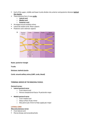
     Each ofthe upper, middle and lower trunks divides into anterior and posterior divisions behind the clavicle.  These divisions form 3 new cords: o Lateral cord o Medial cord o Posterior cord  Arranged around axiallary artery  Lateral & medial cords: flexor aspects  Posterior cord: extensor aspects Roots: posterior triangle Trunks Divisions: behind clavicle Cords: around axillary artery (LMP, cords, blood) TERMINAL NERVES OF THE BRACHIAL PLEXUS: Pectoral nerves:  Lateral pectoral nerve o From lateral cord o Pierces clavipectorial fascia  pectoralis major  Medial pectoral nerve o From medial cord o Deep surface of pec minor o Also pierce pec minor to help supply pec major LATERAL CORD Musculocutaneous nerve:  From lateral cord  Pierces biceps and coracobrachialis
  • 20.
     Lateral cordcontinues as median nerve (+ also recieves contribution from medial cord)  Medial cord supplies flexor aspect below elbow. MEDIAL CORD Ulnar nerve  Continuation of medial cord  Supplies flexor below elbow Medial cutaneous nerve of arm Medial cutaneous nerve of forearm  Arise from medial cord Contributes to median nerve POSTERIOR CORD  Extensor aspects of limb  Continues as the radial nerve Subscapular nerve:  Supply subscapularis and teres major Thoracodorsal nerve:  Latissiumus dorsi Axillary nerve:  Between subscapularis and teres major  Curls round back of humerus  Lies deep to deltoid  Supplies deltoid (damage due to dislocation of shoulder joint can paralyse deltoid and desensitise skin above deltoid tuberosity) AXILLARY ARTERY AND VEIN:  Supply upper limb  Continuation of subclavian artery beyond 1st rib  Enters the apex of axilla  Leaves at lower border of teres major – renamed ‘brachial artery’ Subclavian – 1st RIB – axillary – TERES MAJOR - brachial Axillary artery:  Deep to clavipectoral fascia  deep to pectoralis minor  Brachial plexus aranged around it (cords)  Below level of pec minor, the axillary artery is surrounded by terminal branches of cords of brachial plexus. Axillary vein:  Lies on meidal side of neurovascular complex  Becomes subclavian vein when it crosses over the front of the first rib.
  • 21.
    Artery – behindanterior scalene Vein – in front of anterior scalene Both – behind pec minor ARRANGEMENT OF PLEXUS AROUND AXILLARY ARTERY:  Contribution of median artery from medial cord passes in front of artery  Ulnar nerve (medial cord) and median nerve (lateral cord) lie either side of the artery.  The posterior cord (radial nerve) lies behind the artery.
  • 23.
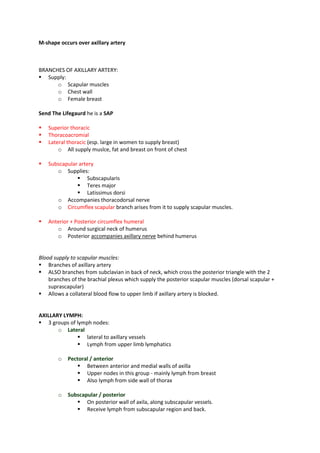
    M-shape occurs overaxillary artery BRANCHES OF AXILLARY ARTERY:  Supply: o Scapular muscles o Chest wall o Female breast Send The Lifegaurd he is a SAP  Superior thoracic  Thoracoacromial  Lateral thoracic (esp. large in women to supply breast) o All supply muslce, fat and breast on front of chest  Subscapular artery o Supplies:  Subscapularis  Teres major  Latissimus dorsi o Accompanies thoracodorsal nerve o Circumflex scapular branch arises from it to supply scapular muscles.  Anterior + Posterior circumflex humeral o Around surgical neck of humerus o Posterior accompanies axillary nerve behind humerus Blood supply to scapular muscles:  Branches of axillary artery  ALSO branches from subclavian in back of neck, which cross the posterior triangle with the 2 branches of the brachial plexus which supply the posterior scapular muscles (dorsal scapular + suprascapular)  Allows a collateral blood flow to upper limb if axillary artery is blocked. AXILLARY LYMPH:  3 groups of lymph nodes: o Lateral  lateral to axillary vessels  Lymph from upper limb lymphatics o Pectoral / anterior  Between anterior and medial walls of axilla  Upper nodes in this group - mainly lymph from breast  Also lymph from side wall of thorax o Subscapular / posterior  On posterior wall of axila, along subscapular vessels.  Receive lymph from subscapular region and back.
  • 24.
     Join withcentral nodes in the fat of the axilla  Central nodes then drain into apical nodes – at apex of axilla  Apical nodes  subclavian lymph trunk  subclavian vein.  In the pectoral region: o Infraclavicular nodes lie on clavipectoral fascia in groove between deltoid and pec major. o  drain into apical nodes.  Axillary nodes are important spread of cancer in breast carcinoma. BREAST LOCATION: • Lies in the fat of the pectoral region, on the deep fascia of the pectoralis major • Extends from the 2nd to the 6th rib • At lower extremity (6th rib), it lies on the origin of the external oblique muscle. • Extends from side of sternum to edge of pectoralis major • Some glandular tissue extends beyond edge of muscle to lie on the medial wall of the axilla – an extension reaches high into the axilla and is called the axillary tail. STRUCTURE: • Consists of glandular tissue embedded in fat, separated into lobes by connective tissue. • During lactation, glandular part enlarges much more than fatty part. • Gland has no fibrous capsule  cancer can easily invade underlying fascia and pectoral muscle • Nipple is surrounded by dark coloured skin – the areolar (darkens after first pregnancy) • Surface of the areolar is irregular due to the presence of Montgomery’s glands (specialised sebaceous glands). • Glandular tissues is divided by fibrous septa (suspensory ligaments) which pass through the fat of the breast from skin to pectoral fascia. • Divisions of the gland are called lobes – 15+ lobes in each breast. • Lobes are further divided into lobules. • Milk produced in each lobe are passed towards nipple by the lactiferous duct. One lactiferous duct arises from each lobe. • Before opening on to the nipple, the lactiferous duct dilates to form the lactiferous sinus. BLOOD SUPPLY TO THE BREAST: 1. Perforating branches of the internal thoracic artery – especially at 2nd , 3rd and 4th intercostals spaces. 2. Thoracic branches of the axillary artery: • Superior thoracic artery • Thoracoacromial artery • Lateral thoracic artery. 3. Lateral mammary branches of cutaneous branches of the posterior intercostals arteries. • These vessels all branch and anastosome with one another.
  • 25.
    • Venous drainagecorresponds to arterial supply. LYMPHATIC DRAINAGE OF BREAST: • Most of the lymph drains to the axillary lymph nodes: o Lateral group (lies on axillary vessels) o Pectoral / anterior group o Subcapsular / posterior group o  lymph from the above groups drain into the  o Central nodes (in the fat of the axilla) o  lymph drains from the central nodes into the  o Apical nodes (in apex of axilla). o  lymph drains from the apical nodes into the subclavian lymph trunk o  lymph then drains into subclavian vein. • From deep parts of the gland, lymphatics pierce the intercostals space and follows the perforating branches of the internal thoracic artery to nodes which lie alongside the internal thoracic artery – the internal thoracic nodes. •  lymph from internal thoracic nodes follow intercostals arteries back to region of aorta where they drain into para-aortic nodes. • Lymphatic drainage is major route of spread of breast carcinoma. • Often leads to metastasis in the axillary lymph nodes – hard lump in the axilla. • By draining to the para-aortic nodes, cancer can spread to the thorax. • Some lymph from lower breast spreads downwards through abdominal wall to spread cancer to the abdominal cavity. • Skin lymphatics link the two breasts across the midline, and thus allow spread of cancer from one breast to the other. • Lymph from the nipple and areola drains into the sub-areolar plexus, which eventually drains into the axillary nodes.
  • 26.
  • 27.
    STRUCTURE OF POSTERIORSCAPULA:  Scapula spine divides suprasinous fossa and infraspinous fossa  Scapula spine becomes acromion laterally  Scapular notch in upper border of scapula  Scapular notch becomes scapular foramen due to suprascapular ligament.  Spinoglenoid notch allows the 2 fossa to communicate with one another. MUSCLES OF THE POSTERIOR SCAPULAR:  Supraspinatus  Action: initiation of abduction of arm & stability.
  • 28.
     Origin: supraspinousfossa  Insertion: Passes beneath overhanging acromion, with tendon inserting into greater tubercle of humerus.  Some fibres fuse with capsule of shoulder joint.  Tendon is prone to:  Rupture in injury  Calcific degeneration.  Suprascapular nerve passes through the scapular notch from brachial plexus to supply the supraspinatus.  Branch of subclavian artery also passes through  Infraspinatus  Action: lateral rotation of the arm (opposite to subscapularis) & stability.  Origin: infraspinatus fossa  Insertion: tendon inserting into greater tubercle of humerus.  Supplied by same vascular bundle as supplies the supraspinatus, via the scapular notch and then the spinoglenoid notch.  Its position corresponds with subscapularis muscle on anterior of scapular.  Teres minor  Small, at lower edge of infraspinatus  Action: initiation of abduction of arm & stability.  Origin: infraspinatus fossa  Insertion: tendon inserting into greater tubercle of humerus.  Supplied by axillary nerve (which also supplies deltoid) – a branch of the posterior cord of the brachial plexus. Posterior muscles which suspend scapular from vertebral column:  Allow rotation of scapula on chest wall  Levator scapulae  Rhomboid  Trapezius Levator scapulae & rhomboid:  Action: draw scapula towards midline and rotate glenoid downwards.  Origin: vertebral column  Insertion: medial edge of scapula  Supplied by dorsal scapular nerve (branch of brachial plexus in neck) Trapezius:  Action: o Upper and lower fibres together: rotate so glenoid faces upwards o Upper fibres only: elevate scapula o Middle fibres only: brace shoulders backwards  Origin: from long linear origin: o Back of scull
  • 29.
    o Ligamentum nuchae oC & T spines and supraspinous ligaments.  Insertion (similar to origin of deltoid): o Upper fibres: lateral 3rd of clavicle o Lower fibres: acromion and spine of scapula  Trapezius supplied by spinal accessory nerve (cranial XI).
  • 30.
     Muscles whichrotate glenoid cavity downwards: o Levator scapulae o Rhomboids  Muscles which rotate glenoid cavity upwards: o Upper and lower fibres of trapezius acting together. SHOULDER JOINTS • 3 main joints: o Sternoclavicular o Acromioclavicular o Shoulder joint STERNOCLAVICULAR: • Only articulation between pectoral girdle and trunk • Synovial • Ball and socket: o Ball: medial end of clavicle o Socket: manubrium and upper border of 1st rib (joined by 1o cartilaginous) • Strong capsule • Synovial cavity divided in two by intra-articular disc; attached to clavicle above and 1st rib below – prevents extreme upwards displacement of medial clavicle. • Clavicle is on a see-saw: o Medial end swings forwards when shoulders braced backwards, and vice versa. • Displacement of joint prevented by: o Several strong ligaments o Capsule o Intra-articular disk ACROMIOCLAVICULAR JOINT: • Between scapcular and lateral clavicle. • Synovial • Limited gliding movement
  • 31.
     The weightof the upper limb is supported mainly by: o Coracoclavicular ligaments – between coracoid process and lateral clavicle o Levator scapulae & trapezius muscles – suspend from head, neck and trunk. SHOULDER JOINT: • Synovial • Ball and socket: o Ball: head of humerus (spherical except for thick anatomical neck) covered with articular cartilage. o Socket: glenoid cavity – pear shape and slightly concave. • Articular surface of glenoid cavity is deepened by rim of fibrous tissue – labrum glenoidale CAPSULE OF SHOULDER JOINT: • Capsule is attached to both humerus and glenoid cavity at their margins: anatomical neck of humerus and labrum glenoidale. • Capsule passes over intertubercular groove, where it is thickened to form transverse ligament. • NOTE that inferiorly, capsule attaches further down, below anatomical neck. HOLES IN THE CAPSULE: • Subscapular bursa: o On the front of joint o Balooning of synovial membrane through hole in capsule • Corresponding bursa often found on the back of the joint, below the infraspinatus muscle. • Synovial membrane also emerges beneath bridge of transverse ligament. • This synovial membrane covers the tendon of long head of the biceps brachii muscle, which travels from above the glenoid in the scapula, into the intertubercular groove of the humerus within the capsule.
  • 32.
    LIGAMENTS WHICH STENGTHENCAPSULE: • Glenohumeral ligaments • Coracohumeral ligament: o Fibrous tissue: coracoid process  greater tubercle o Prevents excessive external rotation • Coracoacromial ligament: o Arch between coracoid process and acromion o Prevents upwards displacement of the joint • Tendon of long head of the biceps brachii muscle: o Intracapsular o Stabilises NATURE OF SYNOVIAL MEMBRANE: • SM lines all intracapsular surfaces except those covered with articular cartilage. • ∆ in shoulder joint, SM covers: o Inside of capsule o Tendon of long head of biceps brachii o Forms subscapular bursa • Subacromial bursa: o NOT in communication with joint SM o Separate bag of SM o Covers:  Upper part of joint capsule  Supraspinatus tendon (on its way to greater tubercle) o Seperates these 2 from the coracoacromial arch and deltoid, so they can move freely.
  • 34.
    • Joint capsuleis surrounded by muscle on all sides except below: o Above: supraspinatus o Behind: infraspinatus & teres minor o Front: subscapularis • Tendons of these muscles fuse with the capsule – forms a sleeve called rotator cuff • Below, because no supporting muscle, capsule falls below between subscapularis and infraspinatus. • Humerus usually dislocates through here in shoulder dislocation – can damage the axillary nerve in doing so.
  • 35.
    MOBILITY OF THESHOULDER JOINT: • Most mobile joint in the body: o Large head of humerus, shallow glenoid cavity o Loose capsule • What stops dislocation then? o Glenoid labrum – deepens socket o Rotator cuff tendons – hold head in glenoid o Long head of biceps which runs intracapuslarly o Ligaments:  Glenohumeral  Coracohumeral  Coracoaromial o Atmospheric pressure (but dislocation is sliding, so this is not a big factor) Movements: • Flexion (forward in saggital plane) o Anterior fibres of deltoid o Pec major • Extension (backwards in saggital plane) o Posterior fibres of deltoid o Latissimus dorsi • Abduction (arm lifted away from body) o Initiated: supraspinatus o Continued: deltoid  If supraspinatus is torn, abduction cannot be initiated – patients must swing arms passively to get initial momentum.  At 90o abduction, articular surface of humerus head is used up  Head must then be rotated laterally to give more articular surface. • Adduction (arm brought inwards to body) o Pec major o Latissimus dorsi • Circumduction • Rotation (humerus rotates in long axis) o Subscapularis o Teres major o Infraspinatus NOTE all 3 joints move in any movement of the shoulder E.g. abduction:
  • 36.
    o Scapular rotatesat the same time as the shoulder joint moves o Trapezius & serratus anterior rotate glenoid upwards o  passive depression of sternoclavicular joint APPLIED ANATOMY Shoulder joint is prone to fracture / dislocation Breast cancer is a common site of carcinoma – must palpate both breast and axilla. Bones and joint injuries: • Make comparison between 2 shoulders in examination Clavicular fracture • Fractured clavicle is common – esp. medial to coracoclavicular ligament (2/3 way along clavicle) • Pectorial girdle and lateral 1/3 of clavicle fall • Bandage used to brace shoulders back • Clavicular fracture unites readily. Humerus fracture • Common at surgical neck, but heals quickly Dislocation: • Humerus head leaves glenoid inferiorly: pulled medially, below and infront of glenoid fossa. • May damage axillary nerve • Most lateral boney landmark becomes to acromion. • Subscapularis spasms – medially rotates the humerus; elbow lies away from body • Reducing the shoulder: o Flex elbow o Rotate forearm outwards o Bring elbow medially across trunk • Should then test function of deltoid, and sensitivity of skin overlying deltoid tuberosity – to check for axillary nerve damage. Supraspinatus tendon:
  • 37.
    • Injured duringfalls, or torn at insertion • Must be repaired surgically. • Also prone to degenerative processes – calcification: o Pain on abduction o Inflamation of the subacromial bursa. Injuries to brachial plexus: • Cervical rib / birth / road traffic accident • RTA: Force hitting shoulder pulls nerve roots from neck • Cervical rib: pressure on T1 contribution, and blood vessels to upper limb. • Birth: traction to head/neck – damage to brachial plexus: o Erb-Duchenne syndrome - C5-C6 & suprascapular branch normally injured  upper limb deformity o C7 – paralysis of muscles supplied by radial nerve o C8 & T1 – paralysis of small muscles of hand (Klumpke syndrome).