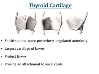
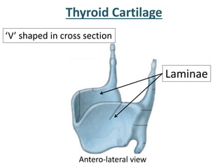
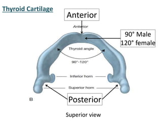
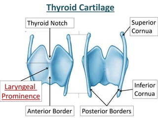
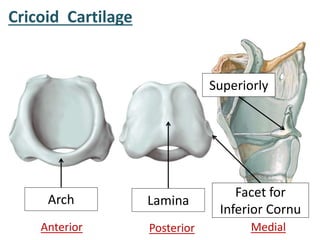
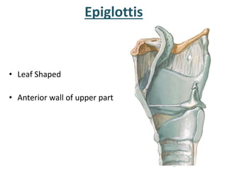
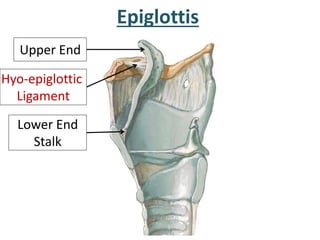
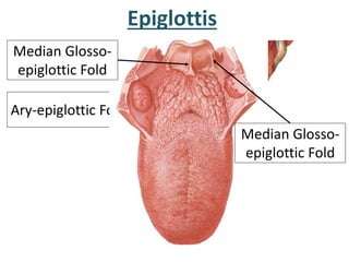
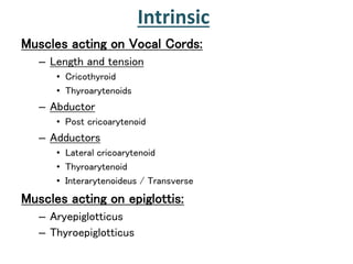
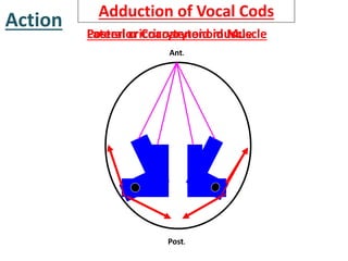
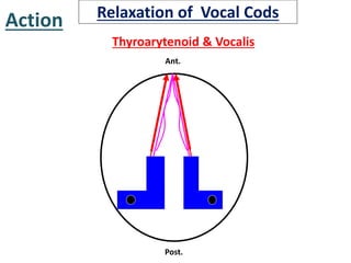
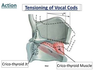
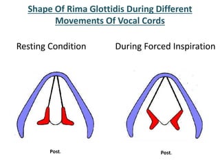
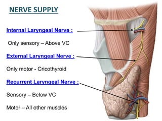
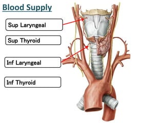
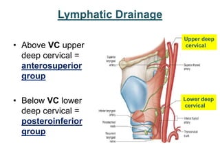
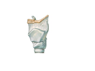
LARYNX
• Introduction
• Situation & Extent
• Size
• Skeletal framework
• Subdivison of larynx
• Muscles
• Movements of vocal cords
• Histology
• Blood supply
• Nerve supply
• Lymphatic drainage
• Applied anatomy
Objectives
Introduction
• Organ of phonation
• Part of air passage
– Regulates passage of air in
inspiration and expiration
• Act as Sphincter at Inlet of
Lower Respiratory Tract
• Upper Respiratory tract?
Situation & Extent
• Lies in anterior midline of neck
• From root of Tongue to Trachea
• From laryngeal inlet up to
lower border of cricoid cartilage
• Opp to C3 to C6 vertebrae
in men
• Slightly higher in female &
children
Measurements
In Males In Females
Length 44 mm. 36 mm.
Transverse diameter 43 mm. 41 mm.
A-P diameter 36 mm 26 mm.
Circumference 136 mm. 112 mm.
Until puberty there is little difference b/w male & female
larynx.
After puberty:- Male larynx undergoes considerable increase;
thyroid cartilage becomes prominent – Adam’s Apple
Constitution of larynx
• 9 Cartilages:
– Connected by
• Joints
• Ligaments
• Membranes
– Moved by
• 8 Muscles(Intrinsic)
• Cavity – Mucous membrane
Cartilages
3 Paired
• Arytenoid
• Corniculate
• Cuneiform
3 Unpaired
• Thyroid
• Cricoid
• Epiglottis
Cartilages
Thyroid
Cricoid
Epiglottis
ArytenoidCorniculate
Anterior Posterior
Cuniform
• Shield shaped, open posteriorly, angulated anteriorly
• Largest cartilage of larynx
• Protect larynx
• Provide an attachment to vocal cords
Thyroid Cartilage
Thyroid Cartilage
Antero-lateral view
‘V’ shaped in cross section
Laminae
Thyroid Cartilage
Superior view
90° Male
120° female
Posterior
Anterior
Thyroid Cartilage
Anterior Border
Laryngeal
Prominence
Thyroid Notch
Posterior Borders
Superior
Cornua
Inferior
Cornua
Thyroid Cartilage
Lateral Thyro-hyoid Ligament
Crico-thyroid Joint
Cartilago Triticia
Thyroid Cartilage
Conus
Elasticus
Inferior Border
Thyroid CartilageOuter Surface
Oblique Line
Superior Thyroid Tubercle
Inferior Thyroid Tubercle
Cricoid Cartilage
• Encircle larynx below thyroid
• Thicker & Stronger than Thyroid
Cricoid Cartilage
Anterior Posterior Medial
Arch Lamina
Superiorly
Facet for
Inferior Cornu
Epiglottis
• Leaf Shaped
• Anterior wall of upper part
Epiglottis
Upper End
Lower End
Stalk
Hyo-epiglottic
Ligament
Ary-epiglottic Fold
Epiglottis
Median Glosso-
epiglottic Fold
Median Glosso-
epiglottic Fold
Arytenoid Cartilage
• Pyramidal Shaped
• On upper border
cricoid cartilage
Apex
Base
Processes of Arytenoid Cartilage
Vocal Process
Muscular
Process
Corniculate And
Cuneiform Cartilage
Corniculate cartilage
of Santorini
Cunieform cartilage
of Wrisberg
Cartilage
Tritiate
Tritiate Cartilage
• Thyroid,
• Cricoid
• Base of Arytenoids
Histology of Laryngeal Cartilages:
• Hyaline Cartilage
• May ossify after 25 yrs
of age
• Epiglottis,
• Corniculate,
• Cuneiform
• Processes of Arytenoid
• Elastic
• They do not ossify
Joints:
Rotatory Movements
Around Vertical Axis
Crico-thyroid Joint Crico-arytenoid Joint
Rotatory Movements
Around Transverse Axis
Synoial
MEMBRANES & LIGAMENTS
• Extrinsic
• Thyrohyoid
• Cricotracheal
• Hyoepiglottic
• Intrinsic
• Quadrangular membrane &
vestibular ligament
• Crico-vocal membrane & vocal
ligament
EXTRINSIC Thyrohyoid Membrane
Superior Laryngeal
Vessels
Internal Laryngeal
Nerve
EXTRINSIC
Hyo-epiglottic
Ligament
Crico-Tracheal
Ligament
Intrinsic
• Part of Fibroelastic Membrane
• Lies just outside of mucous
membrane
Fibroelastic Membrane
• Divided by
Sinus of Larynx
Quadrate Membrane
Crico-vocal Membrane
Conus Elasticus
Quadrate Membrane
Arytenoid Cartilage
Epiglottis
Ary-epiglottic Ligament
(Fold)
Vestibular Ligament / Fold
(False Vocal Cord)
Crico-Vocal Membrane
Arch of Cricoid Cartilage
Cricothyroid Ligament
Vocal Ligament / Fold
(True Vocal Cord)
Vocal Ligament
Middle of Angle of
Thyroid Cartilage
Vocal Process of
Arytenoioid Cartilage
True Vocal Cord
Anterior
Posterior
Laryngeal Cavity
Coronal section Posterior aspect
From Inlet of Larynx
to
Lower border of
Cricoid Cartilage
Laryngeal Inlet
Above & In Front
Upper Margin of
Epiglottis
Below & Behind
Inter-arytenoid fold
of Mucous Memb
On Each Side
Ary-epiglottic Fold
Closure by apposition
of Ary-epiglottic Fold
Laryngeal Cavity
Vestibular Fold
Vocal Fold
Laryngeal Cavity
Rima Vestibuli
Rima Glottidis
Anterior
Posterior
Rima Glottidis
Anterior 3/5th
Inter-membraneous Part
Posterior 2/5th
Inter-cartilageous Part
Laryngeal Cavity
Supraglottic Part / Vestibule
Above Vestibule
Glottic Part / Sinus / Ventricle
Between Vestibular & Vocal Folds
Infraglottic Part
Below Vestibule
Sinus / Ventricle of Larynx
Vestibular Fold
Sinus Of Morgagni
Lamina of Thyroid Cartilage
Saccule Of Larynx
Mucous Glands
Lubrication of Vocal Cords
Extrinsic Muscles
Extrinsic attached to hyoid
(Depressors)
– Sternothyroid
– Sternohyoid
– Thyrohyoid
Extrinsic attached to thyroid
(Elevators )
– Palatopharyngeus
– Stylopharyngeus
Intrinsic
Muscles acting on Vocal Cords:
– Length and tension
• Cricothyroid
• Thyroarytenoids
– Abductor
• Post cricoarytenoid
– Adductors
• Lateral cricoarytenoid
• Thyroarytenoid
• Interarytenoideus / Transverse
Muscles acting on epiglottis:
– Aryepiglotticus
– Thyroepiglotticus
Have 2 fiber types
• Oblique
• Vertical
Action: lengthens and tenses VF
Cricothyroid
Antero-Lat
Action:
Abduction of Vocal Cord
Posterior Cricoarytenoid
Post.
Action:
Adduction of Vocal Cord
Lateral Cricoarytenoid
Lat, Thyroid lamina removed
Action:
Close Laryngeal Inlet
Oblique Arytenoid
Ary-epiglotticus
Post.
Action:
Adduction
Transverse Arytenoid
Only Unpaired
Post.
Action:
Relax Vocal Cords
ThyroArytenoid
Thyro-epiglotticus
Action:
Open Laryngeal Inlet
Post-lat, Thyroid lamina removed
Action:
Relax Vocal Cords
ThyroArytenoid
Vocalis
Post.
Action Abduction of Vocal CodsAdduction of Vocal Cods
Vocal Process
Muscular Process Post.
Action
Posterior Cricoarytenoid MuscleLateral cricoarytenoid muscle
Ant.
Post.
Abduction of Vocal CodsAdduction of Vocal Cods
Ant.
Post.
Action
Thyroarytenoid & Vocalis
Relaxation of Vocal Cods
Action Rotate Thyroid ForwardTensioning of Vocal Cods
Crico-thyroid Jt Med Crico-thyroid Muscle
Shape Of Rima Glottidis During Different
Movements Of Vocal Cords
Resting Condition During Forced Inspiration
Post. Post.
During Phonation During Whispering
Post. Post.
NERVE SUPPLY
Internal Laryngeal Nerve :
Only sensory – Above VC
External Laryngeal Nerve :
Only motor - Cricothyroid
Recurrent Laryngeal Nerve :
Sensory – Below VC
Motor – All other muscles
Blood Supply
Sup Laryngeal
Sup Thyroid
Inf Laryngeal
Inf Thyroid
Lymphatic Drainage
• Above VC upper
deep cervical =
anterosuperior
group
• Below VC lower
deep cervical =
posteroinferior
group
Upper deep
cervical
Lower deep
cervical
Applied Anatomy
 Requires laryngeal mirror and
head mirror
 Structures seen……………
Laryngoscopy-
Indirect Laryngoscopy
Done under GA
Before endotracheal
intubation
Direct Laryngoscopy
Singers / Screamers/ Clergymens
Nodules / Teacher’s Nodule
Reinke’s Edema
Classification Of Laryngeal Paralysis
• May be unilateral or
bilateral and may involve
1. Recurrent laryngeal nerve
2. Superior laryngeal nerve
a. External LN
b. Internal LN
RLN Paralysis
• Moderate trauma produces only abductor
paralysis.
• Severe injury or section of nerve produces
both abductor and adductor paralysis.
• Vocal cords lies in cadaveric position – Bet
Abducted & Adducted position
• Phonation is completely lost
• Semon's law: Progressive moderate lesion
– Abductor: first to paralyzed & last to
recover
Unilateral Abductor Paralysis
 Paralysis of recurrent laryngeal nerve.
 Vocal cord lies in median or paramedian position, doesn’t move
on deep inspiration.
 Initial hoarseness.
 No regurgitation.
 Vocal cord compensation occurs leading
to improvement of voice.
Laryngeal Obstruction
• Produce severe protective
cough reflex
• If Internal laryngeal nerve
is damaged
• Break reflex arc
• Allow entry of foreign
body
• Obstructiom
Laryngotomy
Inflammation Of Larynx
• Produce Hoarseness of Voice
Malignancy Of Larynx
• Supraglottic
• Glottic
• Subglottic
Larynx Anatomy
Larynx Anatomy

Larynx Anatomy