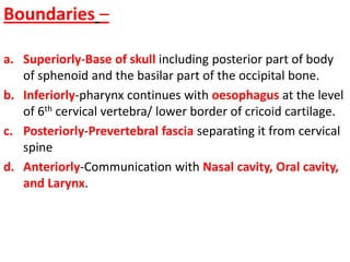
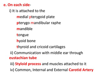
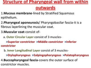
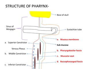
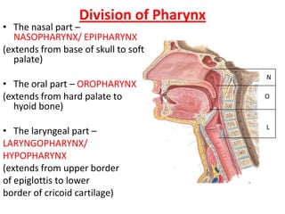
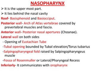
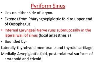
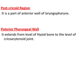
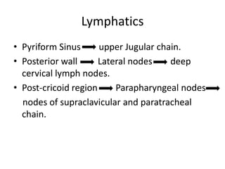
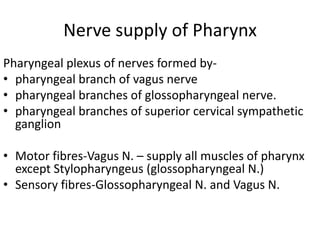
Anatomy Of Pharynx
Boundaries –
a. Superiorly-Base of skull including posterior part of body
of sphenoid and the basilar part of the occipital bone.
b. Inferiorly-pharynx continues with oesophagus at the level
of 6th cervical vertebra/ lower border of cricoid cartilage.
c. Posteriorly-Prevertebral fascia separating it from cervical
spine
d. Anteriorly-Communication with Nasal cavity, Oral cavity,
and Larynx.
e. On each side-
i) It is attached to the
medial pterygoid plate
pterygo mandibular raphe
mandible
tongue
hyoid bone
thyroid and cricoid cartilages
ii) Communication with middle ear through
eustachian tube
iii) Styloid process and muscles attached to it
iv) Common, Internal and External Carotid Artery
Structure of Pharyngeal wall from within
outwards
1.Mucous membrane-lined by Stratified Squamous
epithelium.
2.Pharyngeal aponeurosis/ Pharyngobasilar fascia-it is a
fibrous layerlining the muscular coat.
3.Muscular coat-consist of-
a. Outer Circular Layer consist of 3 muscles-
>Superior constrictor >Middle constrictor >Inferior
constrictor
b. Inner Longitudinal Layer consist of 3 muscles-
>Stylopharyngeus >Salpingopharyngeus >Palatopharyngeus
4.Buccopharyngeal fascia-covers the outer surface of
constrictor muscles.
STRUCTURE OF PHARYNX-
Base of skull
Eustachian tube
a. Mucous membrane
Sub mucosa
b. Pharyngobasilar fascia
c. Muscular coat
d. Buccopharyngeal fascia
a. Superior Constrictor
b. Middle Constrictor
c. Inferior Constrictor
Venous Plexus
Sinus of
Morgagni
Division of Pharynx
• The nasal part –
NASOPHARYNX/ EPIPHARYNX
(extends from base of skull to soft
palate)
• The oral part – OROPHARYNX
(extends from hard palate to
hyoid bone)
• The laryngeal part –
LARYNGOPHARYNX/
HYPOPHARYNX
(extends from upper border
of epiglottis to lower
border of cricoid cartilage)
N
O
L
NASOPHARYNX
 It is the upper most part.
 It lies behind the nasal cavity
Roof- Basisphenoid and Basiocciput.
Posterior wall- Arch of Atlas vertebrae covered by
prevertebral muscles and fascia.
Anterior wall- Posterior nasal apertures (Choanae).
Lateral wall on both sides
- Opening of Eustachian Tube
-Tubal opening bounded by Tubal elevation/Torus tubarius
-Salpingopharyngeal fold raised by Salpingopharyngeus
muscle
-Fossa of Rosenmuller or Lateral/Pharyngeal Recess
Inferiorly- It communicates with oropharynx
1. Pharyngeal/ Nasopharyngeal Tonsil-it is a
subepithelial collection of lymphoid tissue
opposite the basiocciput.
2. Pharyngeal Bursa-it is the small median recess in
the mucosa covering the tonsil.
3. Rathke’s pouch-it is reminiscent of buccal mucosal
invagination, to form anterior lobe of pituitary.
4. Tubal tonsil-collection of subepithelial lymphoid
tissue situated at the tubal elevation.
5. Sinus of Morgagni-space between skull and upper
free border of superior constrictor muscle.
Structures passing through this gap-
-Levator veli palatini
-Ascending palatine artery
-Tensor veli palatini
-Eustachian tube
6. Passavant’s Ridge-mucosal ridge raised by fibres of
Palatopharyngeus. It encircles posterior and lateral walls
of nasopharyngeal isthmus. Soft palate during its
contraction makes firm contact with this ridge to cut off
nasopharynx from oropharynx during deglutition or
speech
• Nasopharynx is lined by Pseudostratified
Ciliated Columnar epithelium.
• Lymphatic drainage-
Deep cervical nodes
Spinal accessory chain of nodes
Contralateral lymph nodes
OROPHARYNX
It is the middle part of the pharynx situated behind
the oral cavity.
Soft Palate
Palatoglossal arch
Palatine Tonsil
Posterior
Pharyngeal Wall
Uvula
Palatopharyngeal
Arch
Anterior Pillar or
Posterior Pillar or
Boundaries
• Above- communicates with nasopharynx through
nasopharyngeal isthmus.
• Below- opens into laryngopharynx.
• Anterior wall-
upper part- deficient, oropharynx communicates with oral
cavity through Oropharyngeal isthmus/ Isthmus of Fauces.
lower part- base of tongue
- lingual tonsils
- valleculae
• Posterior wall- Third cervical vertebrae.
• Lateral wall- Palatine/Faucial tonsil
- Anterior pillar (Palatoglossus muscle)
- Posterior pillar (Palatopharyngeus muscle)
Lymphatic drainage
Oropharynx- Into Upper Jugular chain
particularly Jugulodigastric (tonsillar) node.
Soft palate, Lateral and Posterior pharyngeal
walls and base of Tongue- into
retropharyngeal and parapharyngeal nodes.
In relation to Oropharyngeal Isthmus there are several
aggregations of Lymphoid Tissue that constitute
WALDEYER’S LYMPHATIC RING.
Retropharyngeal
nodes Nasopharyngeal Tonsil
Jugulodigastric Tubal Tonsil
Node
Jugular chain of Palatine Tonsil
Node
Lingual Tonsil
Submandibular nodes Submental nodes
Laryngopharynx
Lower part of pharynx situated behind and
partly on sides of the larynx.
Hypopharynx lies opposite to 3rd, 4th, 5th, 6th
cervical vertebrae.
Clinically it is divided into 3 regions-
1. Pyriform Sinus (fossa)
2. Post-cricoid region
3. Posterior Pharyngeal wall
Pyriform Sinus
• Lies on either side of larynx.
• Extends from Pharyngoepiglottic fold to upper end
of Oesophagus.
• Internal Laryngeal Nerve runs submucosally in the
lateral wall of sinus (local anaesthesia)
• Bounded by-
Laterally-thyrohyoid membrane and thyroid cartilage
Medially-Aryepiglottic fold, posterolateral surfaces of
arytenoid and cricoid.
Post-cricoid Region
It is a part of anterior wall of laryngopharynx.
Poterior Pharyngeal Wall
It extends from level of Hyoid bone to the level of
cricoarytenoid joint.
Lymphatics
• Pyriform Sinus upper Jugular chain.
• Posterior wall Lateral nodes deep
cervical lymph nodes.
• Post-cricoid region Parapharyngeal nodes
nodes of supraclavicular and paratracheal
chain.
Nerve supply of Pharynx
Pharyngeal plexus of nerves formed by-
• pharyngeal branch of vagus nerve
• pharyngeal branches of glossopharyngeal nerve.
• pharyngeal branches of superior cervical sympathetic
ganglion
• Motor fibres-Vagus N. – supply all muscles of pharynx
except Stylopharyngeus (glossopharyngeal N.)
• Sensory fibres-Glossopharyngeal N. and Vagus N.
Killian’s Dehiscence
Inferior constrictor muscle has 2 parts:
1. Thyropharyngeus: has oblique fibres
2. Cricopharyngeus: has transverse fibres
Potential gap between these 2 parts is KILLIAN’S
DEHISCENCE or Gateway of Tears.
This is the site for herniation of pharyngeal
mucosa in cases of pharyngeal pouch.
Pharyngeal Spaces
Potential spaces in relation to pharynx where
abscess can form-
1. Retropharyngeal space- lies behind pharynx,
extend from base of skull to bifurcation of
trachea.
2. Parapharyngeal space- present onon side of
pharynx,
contains (a)Carotid vessels, (b)Jugular vein,
(c)last 4 Cranial Nerves (d)Sympathetic chain
THANK YOU

Anatomyofpharynx 120310220054-phpapp02

  • 1.
  • 2.
    Boundaries – a. Superiorly-Baseof skull including posterior part of body of sphenoid and the basilar part of the occipital bone. b. Inferiorly-pharynx continues with oesophagus at the level of 6th cervical vertebra/ lower border of cricoid cartilage. c. Posteriorly-Prevertebral fascia separating it from cervical spine d. Anteriorly-Communication with Nasal cavity, Oral cavity, and Larynx.
  • 3.
    e. On eachside- i) It is attached to the medial pterygoid plate pterygo mandibular raphe mandible tongue hyoid bone thyroid and cricoid cartilages ii) Communication with middle ear through eustachian tube iii) Styloid process and muscles attached to it iv) Common, Internal and External Carotid Artery
  • 4.
    Structure of Pharyngealwall from within outwards 1.Mucous membrane-lined by Stratified Squamous epithelium. 2.Pharyngeal aponeurosis/ Pharyngobasilar fascia-it is a fibrous layerlining the muscular coat. 3.Muscular coat-consist of- a. Outer Circular Layer consist of 3 muscles- >Superior constrictor >Middle constrictor >Inferior constrictor b. Inner Longitudinal Layer consist of 3 muscles- >Stylopharyngeus >Salpingopharyngeus >Palatopharyngeus 4.Buccopharyngeal fascia-covers the outer surface of constrictor muscles.
  • 5.
    STRUCTURE OF PHARYNX- Baseof skull Eustachian tube a. Mucous membrane Sub mucosa b. Pharyngobasilar fascia c. Muscular coat d. Buccopharyngeal fascia a. Superior Constrictor b. Middle Constrictor c. Inferior Constrictor Venous Plexus Sinus of Morgagni
  • 6.
    Division of Pharynx •The nasal part – NASOPHARYNX/ EPIPHARYNX (extends from base of skull to soft palate) • The oral part – OROPHARYNX (extends from hard palate to hyoid bone) • The laryngeal part – LARYNGOPHARYNX/ HYPOPHARYNX (extends from upper border of epiglottis to lower border of cricoid cartilage) N O L
  • 7.
    NASOPHARYNX  It isthe upper most part.  It lies behind the nasal cavity Roof- Basisphenoid and Basiocciput. Posterior wall- Arch of Atlas vertebrae covered by prevertebral muscles and fascia. Anterior wall- Posterior nasal apertures (Choanae). Lateral wall on both sides - Opening of Eustachian Tube -Tubal opening bounded by Tubal elevation/Torus tubarius -Salpingopharyngeal fold raised by Salpingopharyngeus muscle -Fossa of Rosenmuller or Lateral/Pharyngeal Recess Inferiorly- It communicates with oropharynx
  • 8.
    1. Pharyngeal/ NasopharyngealTonsil-it is a subepithelial collection of lymphoid tissue opposite the basiocciput. 2. Pharyngeal Bursa-it is the small median recess in the mucosa covering the tonsil. 3. Rathke’s pouch-it is reminiscent of buccal mucosal invagination, to form anterior lobe of pituitary. 4. Tubal tonsil-collection of subepithelial lymphoid tissue situated at the tubal elevation.
  • 9.
    5. Sinus ofMorgagni-space between skull and upper free border of superior constrictor muscle. Structures passing through this gap- -Levator veli palatini -Ascending palatine artery -Tensor veli palatini -Eustachian tube 6. Passavant’s Ridge-mucosal ridge raised by fibres of Palatopharyngeus. It encircles posterior and lateral walls of nasopharyngeal isthmus. Soft palate during its contraction makes firm contact with this ridge to cut off nasopharynx from oropharynx during deglutition or speech
  • 10.
    • Nasopharynx islined by Pseudostratified Ciliated Columnar epithelium. • Lymphatic drainage- Deep cervical nodes Spinal accessory chain of nodes Contralateral lymph nodes
  • 11.
    OROPHARYNX It is themiddle part of the pharynx situated behind the oral cavity. Soft Palate Palatoglossal arch Palatine Tonsil Posterior Pharyngeal Wall Uvula Palatopharyngeal Arch Anterior Pillar or Posterior Pillar or
  • 12.
    Boundaries • Above- communicateswith nasopharynx through nasopharyngeal isthmus. • Below- opens into laryngopharynx. • Anterior wall- upper part- deficient, oropharynx communicates with oral cavity through Oropharyngeal isthmus/ Isthmus of Fauces. lower part- base of tongue - lingual tonsils - valleculae • Posterior wall- Third cervical vertebrae. • Lateral wall- Palatine/Faucial tonsil - Anterior pillar (Palatoglossus muscle) - Posterior pillar (Palatopharyngeus muscle)
  • 13.
    Lymphatic drainage Oropharynx- IntoUpper Jugular chain particularly Jugulodigastric (tonsillar) node. Soft palate, Lateral and Posterior pharyngeal walls and base of Tongue- into retropharyngeal and parapharyngeal nodes.
  • 14.
    In relation toOropharyngeal Isthmus there are several aggregations of Lymphoid Tissue that constitute WALDEYER’S LYMPHATIC RING. Retropharyngeal nodes Nasopharyngeal Tonsil Jugulodigastric Tubal Tonsil Node Jugular chain of Palatine Tonsil Node Lingual Tonsil Submandibular nodes Submental nodes
  • 15.
    Laryngopharynx Lower part ofpharynx situated behind and partly on sides of the larynx. Hypopharynx lies opposite to 3rd, 4th, 5th, 6th cervical vertebrae. Clinically it is divided into 3 regions- 1. Pyriform Sinus (fossa) 2. Post-cricoid region 3. Posterior Pharyngeal wall
  • 16.
    Pyriform Sinus • Lieson either side of larynx. • Extends from Pharyngoepiglottic fold to upper end of Oesophagus. • Internal Laryngeal Nerve runs submucosally in the lateral wall of sinus (local anaesthesia) • Bounded by- Laterally-thyrohyoid membrane and thyroid cartilage Medially-Aryepiglottic fold, posterolateral surfaces of arytenoid and cricoid.
  • 17.
    Post-cricoid Region It isa part of anterior wall of laryngopharynx. Poterior Pharyngeal Wall It extends from level of Hyoid bone to the level of cricoarytenoid joint.
  • 18.
    Lymphatics • Pyriform Sinusupper Jugular chain. • Posterior wall Lateral nodes deep cervical lymph nodes. • Post-cricoid region Parapharyngeal nodes nodes of supraclavicular and paratracheal chain.
  • 19.
    Nerve supply ofPharynx Pharyngeal plexus of nerves formed by- • pharyngeal branch of vagus nerve • pharyngeal branches of glossopharyngeal nerve. • pharyngeal branches of superior cervical sympathetic ganglion • Motor fibres-Vagus N. – supply all muscles of pharynx except Stylopharyngeus (glossopharyngeal N.) • Sensory fibres-Glossopharyngeal N. and Vagus N.
  • 20.
    Killian’s Dehiscence Inferior constrictormuscle has 2 parts: 1. Thyropharyngeus: has oblique fibres 2. Cricopharyngeus: has transverse fibres Potential gap between these 2 parts is KILLIAN’S DEHISCENCE or Gateway of Tears. This is the site for herniation of pharyngeal mucosa in cases of pharyngeal pouch.
  • 21.
    Pharyngeal Spaces Potential spacesin relation to pharynx where abscess can form- 1. Retropharyngeal space- lies behind pharynx, extend from base of skull to bifurcation of trachea. 2. Parapharyngeal space- present onon side of pharynx, contains (a)Carotid vessels, (b)Jugular vein, (c)last 4 Cranial Nerves (d)Sympathetic chain
  • 22.