
Recommended
More Related Content
What's hot
What's hot (20)
Similar to Anatomy and Visual field defects of optic nerve and chiasma
Similar to Anatomy and Visual field defects of optic nerve and chiasma (20)
Recently uploaded
Recently uploaded (20)
Anatomy and Visual field defects of optic nerve and chiasma
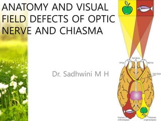
- 1. ANATOMY AND VISUAL FIELD DEFECTS OF OPTIC NERVE AND CHIASMA Dr. Sadhwini M H
- 2. • VISUAL PATHWAY – refers to neural network that extends from retina to visual cortex. • VISUAL FIELD - Each eye sees a part of the visual space that defines its visual field. The visual fields of both eyes overlap extensively to create a binocular visual field. The total visual field is the sum of the right and left hemifields and consists of a binocular zone and two monocular zones.
- 3. PARTS OF VISUAL PATHWAY
- 4. EMBRYOLOGY •Neural plate destined to form prosencephalon depresses to form optic sulcus •Optic sulcus deepens and prosencephalon bulge out to form optic vesicle •Proximal part of optic vesicle becomes constricted and enlongated to form optic stalk •Optic stalk – optic nerve •Optic vesicle – eyeball •Surface ectoderm destined to form lens gets invaginated into optic cup •A notch develops on ventral side of optic stalk and cup – fetal fissure – permits entry of blood vessels
- 5. Week of Gestation Developing events 4 Short optic stalk 5 Development of hyaloid vasculature and primitive retina 7 RGCs differentiate, optic nerve fibres begin to fill optic stalk and these af ferent neaurons reach the chiasm 8 optic nerve vascularization starting to form, optic nerve head starting to form 11 Vascular-connective septa invade the nerve 12 Pia mater, arachnoid, and dura mater distinguishable, glial filaments appear 14 Subarachnoid spaces appears 15 Physiological cup starts to form 18 Vascularization of the optic nerve completed 23 Myelinization starts
- 6. OPTIC NERVE • 2nd cranial nerve. • 47-50 mm in length. • Starts from optic disc & extends upto optic chias ma • Consists of axons originating from ganglion Cells t hat pass out of lamina cribrosa • Contains the afferent fibres of light reflex • Fibres of optic nerve are very thin(2-10um in diam eter)& are million in number • Has 4 parts : intraocular (1mm) intraorbital (30mm) intracanalicular (6-9mm) intracranial (10mm)
- 7. INTRAOCULAR PART • About 1mm in size, 1.5mm in diameter, passe s through sclera, choroid & finally appears in eye as optic disc. • Divided into 4 parts – Surface nerve fibre layer – Prelaminar region – Lamina cribrosa – Retrolaminar region
- 8. •SURFACE NERVE FIBRE LAYER •Composed of axonal bundles from retina which converge on optic disc •Disc is covered by a thin layer of astrocytes, ILM which is continuous •PRELAMINAR REGION – just distal to optic nerve head •Has neurons and significant increased quantity of astroglial tissues •Separated from choroid by Border tissue of Jacobie •LAMINA CRIBROSA •Fibrillar sieve like structure made of fenestrated sheets of connective tissue lined by glial tissue •Nerve fibres leave the eye through these fenestrations •Separated from choroid by Border tissue of Elschnig •RETROLAMINAR REGION •Axons here acquire myelin sheath, supplied by oligodendrocytes and doubles the nerve diametr to 3mm •Optic nerve is analogous to white matter tracts of brain. Hence susceptible to diseases of CNS tracts like MS
- 9. INTRAORBITAL PART • Extends from back of eyeball to optic foramina. • Slightly sinuous & allows for eyemovements. • Surrounded by all 3 layers of meninges & subarachnoi d space • The CRA & CRV enter the subarachnoid space to enter the nerve on its inferomedial aspect, about 10mm fro m eyeball. • Near optic foramina, it is closely surronded by annulus of zinn & the origin of four recti muscles. Some fibres of superior & medial rectus are adherent to its sheath – painful ocular movementa in retrobulbar neuritis
- 10. •Between the nerve & lateral rectus – ciliary ganglion, division of occulomotor nerve, nasociliary nerve, sympathetic nerve, abducent nerve. •This close proximity of the structures around the nerve accounts for the symptom complex of orbital apex syndrome - proptosis, painful ophthalmoplegia (3rd, 4th, 6th nerve palsy) and loss of vision. •Ophthalmic artery, superior ophthalmic vein and the nasociliary nerve cross the optic nerve superiorly from the lateral to the medial side.
- 11. INTRACANALICULAR PART •5-7mm •Limited space for expanion •Most frequently affected in traumatic optic neuropathies •Ophthalmic Artery crosses the nerve inferiorly from medial to lateral side in dural sheath. Sphenoid & posterior ethmoidal sinuses lie medial to it & seperated by thin bony lamina, this relation accounts for retrobulbar neuritis following infection of sinusitis
- 12. INTRACRANIAL PART • About 10mm • Lies above cavernous sinus & convergeswith its fellow to fo rm chiasma. • Ensheathed in pia mater. • Internal carotid artery runs b elow then lateral to it & give s off ophthalmic artery below it. • Superiorly, related to anterior perforated substance, medial root of olfactory tract, anteri or cerebral artery
- 13. OPTIC CHIASMA • Dorsoventrally flattened structure • Results from cross over of 2 opti c nerves • Lies in suprasellar subarachnoid cistern • Ensheathed by pia & surrounded by CSF • Anteroposterior – 8mm • Horixontal – 15mm • Height – 4mm
- 14. •RELATIONS OF CHIASMA •Anterior - anterior cerebral arteries & its communicating arteries. •Posterior- tuber cinereum, infundibulum ,pitutary body ,posterior perforated substance. •Superior- third ventricle. •Inferior- hypophysis •Lateral- extra cavernous part of internal carotid artery& anterior perforated substance.
- 15. Anatomical variations in position of chiasma CENTRAL : lies directly over sella, expanding pituitary tumor involves chiasma first PREFIXED : lies more anteriorly over tuberculum sellae, pituitary tumor involves optic tract first POSTFIXED : lies more posterior over Dorsum sellae,pituitary tumor damage optic nerve first
- 16. ARRANGEMENT OF FIBRES IN RETINA SAF IAF PMB SRF IRF
- 17. ARRANGEMENT OF FIBRES IN OPTIC NERVE HEAD •Peripheral fibers - deep in Retina - superficially in optic nerve •Fibers close to optic nerve head – superficial in retina - central in optic nerve
- 18. ARRANGEMENT OF FIBRES IN DISTAL REGION OF OPTIC NERVE MACULA SRF SAF IRF IAF
- 19. ARRANGEMENT OF FIBRES IN PROXIMAL REGION OF OPTIC NERVE
- 20. ARRANGEMENT OF FIBRES IN OPTIC CHIASMA
- 21. Blood supply of Optic Nerve • Peripapillary choroidal vesselsPrelaminar • Posterior choroidal vessels Lamina cribrosa • Centrifugal branches from central retinal artery • Centripetal branches from pial vessels Retrolaminar INTRAOCULAR PART
- 22. INTRAORBITAL PART • Derived from 6 branches of ICA : ophthalmic, long & short posterior ciliary artery. • Lacrimal artery. • Central artery of retina. PERIAXIAL SYSTEM OF VESSELS • Intraneural b/o central retinal artery. • Central collateral b/o central retinal artery. • Central artery of optic nerve AXIAL SYSTEM OF VESSELS
- 23. •Intracanalicular part : periaxial system of vessels. •Intracranial part : Pial system of vessels
- 24. VENOUS DRAINAGE • Central retinal veinONH • Peripheral pial plexus • Central retinal vein ORBITAL PART • Pial plexus which ends in anterior cerebral & basal vein INTRACRANI AL PART
- 25. BLOOD SUPPLY OF OPTIC CHIASMA • B/o anterior cerebral & anterior communicating arterySUPERIOR ASPECT • B/o internal carotid artery, posterior communicating artery ,anterior superior hypophyseal artery INFERIOR ASPECT
- 26. VENOUS DRAINAGE OF CHIASMA • Superior chiasmal vein drains into anterior cerebral vein SUPERIOR ASPECT • Pre-infundibular vein draining into basilar vein INFERIOR ASPECT
- 28. LESIONS OF OPTIC NERVE • Characterised by : complete blindness in affected eye wit h loss of both direct on i/l & concensual light reflex on c/l side. • Causes : optic atrophy, indir ect optic neuropathy, acute optic neuritis, traumatic avul sion of optic nerve.
- 29. ARCUATE CENTRAL
- 30. ALTITUDINAL
- 32. •U/L or Binasal Hemianopias : •No connection with blind spot •Respects vertical midline •Caused by damage to lateral aspect of optic nerve fibres •Pituitary adenoma compressing lateral aspect of nerve •Intracranial tumors between optic nerve pushing them laterally •Primary hydrocephalus
- 33. CHIASMAL LESIONS • Unilateral or Bilateral. • Junctional scotoma. • Bitemporal defect. • Homonymous defects
- 34. CAUSES OF CHIASMAL SYNDROME • Pituitary adenoma • Suprasellar meningiomas • Supraclinoid internal carotid artery aneurysms • Craniopharyngiomas • Optic nerve gliomas • Uncommon : Optic nerve or chiasmal neuritis, Pachymeningitis , Trauma ,Inflammatory (e.g.,sa rcoidosis)
- 35. LESIONS OF PROXIMAL PART OF 1 OPTIC N ERVE AND ANTERIOR ANGLE OF CHIASMA • Lesion at the angle – Traquair’s junctiona l scotoma – i/l monoocular visual loss & c/l defect in ST area
- 36. •Small lesion damaging only crossing fibres of homolateral eye – monoocular temporal field defect respecting midline. •Only macular crossed fibres of 1 eye damaged – monoocular temporal field defect, but scotomatous and located temporally •Only Wilbrands knee involved – c/l superior temporal field defect
- 37. LESIONS OF BODY OF CHIASM • BITEMPORAL field defct – quandrantic/hemian opic • If lesion compresses chiasm from below – typic al bitemporal fiel defect
- 38. •Pituitary adenoma – field defects are typical •Peripheral fibres are initially affected, usually commence in outer upper quadrants of both eyes •Right eye – clockwise direction •Left eye – anticlockwise direction •Defects may be unequal in both eyes – so one eye may be almost blind
- 39. LESIONS OF POSTERIOR ANGLE OF CHIASM • 90% of chiasmal fibers have macular orig in (superior and posterior portions of chi asm) – bitemporal hemianopic scotomas •Posterior lesions may also involve the optic tract and cause a contralateral homonymous hemianopia
- 40. LESIONS OF LATERAL ASPECT OF CHIASM • Binasal hemianopia - rarely • Distension of 3rd ventricle causing pressure on each si de of optic chiasma • Atheroma of carotids & posterior communicating arter y. • Various tumors, pressure from supraclinoid portion of I CA – damage both uncrossed temporal fibres of i/l eye and crossed nasal fibres of c/l eye – c/l homonymous hemianopic defect
- 41. •If lesion extends from optic nerve or optic tract to chiasm – blind eye is always on the side of the lesion •If chiasmal lesion extends to optic nerve or tract – blind eye is always on the side of extension
- 42. BAND ATROPHY
- 43. REFERENCES • Walsh and Hoyt’s clinical neuroophthalm ology – 5th edition • A.k.Khurana Anatomy and physiology of the eye- 3rd edition • Yanoff & Duker ophthalmology – 4th editi on • Postgraduate ophthalmology – zia chaud huri • Wolff’s anatomy of eye -8th edition