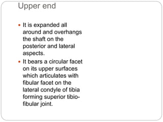
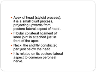
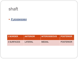
SM BADAR HAYAT
TUTOR
DEPARTMENT OF ANATOMY
NMCH, JAMUHAR
FIBULA & PATELLA
CONTENTS
 Introduction
 Anatomical position
 Side determination
 Parts
 Attachments
 Ossification
 Clinical anatomy
Introduction
 It is the lateral bone of
leg and is long but very
slender.
 Its homologous with
the ulna.
 It takes no part in
transmission of body
weight.
 In latin, the term fibula
means “pin”.
Anatomical position
 Bone is vertically placed.
Side determination
 Head lies above.
 Lower end is expanded
antero-posteriorly but flat
from side to side.
 Medial surface of the lower
end has a triangular facet at
its anterior part while the
malleolar fossa lies below
and behind the facet.
Parts
I. Upper end (head)
II. Shaft
III. Lower end (lateral malleolus)
Upper end
 It is expanded all
around and overhangs
the shaft on the
posterior and lateral
aspects.
 It bears a circular facet
on its upper surfaces
which articulates with
fibular facet on the
lateral condyle of tibia
forming superior tibio-
fibular joint.
 Apex of head (styloid process):
it is a small blunt process,
projecting upwards from
postero-lateral aspect of head .
 Fibular collateral ligament of
knee joint is attached just in
front of the apex
 Neck: the slightly constricted
part just below the head
 It is related on its postero-lateral
aspect to common peroneal
nerve.
shaft
 It possesses
3 BORDER ANTERIOR INTEROSSEOUS POSTERIOR
3 SURFACES LATERAL MEDIAL POSTERIOR
Borders
1. Anterior :
 Extends from anterior
aspect of the upper end to
the apex of an elongated
triangular area which is
continuous below with the
lateral surface of lateral
malleolus.
 Below, it divides into two
lines between which is the
triangular area.
 To its upper 3/4th is attaced
anterior intermuscular
septum of the leg.
Interosseous
 Lies on medial side of
anterior border; in
upper 2/3rd the two
borders lies very close
to each other.
 It divides into two limbs
at the lower fourth to
enclose a rough
triangular area.
Posterior
 Begins above from lower part
of the posterior aspect of the
head and below it is
continuous with medial
border of the groove on the
back of lateral malleolus.
 Its upper part is rounded and
not easily recognisable.
 Gives attachment to posterior
intermuscular septum of leg
in its upper 3/4th part.
Surfaces
1. Lateral (peroneal) :
 Lies between anterior
and posterior borders.
 Its lower fourth becomes
continuous with the
groove on the back of
lateral malleolus.
Medial (extensor)
 Lies between anterior
and interosseous
borders.
 It is very narrow in its
upper part but is wider
below.
Posterior
 Lies between interosseous and posterior
borders and is the widest of all surfaces.
 Its faces backwards and medially.
 Medial crest- It is prominent vertical ridge
dividing upper 2/3rd of this surface into
two areas.
 It gives attachment to deep intermuscular
septum.
 Lower part of this surface faces medially
and fits into the fibular notch of tibia.
 It gives attachment to interosseous tibio-
fibular ligament.
 A nutrient foramen lies near the middle of
this surface between the medial crest and
interosseous border.
 It is directed downwards, therefore upper
end of the bone is the growing end.
 The nutrient artery is a branch of peroneal
artery.
 Anterior & posterior borders of the rough
triangular area give attachment to anterior
& posterior tibiofibular ligaments.
Lower end (lateral malleolus)
 It projects downwards to a lower level
than the medial malleolus and
possesses:
1. Lateral surface : It is subcutaneous,
smooth and convex.
 It forms a bony projection on lateral
aspect of ankle joint.
2. Posterior surface: It has a broad
groove which lodges tendons of
peroneus longus & brevis.
 Lateral border of the groove gives
attachment to superior peroneal
retinaculum.
3. Anterior surface: It is continuous below with the
inferior border and gives attachment to anterior talo-
fibular ligament.
4. Medial surface: It has a triangular facet in front, with its
apex downwards, which articulates with lateral surface
of talus in forming ankle joint.
 Malleolar fossa: It is a rough depressed area behind
the facet. It bears many vascular foramina.
 Upper part of the malleolar fossa gives attachment to
posterior tibio-fibular ligament.
5. Inferior border: It has a small notch in front which
gives attachment to calcaneo-fibular ligament.
Fibula & patella

Fibula & patella

  • 1.
    SM BADAR HAYAT TUTOR DEPARTMENTOF ANATOMY NMCH, JAMUHAR FIBULA & PATELLA
  • 2.
    CONTENTS  Introduction  Anatomicalposition  Side determination  Parts  Attachments  Ossification  Clinical anatomy
  • 3.
    Introduction  It isthe lateral bone of leg and is long but very slender.  Its homologous with the ulna.  It takes no part in transmission of body weight.  In latin, the term fibula means “pin”.
  • 4.
    Anatomical position  Boneis vertically placed.
  • 5.
    Side determination  Headlies above.  Lower end is expanded antero-posteriorly but flat from side to side.  Medial surface of the lower end has a triangular facet at its anterior part while the malleolar fossa lies below and behind the facet.
  • 6.
    Parts I. Upper end(head) II. Shaft III. Lower end (lateral malleolus)
  • 7.
    Upper end  Itis expanded all around and overhangs the shaft on the posterior and lateral aspects.  It bears a circular facet on its upper surfaces which articulates with fibular facet on the lateral condyle of tibia forming superior tibio- fibular joint.
  • 8.
     Apex ofhead (styloid process): it is a small blunt process, projecting upwards from postero-lateral aspect of head .  Fibular collateral ligament of knee joint is attached just in front of the apex  Neck: the slightly constricted part just below the head  It is related on its postero-lateral aspect to common peroneal nerve.
  • 9.
    shaft  It possesses 3BORDER ANTERIOR INTEROSSEOUS POSTERIOR 3 SURFACES LATERAL MEDIAL POSTERIOR
  • 10.
    Borders 1. Anterior : Extends from anterior aspect of the upper end to the apex of an elongated triangular area which is continuous below with the lateral surface of lateral malleolus.  Below, it divides into two lines between which is the triangular area.  To its upper 3/4th is attaced anterior intermuscular septum of the leg.
  • 11.
    Interosseous  Lies onmedial side of anterior border; in upper 2/3rd the two borders lies very close to each other.  It divides into two limbs at the lower fourth to enclose a rough triangular area.
  • 12.
    Posterior  Begins abovefrom lower part of the posterior aspect of the head and below it is continuous with medial border of the groove on the back of lateral malleolus.  Its upper part is rounded and not easily recognisable.  Gives attachment to posterior intermuscular septum of leg in its upper 3/4th part.
  • 13.
    Surfaces 1. Lateral (peroneal):  Lies between anterior and posterior borders.  Its lower fourth becomes continuous with the groove on the back of lateral malleolus.
  • 14.
    Medial (extensor)  Liesbetween anterior and interosseous borders.  It is very narrow in its upper part but is wider below.
  • 15.
    Posterior  Lies betweeninterosseous and posterior borders and is the widest of all surfaces.  Its faces backwards and medially.  Medial crest- It is prominent vertical ridge dividing upper 2/3rd of this surface into two areas.  It gives attachment to deep intermuscular septum.  Lower part of this surface faces medially and fits into the fibular notch of tibia.  It gives attachment to interosseous tibio- fibular ligament.
  • 16.
     A nutrientforamen lies near the middle of this surface between the medial crest and interosseous border.  It is directed downwards, therefore upper end of the bone is the growing end.  The nutrient artery is a branch of peroneal artery.  Anterior & posterior borders of the rough triangular area give attachment to anterior & posterior tibiofibular ligaments.
  • 17.
    Lower end (lateralmalleolus)  It projects downwards to a lower level than the medial malleolus and possesses: 1. Lateral surface : It is subcutaneous, smooth and convex.  It forms a bony projection on lateral aspect of ankle joint. 2. Posterior surface: It has a broad groove which lodges tendons of peroneus longus & brevis.  Lateral border of the groove gives attachment to superior peroneal retinaculum.
  • 18.
    3. Anterior surface:It is continuous below with the inferior border and gives attachment to anterior talo- fibular ligament. 4. Medial surface: It has a triangular facet in front, with its apex downwards, which articulates with lateral surface of talus in forming ankle joint.  Malleolar fossa: It is a rough depressed area behind the facet. It bears many vascular foramina.  Upper part of the malleolar fossa gives attachment to posterior tibio-fibular ligament. 5. Inferior border: It has a small notch in front which gives attachment to calcaneo-fibular ligament.