SlideShare a Scribd company logo
1 of 10
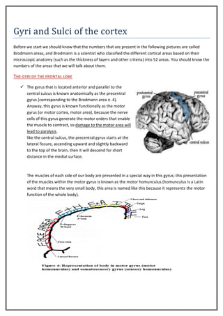
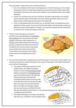
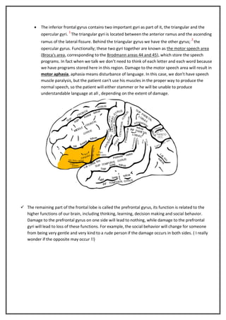
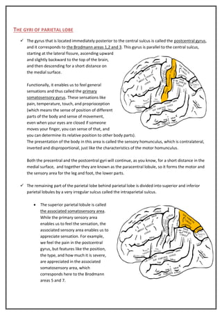
Download to read offline
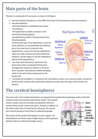
20
Cerebral Cortex (1)

Saleh Bani Hani

3/2/2013

Moath Azmi & Ahmed Hdaib

2

2
Main parts of the brain
The brain is composed of 4 main parts, as shown in the figure;
 the two cerebral hemispheres, In fact 90% of the brain if formed by the cerebral hemispheres.
 two diencephalons:
Each diencephalon is subdivided into 4 parts;
the thalamus,
the hypothalamus (which connects to and
controls the pituitary gland),
the epithalamus (which is formed mainly from
the pineal gland),
and the fourth part is the subthalamus, and as its
name indicates, it is located below the thalamus
but at the same time it is lateral to the
hypothalamus, so we can't see the subthalamus
unless we make a transverse section below the
thalamus, and we expect to see the subthalamus
lateral to the hypothalamus.
 one brain stem (the picture represents half
brain stem), that is composed of three parts
arranged from superior to inferior as the brain
stem, the pons, and lastly the medulla oblongata
which in turn will continue downward as the
spinal cord.
 and finally the cerebellum, it represents the main balance center in our nervous system, the part of
the brain that maintains the coordination between muscles and in this way it will maintain our
balance.

The cerebral hemispheres
The nerve cells in the cerebral hemisphere are organized into superficially located gray matter called the
cerebral cortex and centrally located white matter. The cerebral
cortex is made mainly of cell bodies and dendrites while the
cerebral white matter contains the axons. Anyway, in addition to
the white matter that is formed by the cell axons, the central
part contains groups of nuclei that are called basal ganglia.
The surface of the cerebral hemispheres is very irregular, the
elevated parts are known as gyri (single; Gyrus), and the grooves
between them are called sulci (single; sulcus).
LANDMARKS
In each side we divide the cerebral hemispheres into 4 lobes; frontal lobe, parietal lobe, temporal lobe, and
occipital lobe, in the figure below we can see a lateral aspect to the left and a medial aspect to the right.
Of course, we use landmarks to divide these cerebral hemispheres on the lateral surface into these lobes,
two of these landmarks are true, while the other two are imaginary lines:
 The two true lines are:
1- The lateral fissure : located on the lateral surface as seen in the figure, it has three branches; the
anterior ramus which is directed
forward, the ascending ramus which
ascends upward, in addition to the
posterior ramus that is directed
backward
2- the central sulcus : that is located in
the lateral surface of the brain, in the
center, that is separating the frontal
lobe from the parietal lobe. It
ascends upward and slightly
backward from the lateral fissure to
the superior margin of the brain and
usually continues for a short distance
on the medial surface as you can see.

 The two imaginary lines are:
1- the first imaginary line : passes
upward vertically from the
preoccipital notch, which is a small notch or groove that is located anterior to the occipital lobe, to
the superior margin of the brain.
2- the second imaginary line : that extends from the middle of the first one to the lateral fissure.
The first imaginary line is parallel to a sulcus that is located on the medial surface, this sulcus is called
parieto-occipital sulcus, which –as the name indicates- is located between the parietal lobe and the
occipital lobe.

THE LOBES :
you should know the location of each lobe as the following:
The frontal lobe is located anterior to the central sulcus and above the lateral fissure.
The parietal lobe, is located posterior to the central sulcus, anterior to the first imaginary line , and above
the posterior ramus of the lateral lobe and second imaginary line.
The occipital lobe is located posterior to the first imaginary line.
The temporal lobe is located below the second imaginary line and the lateral fissure, anterior to the first
imaginary line.
Gyri and Sulci of the cortex
Before we start we should know that the numbers that are present in the following pictures are called
Brodmann areas, and Brodmann is a scientist who classified the different cortical areas based on their
microscopic anatomy (such as the thickness of layers and other criteria) into 52 areas. You should know the
numbers of the areas that we will talk about them.

THE GYRI OF THE FRONTAL LOBE
 The gyrus that is located anterior and parallel to the
central sulcus is known anatomically as the precentral
gyrus (corresponding to the Brodmann area n. 4).
Anyway, this gyrus is known functionally as the motor
gyrus (or motor cortex, motor area), because the nerve
cells of this gyrus generate the motor orders that enable
the muscle to contract, so damage to the motor area will
lead to paralysis.
like the central sulcus, the precentral gyrus starts at the
lateral fissure, ascending upward and slightly backward
to the top of the brain, then it will descend for short
distance in the medial surface.

The muscles of each side of our body are presented in a special way in this gyrus; this presentation
of the muscles within the motor gyrus is known as the motor homunculus (homunculus is a Latin
word that means the very small body, this area is named like this because it represents the motor
function of the whole body).
This presentation –motor homunculus- has three features;
 first, it is contralateral, that’s why if a damage occurrs in the left motor gyrus, for example,
the paralysis will be in the right side, because the axons that leave the motor cortex while
descending in the brain stem, will cross the midline to the contralateral side in the medulla
oblongata.
 Second, it is inverted as you can see in the figure, the body is represented in an inverted
manner with the head is represented in the most inferior part of the motor area on the
lateral surface, and the legs are presented in the medial side.
 Third, it is disproportional to the muscles size, on the other hand, it is proportional to the
significance of the muscles. You can see that the hand, fingers and the lips take a wide area,
while the muscles of the abdomen are large in size, but the complexity of their movements
is not that much, so they will occupy a small area.
 Anterior to the motor gyrus we have the
premotor area (corresponding to the Brodmann
area number 6), this area will store the motor
programs of previous motor experience, so
damage to the motor programs will not lead to
paralysis, actually he can move his muscles but
the patient can't use his muscles in the proper
way to produce the normal motor activity
The premotor area continues on the medial
surface what we call supplementary motor
cortex.
 The rest of the frontal lobe is divided by two sulci into three gyri. The two sulci are the superior and
the inferior frontal sulci, and the three gyri are the superior frontal gyrus, the middle frontal gyrus
and the inferior frontal gyrus.
 The superior frontal gyrus continues on the
medial surface, the middle frontal gyrus
contains an area called the frontal eye field
(which corresponds to the Brodmann area
n. 8). The frontal eye field in the right side
moves the two eyes to the left (the
contralateral side) and the frontal eye field
in the left side moves the eyes two the right.
Now, if you have an unconscious patient,
after car accident, for example, suppose
that you saw his eyes are shifted to the left
side, this will indicate that the lesion is in the left side and because the right side is normal,
it shifted the eyes to the left.
 The middle frontal gyrus, the doctor said nothing
about this area. (=Brodmann 46, according to wiki)


The inferior frontal gyrus contains two important gyri as part of it, the triangular and the
1

opercular gyri. The triangular gyri is located between the anterior ramus and the ascending
2

ramus of the lateral fissure. Behind the triangular gyrus we have the other gyrus; the
opercular gyrus. Functionally; these two gyri together are known as the motor speech area
(Broca's area, corresponding to the Brodmann areas 44 and 45), which store the speech
programs. In fact when we talk we don’t need to think of each letter and each word because
we have programs stored here in this region. Damage to the motor speech area will result in
motor aphasia, aphasia means disturbance of language. In this case, we don’t have speech
muscle paralysis, but the patient can't use his muscles in the proper way to produce the
normal speech, so the patient will either stammer or he will be unable to produce
understandable language at all , depending on the extent of damage.

 The remaining part of the frontal lobe is called the prefrontal gyrus, its function is related to the
higher functions of our brain, including thinking, learning, decision making and social behavior.
Damage to the prefrontal gyrus on one side will lead to nothing, while damage to the prefrontal
gyri will lead to loss of these functions. For example, the social behavior will change for someone
from being very gentle and very kind to a rude person if the damage occurs in both sides. ( I really
wonder if the opposite may occur !!)
THE GYRI OF PARIETAL LOBE
 The gyrus that is located immediately posterior to the central sulcus is called the postcentral gyrus,
and it corresponds to the Brodmann areas 1,2 and 3. This gyrus is parallel to the central sulcus,
starting at the lateral fissure, ascending upward
and slightly backward to the top of the brain,
and then descending for a short distance on
the medial surface.
Functionally, it enables us to feel general
sensations and thus called the primary
somatosensory gyrus. These sensations like
pain, temperature, touch, and proprioception
(which means the sense of position of different
parts of the body and sense of movement,
even when your eyes are closed if someone
moves your finger, you can sense of that, and
you can determine its relative position to other body parts).
The presentation of the body in this area is called the sensory homunculus, which is contralateral,
inverted and disproportional, just like the characteristics of the motor homunculus.
Both the precentral and the postcentral gyri will continue, as you know, for a short distance in the
medial surface, and together they are known as the paracentral lobule, so it forms the motor and
the sensory area for the leg and foot, the lower parts.
 The remaining part of the parietal lobe behind parietal lobe is divided into superior and inferior
parietal lobules by a very irregular sulcus called the intraparietal sulcus.


The superior parietal lobule is called
the associated somatosensory area.
While the primary sensory area
enables us to feel the sensation, the
associated sensory area enables us to
appreciate sensation. For example,
we feel the pain in the postcentral
gyrus, but features like the position,
the type, and how much it is severe,
are appreciated in the associated
somatosensory area, which
corresponds here to the Brodmann
areas 5 and 7.


In the inferior parietal lobule contains the supramarginal gyrus and the angular gyrus;
o To identify the supramarginal area in any brain you should follow the lateral fissure,
the posterior ramus until you reach the end of this ramus, this end is called the
margin of the lateral fissure, the gyrus that overlaps the margin is called the
supramarginal gyrus.
The supramarginal gyrus of each side enables us to be aware of the other side; so if
a damage occurs in the left supramarginal gyrus, then the patient will neglect the
right side totally, so if he wants to shave his beard, he will shave the left side but not
the right! and if he will ignore a car that is about to run over him if it is from the
right side. This case is called negligence syndrome.
o The other part of the infraparietal lobule just behind and below the supramarginal,
there is the angular gyrus which enables us to understand what we read, and so if
there is a damage in the angular gyrus this is called visual sensory aphasia (aphasia=
disturbance of language, and in this case we have a problem in understanding the
written language), to be more accurate this is called visual aphasia. If a "Jordanian"
patient has a problem in this region and he is reading the journal, he can see
everything but it is just as he is trying to read a Malaysian journal.
OCCIPITAL LOBE
We will consider in the medial side of the occipital
lobe, and in this surface the occipital lobe is
divided by one sulcus into two gyri. The sulcus is
called calcrine sulcus which will divide the
occipital lobe on the medial side into cuneate
gyrus (cuneus) and lingual gyrus.
Functionally, the part of the cuneate gyrus that is
located immediately above the calcrine sulcus and
the part of the lingual gyrus that is located
immediately below the calcrine together they
represent the primary visual area which enables
us to see, so a damage in the primary visual area
results in loss of vision (for example if I put a
pencil in front of the patient; he won't see it)
The remaining part of the cuneate, and the
remaining part of the lingual gyri on the medial
surface along with their continuation on lateral
surface of the occipital lobe, they form
functionally the associated (secondary) visual area and it enables us to understand or appreciate what we
see, so a damage in the associated area results in loss of the ability to recognize what you see (in the same
example you will see the pencil but you won’t recognize it as a pencil).

TEMPORAL LOBE
On the lateral surface; and it is divided by two sulci (superior &middle temporal sulci, while the inferior
temporal sulcus is located on the inferior surface) into three gyri (superior, middle and inferior temporal
gyri which will continue on the inferior surface of the brain).
 the superior temporal gyrus:
a part of it will run transversely
toward the lateral fissure and that
is why we call this part transverse
temporal gyrus. Functionally it
enables us to hear so we call it the
primary auditory (hearing) area (or
cortex or gyrus).
The remaining part of the superior temporal gyrus is called secondary (associated) auditory
(hearing) area which enables us to understand what we hear.
The most posterior part of the secondary auditory area (corresponding to the Brodmann area n. 22)
enables us to understand the spoken language while the other sounds are understood in the other
parts of the superior temporal gyrus, this area is called wernickes area according to the scientist
wernickes who discoverd it; while its functional name is sensory speech area.
(wernickes= Brodmann #22 = sensory speech area which is the most posterior part of the superior
temporal gyrus that enables us to understand the language that we hear).
So a damage in the primary auditory area results in loss of hearing (the patient can't hear) while a
damage in the sensory speech area results in loss of the ability to understand what you hear (the
patient can hear but can't understand what he hear), and this is one of the sensory aphasia (it is
sensory because the error is in sensation) known as auditory or hearing sensory aphasia.
To sum up "aphasia", we have two main types of aphasia;
1stMotor aphasia caused by damage to the motor speech area (triangular &opercular gyri) and
2ndSensory aphasia which is two types; visual aphasia caused by damage to the angular gyrus where the
patient can't understand what he read &auditory (hearing) aphasia caused by damage to the wernickes
area where the patient can't understand what he hear (so if you ask him the answer won't be related to
the question).

The inferior surface of the cerebral cortex will be covered in the next lecture.
You have to read the hand outs and the lecture, anyone of them is not enough.
Done by:
Ahmad Hdaib
Moath Azmi
‫إن عجبتكم االمحاضرة ادعولنا، و إن ما عجبتكم االمحاضرة ، بعينكم هللا ، كمان ادعولنا‬

More Related Content

What's hot

Anatomy and physiology of brain
Anatomy and physiology of brainAnatomy and physiology of brain
Anatomy and physiology of brainSayali Gujjewar
 
Brain, Neurons, and Spinal Cord
Brain, Neurons, and Spinal CordBrain, Neurons, and Spinal Cord
Brain, Neurons, and Spinal Cordjmorgan80
 
The Hindbrain And Midbrain
The Hindbrain And MidbrainThe Hindbrain And Midbrain
The Hindbrain And Midbrainneurosciust
 
Anatomy and physiology of brain
Anatomy and physiology of brainAnatomy and physiology of brain
Anatomy and physiology of brainRavish Yadav
 
The human-brain-anatomy-1232300552416209-3
The human-brain-anatomy-1232300552416209-3The human-brain-anatomy-1232300552416209-3
The human-brain-anatomy-1232300552416209-3Rebwar Latif
 
Brain anatomy and physiology
Brain  anatomy and physiology Brain  anatomy and physiology
Brain anatomy and physiology AKSHARA V.B
 
The central nervous system
The central nervous systemThe central nervous system
The central nervous systemAhmed Al-Dawoodi
 
The central nervous system
The central nervous systemThe central nervous system
The central nervous systemAhmed Al-Dawoodi
 
envelopes-of-the-brain
envelopes-of-the-brainenvelopes-of-the-brain
envelopes-of-the-brainYoAmoNYC
 
Fore-brain it's structure functions and related Disorders.
Fore-brain it's structure functions and related Disorders.Fore-brain it's structure functions and related Disorders.
Fore-brain it's structure functions and related Disorders.umarawkum
 
8. Central Nervous System (Human Anatomy and Physiology)
8. Central Nervous System (Human Anatomy and Physiology)8. Central Nervous System (Human Anatomy and Physiology)
8. Central Nervous System (Human Anatomy and Physiology)Yogeshwary Bhongade
 
Anatomy of Human brain
Anatomy of  Human brainAnatomy of  Human brain
Anatomy of Human brainddert
 

What's hot (20)

Anatomy and physiology of brain
Anatomy and physiology of brainAnatomy and physiology of brain
Anatomy and physiology of brain
 
Anatomy of brain
Anatomy of brainAnatomy of brain
Anatomy of brain
 
Brain basic
Brain basic Brain basic
Brain basic
 
Brain, Neurons, and Spinal Cord
Brain, Neurons, and Spinal CordBrain, Neurons, and Spinal Cord
Brain, Neurons, and Spinal Cord
 
The Hindbrain And Midbrain
The Hindbrain And MidbrainThe Hindbrain And Midbrain
The Hindbrain And Midbrain
 
Anatomy and physiology of brain
Anatomy and physiology of brainAnatomy and physiology of brain
Anatomy and physiology of brain
 
The human-brain-anatomy-1232300552416209-3
The human-brain-anatomy-1232300552416209-3The human-brain-anatomy-1232300552416209-3
The human-brain-anatomy-1232300552416209-3
 
Neurological disorders
Neurological disordersNeurological disorders
Neurological disorders
 
Anatomy 1st lecture
Anatomy   1st lecture Anatomy   1st lecture
Anatomy 1st lecture
 
Brain anatomy
Brain anatomyBrain anatomy
Brain anatomy
 
Brain anatomy and physiology
Brain  anatomy and physiology Brain  anatomy and physiology
Brain anatomy and physiology
 
Brain structure (2)
Brain structure (2)Brain structure (2)
Brain structure (2)
 
The central nervous system
The central nervous systemThe central nervous system
The central nervous system
 
The central nervous system
The central nervous systemThe central nervous system
The central nervous system
 
Cerebrum
CerebrumCerebrum
Cerebrum
 
envelopes-of-the-brain
envelopes-of-the-brainenvelopes-of-the-brain
envelopes-of-the-brain
 
Fore-brain it's structure functions and related Disorders.
Fore-brain it's structure functions and related Disorders.Fore-brain it's structure functions and related Disorders.
Fore-brain it's structure functions and related Disorders.
 
8. Central Nervous System (Human Anatomy and Physiology)
8. Central Nervous System (Human Anatomy and Physiology)8. Central Nervous System (Human Anatomy and Physiology)
8. Central Nervous System (Human Anatomy and Physiology)
 
Central Nervous system of human body
Central Nervous system of human bodyCentral Nervous system of human body
Central Nervous system of human body
 
Anatomy of Human brain
Anatomy of  Human brainAnatomy of  Human brain
Anatomy of Human brain
 

Viewers also liked

การส่งเสริมเพื่อการศึกษาด้านภาษาอังกฤษ IS2
การส่งเสริมเพื่อการศึกษาด้านภาษาอังกฤษ IS2การส่งเสริมเพื่อการศึกษาด้านภาษาอังกฤษ IS2
การส่งเสริมเพื่อการศึกษาด้านภาษาอังกฤษ IS2Samutprakan School
 
L'i sapuuu - febbraio 2014
L'i sapuuu - febbraio 2014L'i sapuuu - febbraio 2014
L'i sapuuu - febbraio 2014griniric
 
App Store Optimization (SEO apps) - Desayuno ASO IAB MWC
App Store Optimization (SEO apps) - Desayuno ASO IAB MWCApp Store Optimization (SEO apps) - Desayuno ASO IAB MWC
App Store Optimization (SEO apps) - Desayuno ASO IAB MWCPICKASO App Marketing
 
Transforming Front-End Disaster Code™ Into A Maintainable Masterpiece
Transforming Front-End Disaster Code™ Into A Maintainable MasterpieceTransforming Front-End Disaster Code™ Into A Maintainable Masterpiece
Transforming Front-End Disaster Code™ Into A Maintainable MasterpieceDan Gribbin
 
Keseimbangan lingkungan kel. 5
Keseimbangan lingkungan kel. 5   Keseimbangan lingkungan kel. 5
Keseimbangan lingkungan kel. 5 Sebagai Pelajar
 
APPLICATIONS of Biotechnology
APPLICATIONS of BiotechnologyAPPLICATIONS of Biotechnology
APPLICATIONS of BiotechnologySameen Noushad
 
vivanta by taj internship report
vivanta by taj internship report vivanta by taj internship report
vivanta by taj internship report Santosh Pathak
 
Anatomy of brain sulcus and gyrus - Dr.Sajith MD RD
Anatomy of brain sulcus and gyrus - Dr.Sajith MD RDAnatomy of brain sulcus and gyrus - Dr.Sajith MD RD
Anatomy of brain sulcus and gyrus - Dr.Sajith MD RDSajith Selvaganesan
 

Viewers also liked (12)

GNGSOLBERG
GNGSOLBERGGNGSOLBERG
GNGSOLBERG
 
การส่งเสริมเพื่อการศึกษาด้านภาษาอังกฤษ IS2
การส่งเสริมเพื่อการศึกษาด้านภาษาอังกฤษ IS2การส่งเสริมเพื่อการศึกษาด้านภาษาอังกฤษ IS2
การส่งเสริมเพื่อการศึกษาด้านภาษาอังกฤษ IS2
 
L'i sapuuu - febbraio 2014
L'i sapuuu - febbraio 2014L'i sapuuu - febbraio 2014
L'i sapuuu - febbraio 2014
 
Hipaa Compliance
Hipaa Compliance Hipaa Compliance
Hipaa Compliance
 
App Store Optimization (SEO apps) - Desayuno ASO IAB MWC
App Store Optimization (SEO apps) - Desayuno ASO IAB MWCApp Store Optimization (SEO apps) - Desayuno ASO IAB MWC
App Store Optimization (SEO apps) - Desayuno ASO IAB MWC
 
Jacobs Law, LLC
Jacobs Law, LLCJacobs Law, LLC
Jacobs Law, LLC
 
Transforming Front-End Disaster Code™ Into A Maintainable Masterpiece
Transforming Front-End Disaster Code™ Into A Maintainable MasterpieceTransforming Front-End Disaster Code™ Into A Maintainable Masterpiece
Transforming Front-End Disaster Code™ Into A Maintainable Masterpiece
 
Keseimbangan lingkungan kel. 5
Keseimbangan lingkungan kel. 5   Keseimbangan lingkungan kel. 5
Keseimbangan lingkungan kel. 5
 
Animalia kelompok 5
Animalia kelompok 5Animalia kelompok 5
Animalia kelompok 5
 
APPLICATIONS of Biotechnology
APPLICATIONS of BiotechnologyAPPLICATIONS of Biotechnology
APPLICATIONS of Biotechnology
 
vivanta by taj internship report
vivanta by taj internship report vivanta by taj internship report
vivanta by taj internship report
 
Anatomy of brain sulcus and gyrus - Dr.Sajith MD RD
Anatomy of brain sulcus and gyrus - Dr.Sajith MD RDAnatomy of brain sulcus and gyrus - Dr.Sajith MD RD
Anatomy of brain sulcus and gyrus - Dr.Sajith MD RD
 

Similar to Anatomy lec 2

Brain psychology
Brain psychologyBrain psychology
Brain psychologySana butt
 
Structure and Functions of cerebrum
Structure and Functions of cerebrumStructure and Functions of cerebrum
Structure and Functions of cerebrumManeera Gulzar
 
Cerebrum external features
Cerebrum external featuresCerebrum external features
Cerebrum external featuresPrabhakar Yadav
 
Overview of the anatomy of the brain and its physiology
Overview of the anatomy of the brain and its physiologyOverview of the anatomy of the brain and its physiology
Overview of the anatomy of the brain and its physiologyssuser1d880f
 
BRAIN ANATOMY SHOWING THE PARTS OF THE BRAIN
BRAIN ANATOMY SHOWING THE PARTS OF THE BRAINBRAIN ANATOMY SHOWING THE PARTS OF THE BRAIN
BRAIN ANATOMY SHOWING THE PARTS OF THE BRAINJyothiK38
 
7. cerebral cortex functions lec 10 & 11.pptx
7. cerebral cortex functions lec 10 & 11.pptx7. cerebral cortex functions lec 10 & 11.pptx
7. cerebral cortex functions lec 10 & 11.pptxAqsaMushtaq32
 
introduction to neurology (nervous system, areas, motor and sensory systems)
introduction to neurology (nervous system, areas, motor and sensory systems)introduction to neurology (nervous system, areas, motor and sensory systems)
introduction to neurology (nervous system, areas, motor and sensory systems)Mohamed Rizk Khodair
 
PARTS OF THE BRAIN OF HUMAN AND ITS FUNCTION.pptx
PARTS OF THE BRAIN OF HUMAN AND ITS FUNCTION.pptxPARTS OF THE BRAIN OF HUMAN AND ITS FUNCTION.pptx
PARTS OF THE BRAIN OF HUMAN AND ITS FUNCTION.pptxabbygailshhh
 
Neuropsychology.pptx
Neuropsychology.pptxNeuropsychology.pptx
Neuropsychology.pptxAditiPandey48
 
Presentation1.pptx, radiological anatomy of the brain and pituitary gland
Presentation1.pptx, radiological anatomy of the brain and pituitary glandPresentation1.pptx, radiological anatomy of the brain and pituitary gland
Presentation1.pptx, radiological anatomy of the brain and pituitary glandAbdellah Nazeer
 
Functional cortical area & white matter of cerebrum
Functional cortical area & white matter of cerebrumFunctional cortical area & white matter of cerebrum
Functional cortical area & white matter of cerebrumPrabhakar Yadav
 
Human brain understanding the complex structure
Human brain   understanding the complex structureHuman brain   understanding the complex structure
Human brain understanding the complex structureAnupama Saha
 
Psychology Unit 1 The brain
Psychology Unit 1 The brainPsychology Unit 1 The brain
Psychology Unit 1 The brainNatalieKunovic
 
Anatomy of the cerebrum; Anatomy - January 2015
Anatomy of the cerebrum; Anatomy - January 2015Anatomy of the cerebrum; Anatomy - January 2015
Anatomy of the cerebrum; Anatomy - January 2015Kareem Alnakeeb
 
Presentation1.pptx, radiological anatomy of the brain.
Presentation1.pptx, radiological anatomy of the brain.Presentation1.pptx, radiological anatomy of the brain.
Presentation1.pptx, radiological anatomy of the brain.Abdellah Nazeer
 
External Morphology Of The Cerebrum
External Morphology Of The CerebrumExternal Morphology Of The Cerebrum
External Morphology Of The Cerebrumguest098d461
 

Similar to Anatomy lec 2 (20)

Brain psychology
Brain psychologyBrain psychology
Brain psychology
 
Structure and Functions of cerebrum
Structure and Functions of cerebrumStructure and Functions of cerebrum
Structure and Functions of cerebrum
 
Cerebrum
CerebrumCerebrum
Cerebrum
 
Cerebrum external features
Cerebrum external featuresCerebrum external features
Cerebrum external features
 
brain.pptx
brain.pptxbrain.pptx
brain.pptx
 
Overview of the anatomy of the brain and its physiology
Overview of the anatomy of the brain and its physiologyOverview of the anatomy of the brain and its physiology
Overview of the anatomy of the brain and its physiology
 
BRAIN ANATOMY SHOWING THE PARTS OF THE BRAIN
BRAIN ANATOMY SHOWING THE PARTS OF THE BRAINBRAIN ANATOMY SHOWING THE PARTS OF THE BRAIN
BRAIN ANATOMY SHOWING THE PARTS OF THE BRAIN
 
7. cerebral cortex functions lec 10 & 11.pptx
7. cerebral cortex functions lec 10 & 11.pptx7. cerebral cortex functions lec 10 & 11.pptx
7. cerebral cortex functions lec 10 & 11.pptx
 
introduction to neurology (nervous system, areas, motor and sensory systems)
introduction to neurology (nervous system, areas, motor and sensory systems)introduction to neurology (nervous system, areas, motor and sensory systems)
introduction to neurology (nervous system, areas, motor and sensory systems)
 
PARTS OF THE BRAIN OF HUMAN AND ITS FUNCTION.pptx
PARTS OF THE BRAIN OF HUMAN AND ITS FUNCTION.pptxPARTS OF THE BRAIN OF HUMAN AND ITS FUNCTION.pptx
PARTS OF THE BRAIN OF HUMAN AND ITS FUNCTION.pptx
 
Neuropsychology.pptx
Neuropsychology.pptxNeuropsychology.pptx
Neuropsychology.pptx
 
Presentation1.pptx, radiological anatomy of the brain and pituitary gland
Presentation1.pptx, radiological anatomy of the brain and pituitary glandPresentation1.pptx, radiological anatomy of the brain and pituitary gland
Presentation1.pptx, radiological anatomy of the brain and pituitary gland
 
Functional cortical area & white matter of cerebrum
Functional cortical area & white matter of cerebrumFunctional cortical area & white matter of cerebrum
Functional cortical area & white matter of cerebrum
 
Cerebrum
CerebrumCerebrum
Cerebrum
 
Ch11 eoc
Ch11 eocCh11 eoc
Ch11 eoc
 
Human brain understanding the complex structure
Human brain   understanding the complex structureHuman brain   understanding the complex structure
Human brain understanding the complex structure
 
Psychology Unit 1 The brain
Psychology Unit 1 The brainPsychology Unit 1 The brain
Psychology Unit 1 The brain
 
Anatomy of the cerebrum; Anatomy - January 2015
Anatomy of the cerebrum; Anatomy - January 2015Anatomy of the cerebrum; Anatomy - January 2015
Anatomy of the cerebrum; Anatomy - January 2015
 
Presentation1.pptx, radiological anatomy of the brain.
Presentation1.pptx, radiological anatomy of the brain.Presentation1.pptx, radiological anatomy of the brain.
Presentation1.pptx, radiological anatomy of the brain.
 
External Morphology Of The Cerebrum
External Morphology Of The CerebrumExternal Morphology Of The Cerebrum
External Morphology Of The Cerebrum
 

Recently uploaded

microwave assisted reaction. General introduction
microwave assisted reaction. General introductionmicrowave assisted reaction. General introduction
microwave assisted reaction. General introductionMaksud Ahmed
 
Q4-W6-Restating Informational Text Grade 3
Q4-W6-Restating Informational Text Grade 3Q4-W6-Restating Informational Text Grade 3
Q4-W6-Restating Informational Text Grade 3JemimahLaneBuaron
 
Micromeritics - Fundamental and Derived Properties of Powders
Micromeritics - Fundamental and Derived Properties of PowdersMicromeritics - Fundamental and Derived Properties of Powders
Micromeritics - Fundamental and Derived Properties of PowdersChitralekhaTherkar
 
Separation of Lanthanides/ Lanthanides and Actinides
Separation of Lanthanides/ Lanthanides and ActinidesSeparation of Lanthanides/ Lanthanides and Actinides
Separation of Lanthanides/ Lanthanides and ActinidesFatimaKhan178732
 
Concept of Vouching. B.Com(Hons) /B.Compdf
Concept of Vouching. B.Com(Hons) /B.CompdfConcept of Vouching. B.Com(Hons) /B.Compdf
Concept of Vouching. B.Com(Hons) /B.CompdfUmakantAnnand
 
Paris 2024 Olympic Geographies - an activity
Paris 2024 Olympic Geographies - an activityParis 2024 Olympic Geographies - an activity
Paris 2024 Olympic Geographies - an activityGeoBlogs
 
Arihant handbook biology for class 11 .pdf
Arihant handbook biology for class 11 .pdfArihant handbook biology for class 11 .pdf
Arihant handbook biology for class 11 .pdfchloefrazer622
 
Grant Readiness 101 TechSoup and Remy Consulting
Grant Readiness 101 TechSoup and Remy ConsultingGrant Readiness 101 TechSoup and Remy Consulting
Grant Readiness 101 TechSoup and Remy ConsultingTechSoup
 
How to Make a Pirate ship Primary Education.pptx
How to Make a Pirate ship Primary Education.pptxHow to Make a Pirate ship Primary Education.pptx
How to Make a Pirate ship Primary Education.pptxmanuelaromero2013
 
Organic Name Reactions for the students and aspirants of Chemistry12th.pptx
Organic Name Reactions  for the students and aspirants of Chemistry12th.pptxOrganic Name Reactions  for the students and aspirants of Chemistry12th.pptx
Organic Name Reactions for the students and aspirants of Chemistry12th.pptxVS Mahajan Coaching Centre
 
Introduction to ArtificiaI Intelligence in Higher Education
Introduction to ArtificiaI Intelligence in Higher EducationIntroduction to ArtificiaI Intelligence in Higher Education
Introduction to ArtificiaI Intelligence in Higher Educationpboyjonauth
 
_Math 4-Q4 Week 5.pptx Steps in Collecting Data
_Math 4-Q4 Week 5.pptx Steps in Collecting Data_Math 4-Q4 Week 5.pptx Steps in Collecting Data
_Math 4-Q4 Week 5.pptx Steps in Collecting DataJhengPantaleon
 
mini mental status format.docx
mini    mental       status     format.docxmini    mental       status     format.docx
mini mental status format.docxPoojaSen20
 
Introduction to AI in Higher Education_draft.pptx
Introduction to AI in Higher Education_draft.pptxIntroduction to AI in Higher Education_draft.pptx
Introduction to AI in Higher Education_draft.pptxpboyjonauth
 
POINT- BIOCHEMISTRY SEM 2 ENZYMES UNIT 5.pptx
POINT- BIOCHEMISTRY SEM 2 ENZYMES UNIT 5.pptxPOINT- BIOCHEMISTRY SEM 2 ENZYMES UNIT 5.pptx
POINT- BIOCHEMISTRY SEM 2 ENZYMES UNIT 5.pptxSayali Powar
 
Employee wellbeing at the workplace.pptx
Employee wellbeing at the workplace.pptxEmployee wellbeing at the workplace.pptx
Employee wellbeing at the workplace.pptxNirmalaLoungPoorunde1
 

Recently uploaded (20)

microwave assisted reaction. General introduction
microwave assisted reaction. General introductionmicrowave assisted reaction. General introduction
microwave assisted reaction. General introduction
 
TataKelola dan KamSiber Kecerdasan Buatan v022.pdf
TataKelola dan KamSiber Kecerdasan Buatan v022.pdfTataKelola dan KamSiber Kecerdasan Buatan v022.pdf
TataKelola dan KamSiber Kecerdasan Buatan v022.pdf
 
Q4-W6-Restating Informational Text Grade 3
Q4-W6-Restating Informational Text Grade 3Q4-W6-Restating Informational Text Grade 3
Q4-W6-Restating Informational Text Grade 3
 
Micromeritics - Fundamental and Derived Properties of Powders
Micromeritics - Fundamental and Derived Properties of PowdersMicromeritics - Fundamental and Derived Properties of Powders
Micromeritics - Fundamental and Derived Properties of Powders
 
Separation of Lanthanides/ Lanthanides and Actinides
Separation of Lanthanides/ Lanthanides and ActinidesSeparation of Lanthanides/ Lanthanides and Actinides
Separation of Lanthanides/ Lanthanides and Actinides
 
Concept of Vouching. B.Com(Hons) /B.Compdf
Concept of Vouching. B.Com(Hons) /B.CompdfConcept of Vouching. B.Com(Hons) /B.Compdf
Concept of Vouching. B.Com(Hons) /B.Compdf
 
Paris 2024 Olympic Geographies - an activity
Paris 2024 Olympic Geographies - an activityParis 2024 Olympic Geographies - an activity
Paris 2024 Olympic Geographies - an activity
 
Arihant handbook biology for class 11 .pdf
Arihant handbook biology for class 11 .pdfArihant handbook biology for class 11 .pdf
Arihant handbook biology for class 11 .pdf
 
Grant Readiness 101 TechSoup and Remy Consulting
Grant Readiness 101 TechSoup and Remy ConsultingGrant Readiness 101 TechSoup and Remy Consulting
Grant Readiness 101 TechSoup and Remy Consulting
 
Model Call Girl in Tilak Nagar Delhi reach out to us at 🔝9953056974🔝
Model Call Girl in Tilak Nagar Delhi reach out to us at 🔝9953056974🔝Model Call Girl in Tilak Nagar Delhi reach out to us at 🔝9953056974🔝
Model Call Girl in Tilak Nagar Delhi reach out to us at 🔝9953056974🔝
 
How to Make a Pirate ship Primary Education.pptx
How to Make a Pirate ship Primary Education.pptxHow to Make a Pirate ship Primary Education.pptx
How to Make a Pirate ship Primary Education.pptx
 
Organic Name Reactions for the students and aspirants of Chemistry12th.pptx
Organic Name Reactions  for the students and aspirants of Chemistry12th.pptxOrganic Name Reactions  for the students and aspirants of Chemistry12th.pptx
Organic Name Reactions for the students and aspirants of Chemistry12th.pptx
 
Introduction to ArtificiaI Intelligence in Higher Education
Introduction to ArtificiaI Intelligence in Higher EducationIntroduction to ArtificiaI Intelligence in Higher Education
Introduction to ArtificiaI Intelligence in Higher Education
 
_Math 4-Q4 Week 5.pptx Steps in Collecting Data
_Math 4-Q4 Week 5.pptx Steps in Collecting Data_Math 4-Q4 Week 5.pptx Steps in Collecting Data
_Math 4-Q4 Week 5.pptx Steps in Collecting Data
 
Código Creativo y Arte de Software | Unidad 1
Código Creativo y Arte de Software | Unidad 1Código Creativo y Arte de Software | Unidad 1
Código Creativo y Arte de Software | Unidad 1
 
Model Call Girl in Bikash Puri Delhi reach out to us at 🔝9953056974🔝
Model Call Girl in Bikash Puri  Delhi reach out to us at 🔝9953056974🔝Model Call Girl in Bikash Puri  Delhi reach out to us at 🔝9953056974🔝
Model Call Girl in Bikash Puri Delhi reach out to us at 🔝9953056974🔝
 
mini mental status format.docx
mini    mental       status     format.docxmini    mental       status     format.docx
mini mental status format.docx
 
Introduction to AI in Higher Education_draft.pptx
Introduction to AI in Higher Education_draft.pptxIntroduction to AI in Higher Education_draft.pptx
Introduction to AI in Higher Education_draft.pptx
 
POINT- BIOCHEMISTRY SEM 2 ENZYMES UNIT 5.pptx
POINT- BIOCHEMISTRY SEM 2 ENZYMES UNIT 5.pptxPOINT- BIOCHEMISTRY SEM 2 ENZYMES UNIT 5.pptx
POINT- BIOCHEMISTRY SEM 2 ENZYMES UNIT 5.pptx
 
Employee wellbeing at the workplace.pptx
Employee wellbeing at the workplace.pptxEmployee wellbeing at the workplace.pptx
Employee wellbeing at the workplace.pptx
 

Anatomy lec 2

  • 1. 20 Cerebral Cortex (1) Saleh Bani Hani 3/2/2013 Moath Azmi & Ahmed Hdaib 2 2
  • 2. Main parts of the brain The brain is composed of 4 main parts, as shown in the figure;  the two cerebral hemispheres, In fact 90% of the brain if formed by the cerebral hemispheres.  two diencephalons: Each diencephalon is subdivided into 4 parts; the thalamus, the hypothalamus (which connects to and controls the pituitary gland), the epithalamus (which is formed mainly from the pineal gland), and the fourth part is the subthalamus, and as its name indicates, it is located below the thalamus but at the same time it is lateral to the hypothalamus, so we can't see the subthalamus unless we make a transverse section below the thalamus, and we expect to see the subthalamus lateral to the hypothalamus.  one brain stem (the picture represents half brain stem), that is composed of three parts arranged from superior to inferior as the brain stem, the pons, and lastly the medulla oblongata which in turn will continue downward as the spinal cord.  and finally the cerebellum, it represents the main balance center in our nervous system, the part of the brain that maintains the coordination between muscles and in this way it will maintain our balance. The cerebral hemispheres The nerve cells in the cerebral hemisphere are organized into superficially located gray matter called the cerebral cortex and centrally located white matter. The cerebral cortex is made mainly of cell bodies and dendrites while the cerebral white matter contains the axons. Anyway, in addition to the white matter that is formed by the cell axons, the central part contains groups of nuclei that are called basal ganglia. The surface of the cerebral hemispheres is very irregular, the elevated parts are known as gyri (single; Gyrus), and the grooves between them are called sulci (single; sulcus).
  • 3. LANDMARKS In each side we divide the cerebral hemispheres into 4 lobes; frontal lobe, parietal lobe, temporal lobe, and occipital lobe, in the figure below we can see a lateral aspect to the left and a medial aspect to the right. Of course, we use landmarks to divide these cerebral hemispheres on the lateral surface into these lobes, two of these landmarks are true, while the other two are imaginary lines:  The two true lines are: 1- The lateral fissure : located on the lateral surface as seen in the figure, it has three branches; the anterior ramus which is directed forward, the ascending ramus which ascends upward, in addition to the posterior ramus that is directed backward 2- the central sulcus : that is located in the lateral surface of the brain, in the center, that is separating the frontal lobe from the parietal lobe. It ascends upward and slightly backward from the lateral fissure to the superior margin of the brain and usually continues for a short distance on the medial surface as you can see.  The two imaginary lines are: 1- the first imaginary line : passes upward vertically from the preoccipital notch, which is a small notch or groove that is located anterior to the occipital lobe, to the superior margin of the brain. 2- the second imaginary line : that extends from the middle of the first one to the lateral fissure. The first imaginary line is parallel to a sulcus that is located on the medial surface, this sulcus is called parieto-occipital sulcus, which –as the name indicates- is located between the parietal lobe and the occipital lobe. THE LOBES : you should know the location of each lobe as the following: The frontal lobe is located anterior to the central sulcus and above the lateral fissure. The parietal lobe, is located posterior to the central sulcus, anterior to the first imaginary line , and above the posterior ramus of the lateral lobe and second imaginary line. The occipital lobe is located posterior to the first imaginary line. The temporal lobe is located below the second imaginary line and the lateral fissure, anterior to the first imaginary line.
  • 4. Gyri and Sulci of the cortex Before we start we should know that the numbers that are present in the following pictures are called Brodmann areas, and Brodmann is a scientist who classified the different cortical areas based on their microscopic anatomy (such as the thickness of layers and other criteria) into 52 areas. You should know the numbers of the areas that we will talk about them. THE GYRI OF THE FRONTAL LOBE  The gyrus that is located anterior and parallel to the central sulcus is known anatomically as the precentral gyrus (corresponding to the Brodmann area n. 4). Anyway, this gyrus is known functionally as the motor gyrus (or motor cortex, motor area), because the nerve cells of this gyrus generate the motor orders that enable the muscle to contract, so damage to the motor area will lead to paralysis. like the central sulcus, the precentral gyrus starts at the lateral fissure, ascending upward and slightly backward to the top of the brain, then it will descend for short distance in the medial surface. The muscles of each side of our body are presented in a special way in this gyrus; this presentation of the muscles within the motor gyrus is known as the motor homunculus (homunculus is a Latin word that means the very small body, this area is named like this because it represents the motor function of the whole body).
  • 5. This presentation –motor homunculus- has three features;  first, it is contralateral, that’s why if a damage occurrs in the left motor gyrus, for example, the paralysis will be in the right side, because the axons that leave the motor cortex while descending in the brain stem, will cross the midline to the contralateral side in the medulla oblongata.  Second, it is inverted as you can see in the figure, the body is represented in an inverted manner with the head is represented in the most inferior part of the motor area on the lateral surface, and the legs are presented in the medial side.  Third, it is disproportional to the muscles size, on the other hand, it is proportional to the significance of the muscles. You can see that the hand, fingers and the lips take a wide area, while the muscles of the abdomen are large in size, but the complexity of their movements is not that much, so they will occupy a small area.  Anterior to the motor gyrus we have the premotor area (corresponding to the Brodmann area number 6), this area will store the motor programs of previous motor experience, so damage to the motor programs will not lead to paralysis, actually he can move his muscles but the patient can't use his muscles in the proper way to produce the normal motor activity The premotor area continues on the medial surface what we call supplementary motor cortex.  The rest of the frontal lobe is divided by two sulci into three gyri. The two sulci are the superior and the inferior frontal sulci, and the three gyri are the superior frontal gyrus, the middle frontal gyrus and the inferior frontal gyrus.  The superior frontal gyrus continues on the medial surface, the middle frontal gyrus contains an area called the frontal eye field (which corresponds to the Brodmann area n. 8). The frontal eye field in the right side moves the two eyes to the left (the contralateral side) and the frontal eye field in the left side moves the eyes two the right. Now, if you have an unconscious patient, after car accident, for example, suppose that you saw his eyes are shifted to the left side, this will indicate that the lesion is in the left side and because the right side is normal, it shifted the eyes to the left.  The middle frontal gyrus, the doctor said nothing about this area. (=Brodmann 46, according to wiki)
  • 6.  The inferior frontal gyrus contains two important gyri as part of it, the triangular and the 1 opercular gyri. The triangular gyri is located between the anterior ramus and the ascending 2 ramus of the lateral fissure. Behind the triangular gyrus we have the other gyrus; the opercular gyrus. Functionally; these two gyri together are known as the motor speech area (Broca's area, corresponding to the Brodmann areas 44 and 45), which store the speech programs. In fact when we talk we don’t need to think of each letter and each word because we have programs stored here in this region. Damage to the motor speech area will result in motor aphasia, aphasia means disturbance of language. In this case, we don’t have speech muscle paralysis, but the patient can't use his muscles in the proper way to produce the normal speech, so the patient will either stammer or he will be unable to produce understandable language at all , depending on the extent of damage.  The remaining part of the frontal lobe is called the prefrontal gyrus, its function is related to the higher functions of our brain, including thinking, learning, decision making and social behavior. Damage to the prefrontal gyrus on one side will lead to nothing, while damage to the prefrontal gyri will lead to loss of these functions. For example, the social behavior will change for someone from being very gentle and very kind to a rude person if the damage occurs in both sides. ( I really wonder if the opposite may occur !!)
  • 7. THE GYRI OF PARIETAL LOBE  The gyrus that is located immediately posterior to the central sulcus is called the postcentral gyrus, and it corresponds to the Brodmann areas 1,2 and 3. This gyrus is parallel to the central sulcus, starting at the lateral fissure, ascending upward and slightly backward to the top of the brain, and then descending for a short distance on the medial surface. Functionally, it enables us to feel general sensations and thus called the primary somatosensory gyrus. These sensations like pain, temperature, touch, and proprioception (which means the sense of position of different parts of the body and sense of movement, even when your eyes are closed if someone moves your finger, you can sense of that, and you can determine its relative position to other body parts). The presentation of the body in this area is called the sensory homunculus, which is contralateral, inverted and disproportional, just like the characteristics of the motor homunculus. Both the precentral and the postcentral gyri will continue, as you know, for a short distance in the medial surface, and together they are known as the paracentral lobule, so it forms the motor and the sensory area for the leg and foot, the lower parts.  The remaining part of the parietal lobe behind parietal lobe is divided into superior and inferior parietal lobules by a very irregular sulcus called the intraparietal sulcus.  The superior parietal lobule is called the associated somatosensory area. While the primary sensory area enables us to feel the sensation, the associated sensory area enables us to appreciate sensation. For example, we feel the pain in the postcentral gyrus, but features like the position, the type, and how much it is severe, are appreciated in the associated somatosensory area, which corresponds here to the Brodmann areas 5 and 7.
  • 8.  In the inferior parietal lobule contains the supramarginal gyrus and the angular gyrus; o To identify the supramarginal area in any brain you should follow the lateral fissure, the posterior ramus until you reach the end of this ramus, this end is called the margin of the lateral fissure, the gyrus that overlaps the margin is called the supramarginal gyrus. The supramarginal gyrus of each side enables us to be aware of the other side; so if a damage occurs in the left supramarginal gyrus, then the patient will neglect the right side totally, so if he wants to shave his beard, he will shave the left side but not the right! and if he will ignore a car that is about to run over him if it is from the right side. This case is called negligence syndrome. o The other part of the infraparietal lobule just behind and below the supramarginal, there is the angular gyrus which enables us to understand what we read, and so if there is a damage in the angular gyrus this is called visual sensory aphasia (aphasia= disturbance of language, and in this case we have a problem in understanding the written language), to be more accurate this is called visual aphasia. If a "Jordanian" patient has a problem in this region and he is reading the journal, he can see everything but it is just as he is trying to read a Malaysian journal.
  • 9. OCCIPITAL LOBE We will consider in the medial side of the occipital lobe, and in this surface the occipital lobe is divided by one sulcus into two gyri. The sulcus is called calcrine sulcus which will divide the occipital lobe on the medial side into cuneate gyrus (cuneus) and lingual gyrus. Functionally, the part of the cuneate gyrus that is located immediately above the calcrine sulcus and the part of the lingual gyrus that is located immediately below the calcrine together they represent the primary visual area which enables us to see, so a damage in the primary visual area results in loss of vision (for example if I put a pencil in front of the patient; he won't see it) The remaining part of the cuneate, and the remaining part of the lingual gyri on the medial surface along with their continuation on lateral surface of the occipital lobe, they form functionally the associated (secondary) visual area and it enables us to understand or appreciate what we see, so a damage in the associated area results in loss of the ability to recognize what you see (in the same example you will see the pencil but you won’t recognize it as a pencil). TEMPORAL LOBE On the lateral surface; and it is divided by two sulci (superior &middle temporal sulci, while the inferior temporal sulcus is located on the inferior surface) into three gyri (superior, middle and inferior temporal gyri which will continue on the inferior surface of the brain).  the superior temporal gyrus: a part of it will run transversely toward the lateral fissure and that is why we call this part transverse temporal gyrus. Functionally it enables us to hear so we call it the primary auditory (hearing) area (or cortex or gyrus).
  • 10. The remaining part of the superior temporal gyrus is called secondary (associated) auditory (hearing) area which enables us to understand what we hear. The most posterior part of the secondary auditory area (corresponding to the Brodmann area n. 22) enables us to understand the spoken language while the other sounds are understood in the other parts of the superior temporal gyrus, this area is called wernickes area according to the scientist wernickes who discoverd it; while its functional name is sensory speech area. (wernickes= Brodmann #22 = sensory speech area which is the most posterior part of the superior temporal gyrus that enables us to understand the language that we hear). So a damage in the primary auditory area results in loss of hearing (the patient can't hear) while a damage in the sensory speech area results in loss of the ability to understand what you hear (the patient can hear but can't understand what he hear), and this is one of the sensory aphasia (it is sensory because the error is in sensation) known as auditory or hearing sensory aphasia. To sum up "aphasia", we have two main types of aphasia; 1stMotor aphasia caused by damage to the motor speech area (triangular &opercular gyri) and 2ndSensory aphasia which is two types; visual aphasia caused by damage to the angular gyrus where the patient can't understand what he read &auditory (hearing) aphasia caused by damage to the wernickes area where the patient can't understand what he hear (so if you ask him the answer won't be related to the question). The inferior surface of the cerebral cortex will be covered in the next lecture. You have to read the hand outs and the lecture, anyone of them is not enough. Done by: Ahmad Hdaib Moath Azmi ‫إن عجبتكم االمحاضرة ادعولنا، و إن ما عجبتكم االمحاضرة ، بعينكم هللا ، كمان ادعولنا‬