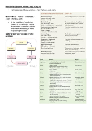
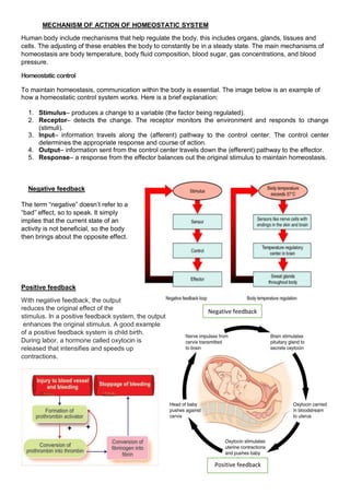
Anatomy and Physiology:
Introduction:-
Anatomy is the branch of biology concerned with the study of the structure of organisms and their parts. It is
concerned with the shape, size, position, structure, blood supply and innervation of different organs.
 “Halophiles” is called the father of anatomy. He was a Greek physician, and was one of the first to
dissect the human body.
CADAVER: - A cadaver is a dead human body that is
used by medical students, physicians and other
scientists to study anatomy, identify disease sites,
determine causes of death, and provide tissue to repair
a defect in a living human being. Students in medical
school study and dissect cadavers as a part of their
education.
Anatomy (Ana- up; -tomy process of cutting)
 Is the science of body structures and the relationships
among them.
 It was first studied by Dissection.
Dissection (dis- apart; section: act of cutting)
 The careful cutting apart of body structures to study their
relationships.
 Today, a variety of imaging techniques also contribute to
the advancement of anatomical knowledge.
LEVEL OF BODY ORGINATION
suku
Anatomical position
• The subject stands erect facing the observer, with the head level and the eyes facing directly forward.
• The feet are flat on the floor and directed forward, and the upper limbs are at the sides with the palms
turned forward.
ANATOMICAL PLANE
Coronal planes
• Oriented vertically and divide the body into anterior and posterior parts.
Sagittal planes
• Oriented vertically, but are at right angles to the coronal planes and divide the body into right and
left parts.
Median sagittal plane.
• The plane that passes through the center of the body dividing it into equal right and left halves
Transverse, horizontal, or axial planes
• Divide the body into superior and inferior parts.
BODY MOVEMENTS
Flexion: Movement in which angle of the joints is reduces.
Extension: Movement in which angle of the joints is increase
Adduction: Movement toward midline of the body in the coronal plane
Abduction: Movement away from the midline of the body in the coronal plane
Medial rotation: Inward rotation
Lateral rotation: Outward rotation
Circumduction: Combined movements of flexion, extension, abduction, adduction, medial and lateral
rotation
Pronation: Rotation of forearm so that the palm is turned backward
Supination: Rotation of the forearm so that the turned forward
Inversion: Movement of the foot so that the sole faces in a medial direction
Eversion: Is the opposite movement of the foot so that the sole faces in a lateral direction
POSITIONS OF BODY
• The supine position of the body is lying on the back.
• The prone position is lying face downward.
• Right Lateral Recumbent is lying on their right side.
• Left Lateral Recumbent is lying on their left side
Body cavity
• It is any space
• Filled by viscera, or fluid
Dorsal body cavity
Cranial cavity: Enclosed by the skull and
contains Brain and lined by Meninges
Vertebral canal: Enclosed by the spine and
contains the spinal cord and lined by meninges
Ventral body cavity
Thoracic cavity: Enclosed by the ribcage
and contains the lungs and heart and lined by
pleura and pericardium
Abdominal-pelvic cavity: Enclosed by the
ribcage and pelvis and contains the kidneys,
ureters, stomach, intestines, liver, gallbladder,
and pancreas and lined by peritoneum.
A. Abdominal cavity: Thoracic diaphragm AND
pelvic inlet
B. Pelvic cavity: Pelvic inlet (the superior opening
of the pelvis) and pelvic floor.
Physiology (fphysio- nature; -logy study of)
• Is the science of body functions—how the body parts work.
Homeostasis ( homeo- sameness; -
stasis -standing still)
• Is the condition of equilibrium
(balance) in the body’s internal
environment due to the constant
interaction of the body’s many
regulatory processes.
COMPONENTS OF HOMEOSTATIC
SYSTEM
MECHANISM OF ACTION OF HOMEOSTATIC SYSTEM
Human body include mechanisms that help regulate the body, this includes organs, glands, tissues and
cells. The adjusting of these enables the body to constantly be in a steady state. The main mechanisms of
homeostasis are body temperature, body fluid composition, blood sugar, gas concentrations, and blood
pressure.
Homeostatic control
To maintain homeostasis, communication within the body is essential. The image below is an example of
how a homeostatic control system works. Here is a brief explanation:
1. Stimulus– produces a change to a variable (the factor being regulated).
2. Receptor– detects the change. The receptor monitors the environment and responds to change
(stimuli).
3. Input– information travels along the (afferent) pathway to the control center. The control center
determines the appropriate response and course of action.
4. Output– information sent from the control center travels down the (efferent) pathway to the effector.
5. Response– a response from the effector balances out the original stimulus to maintain homeostasis.
Negative feedback
The term “negative” doesn’t refer to a
“bad” effect, so to speak. It simply
implies that the current state of an
activity is not beneficial, so the body
then brings about the opposite effect.
Positive feedback
With negative feedback, the output
reduces the original effect of the
stimulus. In a positive feedback system, the output
enhances the original stimulus. A good example
of a positive feedback system is child birth.
During labor, a hormone called oxytocin is
released that intensifies and speeds up
contractions.
Positive feedback
Negative feedback
Body fluid
Total fluid contain in the body along with its solute
Fluid intake and output
I. Daily intake of water
• Water is added to the body by 2major sources
1. It is ingested in form of liquid or water in the food , about 2100ml/day
2. It is synthesized in the body as a result of oxidation of carbohydrate,200ml/day
• Intake of water is highly variable among different people & even same person on different days
II. Daily loss of body water
a. Insensible water loss
• Some of water losses cannot be precisely regulated e.g: there is continuous loss of water by
evaporation from Respiratory tract(350ml/d) & diffusion through skin(350ml/d)
• It is termed so because we are not aware of it even though it occurs continually
b. Fluid loss in sweat gland
• It depends on physical activity & environmental temperature
• Normally:100ml/d, but can increase to 2l/d
c. Water in feces
• Normally:100ml/d, can increase in diarrhea
d. Water loss by kidneys
• In the form of urine,1400ml/d
Body fluid compartment (60% of body weight is fluid)
 Extracellular fluid (20% of body weight, 35-40% of body water)
 All fluids outside cell
 Divided into
a) Interstitial fluid (extravascular compartment)
 It makes up over 3/4
th
of ECF.
b) Plasma
 Makes up almost 1/4th of ECF
 Composition of plasma and interstitial fluid is same except for protein which is high in plasma
c) Transcellular fluid
 There is another small compartment of fluid which includes fluid in synovial, peritoneal, pericardial,
intraocular space as well as CSF
 Intracellular fluid(40% of body weight,60-65% of body water): Fluid inside the cell
ELECTROLYTE
• Electrolytes are the chemical substances capable of breaking into ions and developing electrical
charges when dissolved in solution.
• The predominant positively charged electrolytes in the body are sodium, potassium, calcium, and
magnesium,
• While negatively charged electrolytes include chloride, phosphates, and bicarbonate.
• Balance of concentration of ion is called electrolyte balance.
Composition of Oral
salt/suspension/Solution (ORS)
WHO standard formula
 NaCl = 2.6 g
 KCl = 1.5 g
 Trisodium citrate, dihydrate = 2.9 g
 Glucose, anhydrous = 13.5 g
Total Weight = 20.5 g
(To be dissolved in 1 liter of water)
 Sodium = 75 mM
 Chloride = 65 mM
 Citrate = 10 mM
 Glucose, anhydrous = 75 mM
 Potassium = 20 mM
Total osmolarity = 245 mM

Anatomy and physiology introduction

  • 1.
    Anatomy and Physiology: Introduction:- Anatomyis the branch of biology concerned with the study of the structure of organisms and their parts. It is concerned with the shape, size, position, structure, blood supply and innervation of different organs.  “Halophiles” is called the father of anatomy. He was a Greek physician, and was one of the first to dissect the human body. CADAVER: - A cadaver is a dead human body that is used by medical students, physicians and other scientists to study anatomy, identify disease sites, determine causes of death, and provide tissue to repair a defect in a living human being. Students in medical school study and dissect cadavers as a part of their education. Anatomy (Ana- up; -tomy process of cutting)  Is the science of body structures and the relationships among them.  It was first studied by Dissection. Dissection (dis- apart; section: act of cutting)  The careful cutting apart of body structures to study their relationships.  Today, a variety of imaging techniques also contribute to the advancement of anatomical knowledge. LEVEL OF BODY ORGINATION suku
  • 2.
    Anatomical position • Thesubject stands erect facing the observer, with the head level and the eyes facing directly forward. • The feet are flat on the floor and directed forward, and the upper limbs are at the sides with the palms turned forward.
  • 4.
    ANATOMICAL PLANE Coronal planes •Oriented vertically and divide the body into anterior and posterior parts. Sagittal planes • Oriented vertically, but are at right angles to the coronal planes and divide the body into right and left parts. Median sagittal plane. • The plane that passes through the center of the body dividing it into equal right and left halves Transverse, horizontal, or axial planes • Divide the body into superior and inferior parts.
  • 5.
    BODY MOVEMENTS Flexion: Movementin which angle of the joints is reduces. Extension: Movement in which angle of the joints is increase Adduction: Movement toward midline of the body in the coronal plane Abduction: Movement away from the midline of the body in the coronal plane Medial rotation: Inward rotation Lateral rotation: Outward rotation Circumduction: Combined movements of flexion, extension, abduction, adduction, medial and lateral rotation Pronation: Rotation of forearm so that the palm is turned backward Supination: Rotation of the forearm so that the turned forward Inversion: Movement of the foot so that the sole faces in a medial direction Eversion: Is the opposite movement of the foot so that the sole faces in a lateral direction
  • 6.
    POSITIONS OF BODY •The supine position of the body is lying on the back. • The prone position is lying face downward. • Right Lateral Recumbent is lying on their right side. • Left Lateral Recumbent is lying on their left side Body cavity • It is any space • Filled by viscera, or fluid Dorsal body cavity Cranial cavity: Enclosed by the skull and contains Brain and lined by Meninges Vertebral canal: Enclosed by the spine and contains the spinal cord and lined by meninges Ventral body cavity Thoracic cavity: Enclosed by the ribcage and contains the lungs and heart and lined by pleura and pericardium Abdominal-pelvic cavity: Enclosed by the ribcage and pelvis and contains the kidneys, ureters, stomach, intestines, liver, gallbladder, and pancreas and lined by peritoneum. A. Abdominal cavity: Thoracic diaphragm AND pelvic inlet B. Pelvic cavity: Pelvic inlet (the superior opening of the pelvis) and pelvic floor.
  • 7.
    Physiology (fphysio- nature;-logy study of) • Is the science of body functions—how the body parts work. Homeostasis ( homeo- sameness; - stasis -standing still) • Is the condition of equilibrium (balance) in the body’s internal environment due to the constant interaction of the body’s many regulatory processes. COMPONENTS OF HOMEOSTATIC SYSTEM
  • 8.
    MECHANISM OF ACTIONOF HOMEOSTATIC SYSTEM Human body include mechanisms that help regulate the body, this includes organs, glands, tissues and cells. The adjusting of these enables the body to constantly be in a steady state. The main mechanisms of homeostasis are body temperature, body fluid composition, blood sugar, gas concentrations, and blood pressure. Homeostatic control To maintain homeostasis, communication within the body is essential. The image below is an example of how a homeostatic control system works. Here is a brief explanation: 1. Stimulus– produces a change to a variable (the factor being regulated). 2. Receptor– detects the change. The receptor monitors the environment and responds to change (stimuli). 3. Input– information travels along the (afferent) pathway to the control center. The control center determines the appropriate response and course of action. 4. Output– information sent from the control center travels down the (efferent) pathway to the effector. 5. Response– a response from the effector balances out the original stimulus to maintain homeostasis. Negative feedback The term “negative” doesn’t refer to a “bad” effect, so to speak. It simply implies that the current state of an activity is not beneficial, so the body then brings about the opposite effect. Positive feedback With negative feedback, the output reduces the original effect of the stimulus. In a positive feedback system, the output enhances the original stimulus. A good example of a positive feedback system is child birth. During labor, a hormone called oxytocin is released that intensifies and speeds up contractions. Positive feedback Negative feedback
  • 9.
    Body fluid Total fluidcontain in the body along with its solute Fluid intake and output I. Daily intake of water • Water is added to the body by 2major sources 1. It is ingested in form of liquid or water in the food , about 2100ml/day 2. It is synthesized in the body as a result of oxidation of carbohydrate,200ml/day • Intake of water is highly variable among different people & even same person on different days II. Daily loss of body water a. Insensible water loss • Some of water losses cannot be precisely regulated e.g: there is continuous loss of water by evaporation from Respiratory tract(350ml/d) & diffusion through skin(350ml/d) • It is termed so because we are not aware of it even though it occurs continually b. Fluid loss in sweat gland • It depends on physical activity & environmental temperature • Normally:100ml/d, but can increase to 2l/d c. Water in feces • Normally:100ml/d, can increase in diarrhea d. Water loss by kidneys • In the form of urine,1400ml/d Body fluid compartment (60% of body weight is fluid)  Extracellular fluid (20% of body weight, 35-40% of body water)  All fluids outside cell  Divided into a) Interstitial fluid (extravascular compartment)  It makes up over 3/4 th of ECF. b) Plasma  Makes up almost 1/4th of ECF  Composition of plasma and interstitial fluid is same except for protein which is high in plasma c) Transcellular fluid  There is another small compartment of fluid which includes fluid in synovial, peritoneal, pericardial, intraocular space as well as CSF  Intracellular fluid(40% of body weight,60-65% of body water): Fluid inside the cell
  • 10.
    ELECTROLYTE • Electrolytes arethe chemical substances capable of breaking into ions and developing electrical charges when dissolved in solution. • The predominant positively charged electrolytes in the body are sodium, potassium, calcium, and magnesium, • While negatively charged electrolytes include chloride, phosphates, and bicarbonate. • Balance of concentration of ion is called electrolyte balance. Composition of Oral salt/suspension/Solution (ORS) WHO standard formula  NaCl = 2.6 g  KCl = 1.5 g  Trisodium citrate, dihydrate = 2.9 g  Glucose, anhydrous = 13.5 g Total Weight = 20.5 g (To be dissolved in 1 liter of water)  Sodium = 75 mM  Chloride = 65 mM  Citrate = 10 mM  Glucose, anhydrous = 75 mM  Potassium = 20 mM Total osmolarity = 245 mM