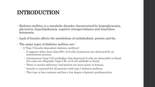
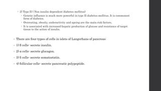
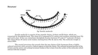
AN INTRODUCTION
TO INSULIN
PRIYESH PANDYA
M.PHARMACY (IP)
INTRODUCTION
• Diabetes mellitus is a metabolic disorder characterised by hyperglycaemia,
glycosuria, hyperlipidaemia, negative nitrogen balance and sometimes
ketonemia.
• Lack of Insulin affects the metabolism of carbohydrate, protein and fat.
• The major types of diabetes mellitus are:-
 1) Type I (insulin dependent diabetes mellitus):
 It appears when more than 90% of ß cells of pancreas are destroyed by an
autoimmune process.
 Autoimmune (type I A) antibodies that destroyed ß cells are detectable in blood,
but some are idiopathic (type I B), no ß cell antibody is found.
 There is insulin deficiency and patient are more prone to ketosis.
 Insulin is essential for all patients with type I diabetes mellitus.
 This type is less common and has a low degree of genetic predisposition.
 2) Type II ( Non insulin dependent diabetes mellitus)
 Genetic influence is much more powerful in type II diabetes mellitus. It is commonest
form of diabetes.
 Overeating, obesity, underactivity and ageing are the main risk factors.
 It is associated with increased hepatic production of glucose and resistance of target
tissue to the action of insulin.
• There are four types of cells in islets of Langerhans of pancreas:
• 1) β cells- secrete insulin.
• 2) ⍺ cells- secrete glucagon.
• 3) δ cells- secrete somatostatin.
• 4) follicular cells- secrete pancreatic polypeptide.
INSULIN:
Introduction:
 Insulin was discovered by Banting and Best.
 It is primary hormone which is responsible for controlling the storage and
utilization of cellular nutrients.
 Insulin is a two chain polypeptide having 51 amino acids and molecular
weight about 6000.
 It has two amino acid chains called α and β chains, which are linked by
disulfide bridges. The α-chain of insulin contains 21 amino acids and β-
chain contains 30 amino acids.
 The biological half-life of insulin is 5 minutes.
 Insulin activates the transport system and the enzymes involved in the
intra cellular utilization and storage of glucose, amino acids and fatty acids.
 Insulin inhibit catabolic processes such as breakdown of glycogen, fat and
protein.
 Deficiency of effective insulin in the body cause diabetes mellitus, which
alter the metabolism of lipids, carbohydrate and proteins.
Structure:
fig: Insulin molecule
Insulin molecule is consist of two peptide chains, A chain and B chain, which are
connected by disulphide bond. The chain A is composed of 21 amino acid residues starting from Gly
and end to Asn. It has one intrachain disulphide bond in between 6th and 11th molecule of chain.
The chain B has 30 amino acid residue starting from Phe & end on Thr. The molecule weight is
about 6000.
The crystal structure also reveals that the two chains of the hormones from a highly
ordered structure with several ⍺- helical regions in both the chains. The carboxyl terminal portion of the
B chain and the amino and carboxyl terminal residue of the A chain from the surface of the molecules which
interact with the receptor.
• The insulin receptor is a large transmembrane glycoprotein composed of two ⍺
subunits and two β subunits. The subunits are linked by disulphide bonds to form β-⍺-⍺-β
hetro-tetramer.
Biosynthesis of insulin:
• Insulin is produced by the β- cells of the islets of Langerhans of pancreas.
• The gene for this protein synthesis is located on chromosome 11.
• The synthesis of insulin involves two precursors namely preproinsulin with 108 amino acid
and proinsulin with 86 amino acid. They are sequentially degraded to form the active
hormone insulin and a connecting peptide ( c- peptide).
• Insulin and c- peptide are produced in equimolar concentration.
• C- peptide has no biological activities but its estimation in the plasma serves as a useful
index for the endogenous production of the insulin.
• In the β cells, Insulin and also proinsulin combines with zinc to form complexes. In this
form insulin is stored in the granules of the cytosol, which is released in response to
various stimuli by exocytosis.
• Insulin circulates in blood as a free monomer.
Regulation of insulin secretion:
• About 40-50 units of insulin (1- 2 mg) is secreted daily by human pancreas.
• The normal insulin concentration in plasma is 20-30 μU/ml.
• The important factors that influence the release of insulin from the β cells of pancreas are-
 1) Factors stimulating insulin secretion:
 a) Glucose: A rise in blood glucose level is a signal for insulin secretion.
 b) Amino acids: protein rich meal cause transient rise in plasma amino acid
concentration. Argenine and leucine are potent stimulator of insulin release.
 C) GI hormones: secretin, gastrin, pancreozymin enhance the secretion of insulin.
 2) Factors inhibit insulin secretion:
 Epinephrine is the most potent inhibitor of insulin release.
 In emergency situations like stress, extreme exercise and trauma, the nervous system
stimulate adrenal medulla to release epinephrine. Epinephrine supress insulin release
and promotes energy metabolism by mobilizing energy yielding compounds glucose from
liver and fatty acid from adipose tissue.
Degradation of Insulin:
• Insulin has a normal half life of 4 to 5 min. A protease enzyme, insulinase (mainly
found in liver and kidney) degrades insulin.
Mechanism of action of insulin:
Insulin binds to specific plasma membrane receptors present on the target
tissue, such as muscle and adipose. There are 3 distinct mechanism of insulin action:
1) The induction of transmembrane signals:
The insulin receptor is synthesized as a single polypeptide and cleaved to ⍺ and
β subunits. The insulin receptor has a half life of 6- 12 hours. As the hormone insulin binds to
the receptor, a conformational change is induced in ⍺ subunits. This result in generation of
signals which are transduce to β subunit. As a result the insulin binding activates tyrosine kinase
activity. This cause the autophosphorylation of tyrosine residue on β subunit. Tyrosine kinase
also phosphorylate insulin receptor substrate, this promotes activation of other protein kinase
and phosphatase, finally leading to biological actions.
2) Insulin mediated glucose transport:
The binding of insulin to insulin receptor signals, the translocation of vesicles containing
glucose transporters from intracellular pool to the plasma membrane.
Official Drugs:
1. Insulin (I.P., B.P.)-
- Obtained from beef or pork pancreases and purified.
- White or almost white powder.
- Insoluble in water practically.
- Kept in air tight container, protect form light & stored at cold temperature.
2. Insulin Injection-
- Available as sterile, acidified or neutral solution of bovine, porcine or human insulin in
water for injection containing 40, 80, 100 or 500 USP insulin units per ml.
- It is colourless, free from turbidity and foreign matter.
3. Lipro / human insulin-
-It is a human insulin analogue of rDNA origin synthesis from a
special non-pathogenic strain of E. coli genetically altered by addition of the
gene for insulin lispro; Lys (B28), pro (B29)
-It is a very rapid acting insulin, injected just prior to a meal.
THANK YOU

An introduction to Insulin

  • 1.
    AN INTRODUCTION TO INSULIN PRIYESHPANDYA M.PHARMACY (IP)
  • 2.
    INTRODUCTION • Diabetes mellitusis a metabolic disorder characterised by hyperglycaemia, glycosuria, hyperlipidaemia, negative nitrogen balance and sometimes ketonemia. • Lack of Insulin affects the metabolism of carbohydrate, protein and fat. • The major types of diabetes mellitus are:-  1) Type I (insulin dependent diabetes mellitus):  It appears when more than 90% of ß cells of pancreas are destroyed by an autoimmune process.  Autoimmune (type I A) antibodies that destroyed ß cells are detectable in blood, but some are idiopathic (type I B), no ß cell antibody is found.  There is insulin deficiency and patient are more prone to ketosis.  Insulin is essential for all patients with type I diabetes mellitus.  This type is less common and has a low degree of genetic predisposition.
  • 3.
     2) TypeII ( Non insulin dependent diabetes mellitus)  Genetic influence is much more powerful in type II diabetes mellitus. It is commonest form of diabetes.  Overeating, obesity, underactivity and ageing are the main risk factors.  It is associated with increased hepatic production of glucose and resistance of target tissue to the action of insulin. • There are four types of cells in islets of Langerhans of pancreas: • 1) β cells- secrete insulin. • 2) ⍺ cells- secrete glucagon. • 3) δ cells- secrete somatostatin. • 4) follicular cells- secrete pancreatic polypeptide.
  • 4.
    INSULIN: Introduction:  Insulin wasdiscovered by Banting and Best.  It is primary hormone which is responsible for controlling the storage and utilization of cellular nutrients.  Insulin is a two chain polypeptide having 51 amino acids and molecular weight about 6000.  It has two amino acid chains called α and β chains, which are linked by disulfide bridges. The α-chain of insulin contains 21 amino acids and β- chain contains 30 amino acids.  The biological half-life of insulin is 5 minutes.  Insulin activates the transport system and the enzymes involved in the intra cellular utilization and storage of glucose, amino acids and fatty acids.  Insulin inhibit catabolic processes such as breakdown of glycogen, fat and protein.  Deficiency of effective insulin in the body cause diabetes mellitus, which alter the metabolism of lipids, carbohydrate and proteins.
  • 5.
    Structure: fig: Insulin molecule Insulinmolecule is consist of two peptide chains, A chain and B chain, which are connected by disulphide bond. The chain A is composed of 21 amino acid residues starting from Gly and end to Asn. It has one intrachain disulphide bond in between 6th and 11th molecule of chain. The chain B has 30 amino acid residue starting from Phe & end on Thr. The molecule weight is about 6000. The crystal structure also reveals that the two chains of the hormones from a highly ordered structure with several ⍺- helical regions in both the chains. The carboxyl terminal portion of the B chain and the amino and carboxyl terminal residue of the A chain from the surface of the molecules which interact with the receptor.
  • 6.
    • The insulinreceptor is a large transmembrane glycoprotein composed of two ⍺ subunits and two β subunits. The subunits are linked by disulphide bonds to form β-⍺-⍺-β hetro-tetramer. Biosynthesis of insulin: • Insulin is produced by the β- cells of the islets of Langerhans of pancreas. • The gene for this protein synthesis is located on chromosome 11. • The synthesis of insulin involves two precursors namely preproinsulin with 108 amino acid and proinsulin with 86 amino acid. They are sequentially degraded to form the active hormone insulin and a connecting peptide ( c- peptide). • Insulin and c- peptide are produced in equimolar concentration. • C- peptide has no biological activities but its estimation in the plasma serves as a useful index for the endogenous production of the insulin. • In the β cells, Insulin and also proinsulin combines with zinc to form complexes. In this form insulin is stored in the granules of the cytosol, which is released in response to various stimuli by exocytosis. • Insulin circulates in blood as a free monomer.
  • 7.
    Regulation of insulinsecretion: • About 40-50 units of insulin (1- 2 mg) is secreted daily by human pancreas. • The normal insulin concentration in plasma is 20-30 μU/ml. • The important factors that influence the release of insulin from the β cells of pancreas are-  1) Factors stimulating insulin secretion:  a) Glucose: A rise in blood glucose level is a signal for insulin secretion.  b) Amino acids: protein rich meal cause transient rise in plasma amino acid concentration. Argenine and leucine are potent stimulator of insulin release.  C) GI hormones: secretin, gastrin, pancreozymin enhance the secretion of insulin.  2) Factors inhibit insulin secretion:  Epinephrine is the most potent inhibitor of insulin release.  In emergency situations like stress, extreme exercise and trauma, the nervous system stimulate adrenal medulla to release epinephrine. Epinephrine supress insulin release and promotes energy metabolism by mobilizing energy yielding compounds glucose from liver and fatty acid from adipose tissue.
  • 8.
    Degradation of Insulin: •Insulin has a normal half life of 4 to 5 min. A protease enzyme, insulinase (mainly found in liver and kidney) degrades insulin. Mechanism of action of insulin: Insulin binds to specific plasma membrane receptors present on the target tissue, such as muscle and adipose. There are 3 distinct mechanism of insulin action: 1) The induction of transmembrane signals: The insulin receptor is synthesized as a single polypeptide and cleaved to ⍺ and β subunits. The insulin receptor has a half life of 6- 12 hours. As the hormone insulin binds to the receptor, a conformational change is induced in ⍺ subunits. This result in generation of signals which are transduce to β subunit. As a result the insulin binding activates tyrosine kinase activity. This cause the autophosphorylation of tyrosine residue on β subunit. Tyrosine kinase also phosphorylate insulin receptor substrate, this promotes activation of other protein kinase and phosphatase, finally leading to biological actions.
  • 9.
    2) Insulin mediatedglucose transport: The binding of insulin to insulin receptor signals, the translocation of vesicles containing glucose transporters from intracellular pool to the plasma membrane. Official Drugs: 1. Insulin (I.P., B.P.)- - Obtained from beef or pork pancreases and purified. - White or almost white powder. - Insoluble in water practically. - Kept in air tight container, protect form light & stored at cold temperature. 2. Insulin Injection- - Available as sterile, acidified or neutral solution of bovine, porcine or human insulin in water for injection containing 40, 80, 100 or 500 USP insulin units per ml. - It is colourless, free from turbidity and foreign matter.
  • 10.
    3. Lipro /human insulin- -It is a human insulin analogue of rDNA origin synthesis from a special non-pathogenic strain of E. coli genetically altered by addition of the gene for insulin lispro; Lys (B28), pro (B29) -It is a very rapid acting insulin, injected just prior to a meal.
  • 11.