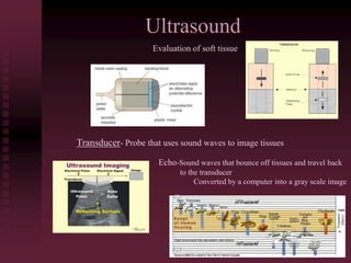
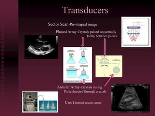
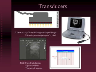
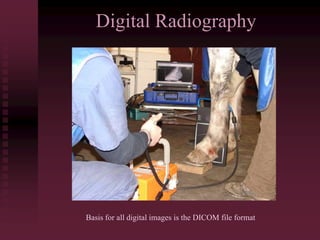
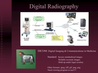
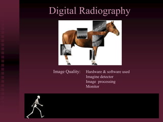
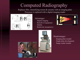
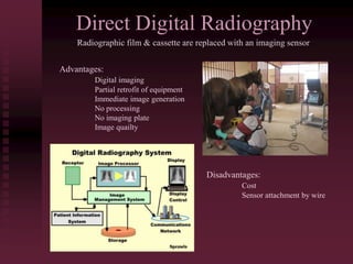
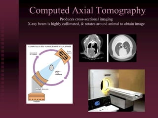
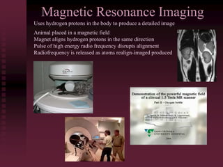
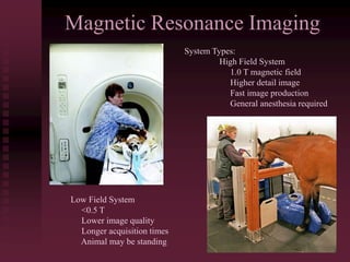
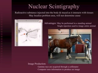
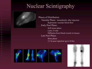
EQUINE MEDICINE
Alternative Imaging Techniques: Ultrasound, MRI, CT Scan
Ultrasound
Evaluation of soft tissue
Transducer- Probe that uses sound waves to image tissues
Echo-Sound waves that bounce off tissues and travel back
to the transducer
Converted by a computer into a gray scale image
Transducers
Sector Scan-Pie-shaped image
Phased Array-Crystals pulsed sequentially
Delay between pulses
Annular Array-Crystals in ring
Pulse directed through crystals
Use: Limited access areas
Transducers
Linear Array Scan-Rectangular-shaped image
Alternate pulse on groups of crystals
Use: Unrestricted areas
Equine tendons
Transrectal imaging
Patient Preparation
Clip or shave area
Scrub area with shampoo & rinse thoroughly
Wipe area with alcohol
Use acoustic coupling gel to eliminate air
Imaging
Digital Radiography
Basis for all digital images is the DICOM file format
Digital Radiography
DICOM: Digital Imaging & Communications in Medicine
Standard: Secure standardized images
Reliable accurate images
Hold up under legal scrutiny
Other formats: jpeg, tiff, gif, png, psg
Need viewing program to use!!!!
Digital Radiography
Image Quality: Hardware & software used
Imagine detector
Image processing
Monitor
Computed Radiography
Replaces film, intensifying screen & cassette with an imaging plate
Processor is replaced with a digital imaging reader
Advantages:
Digital imaging
Retrofit of equipment
Mobile
Relatively inexpensive
Disadvantages:
Imaging plate needed
Single plate development
Image reader needed
Direct Digital Radiography
Radiographic film & cassette are replaced with an imaging sensor
Advantages:
Digital imaging
Partial retrofit of equipment
Immediate image generation
No processing
No imaging plate
Image quailty
Disadvantages:
Cost
Sensor attachment by wire
Computed Axial Tomography
Produces cross-sectional imaging
X-ray beam is highly collimated, & rotates around animal to obtain image
Computed Axial Tomography
Voxel-Density of a small volume within a patient
Projected onto screen or film to produce image
Advantages: Detects small tissue density differences
Highly detailed image produced
Ability to see deep/small changes
Disadvantages: Cost
Requires general anesthesia
Gantry opening may limit patient access
Magnetic Resonance Imaging
Uses hydrogen protons in the body to produce a detailed image
Animal placed in a magnetic field
Magnet aligns hydrogen protons in the same direction
Pulse of high energy radio frequency disrupts alignment
Radiofrequency is released as atoms realign-imaged produced
Magnetic Resonance Imaging
System Types:
High Field System
1.0 T magnetic field
Higher detail image
Fast image production
General anesthesia required
Low Field System
<0.5 T
Lower image quality
Longer acquisition times
Animal may be standing
Magnetic Resonance Imaging
Imaging Protocols
Proton Density-Clear anatomic structure
T2 Weighting-High fluid contrast
T1 Weighting-Good bone definition
Variations
Short Tau Inversion Recovery (STIR)
Fat Suppression (FS)
Water-Excitation (WE)
Imaging
Nuclear Scintigraphy
Radioactive substance injected into the body & traced as it interacts with tissues
May localize problem area, will not determine cause
Advantages: May be performed in a standing animal
Single injection used to image entire animal
Image Production:
Gamma rays are acquired through a collimator
Computer uses information to produce an image
Nuclear Scintigraphy
Phases of Distribution:
Vascular Phase: Immediately after injection
Represents vascular blood flow
Early Pool Phase:
Soft tissue phase
Lasts 15 min
Diffusion from blood vessels to tissues
Late Pool Phase:
Bone phase
2-3 hr post injection up to 24 hrs

Alternative Imaging Techniques

  • 1.
    EQUINE MEDICINE Alternative ImagingTechniques: Ultrasound, MRI, CT Scan
  • 2.
    Ultrasound Evaluation of softtissue Transducer- Probe that uses sound waves to image tissues Echo-Sound waves that bounce off tissues and travel back to the transducer Converted by a computer into a gray scale image
  • 3.
    Transducers Sector Scan-Pie-shaped image PhasedArray-Crystals pulsed sequentially Delay between pulses Annular Array-Crystals in ring Pulse directed through crystals Use: Limited access areas
  • 4.
    Transducers Linear Array Scan-Rectangular-shapedimage Alternate pulse on groups of crystals Use: Unrestricted areas Equine tendons Transrectal imaging
  • 5.
    Patient Preparation Clip orshave area Scrub area with shampoo & rinse thoroughly Wipe area with alcohol Use acoustic coupling gel to eliminate air
  • 6.
  • 7.
    Digital Radiography Basis forall digital images is the DICOM file format
  • 8.
    Digital Radiography DICOM: DigitalImaging & Communications in Medicine Standard: Secure standardized images Reliable accurate images Hold up under legal scrutiny Other formats: jpeg, tiff, gif, png, psg Need viewing program to use!!!!
  • 9.
    Digital Radiography Image Quality:Hardware & software used Imagine detector Image processing Monitor
  • 10.
    Computed Radiography Replaces film,intensifying screen & cassette with an imaging plate Processor is replaced with a digital imaging reader Advantages: Digital imaging Retrofit of equipment Mobile Relatively inexpensive Disadvantages: Imaging plate needed Single plate development Image reader needed
  • 11.
    Direct Digital Radiography Radiographicfilm & cassette are replaced with an imaging sensor Advantages: Digital imaging Partial retrofit of equipment Immediate image generation No processing No imaging plate Image quailty Disadvantages: Cost Sensor attachment by wire
  • 12.
    Computed Axial Tomography Producescross-sectional imaging X-ray beam is highly collimated, & rotates around animal to obtain image
  • 13.
    Computed Axial Tomography Voxel-Densityof a small volume within a patient Projected onto screen or film to produce image Advantages: Detects small tissue density differences Highly detailed image produced Ability to see deep/small changes Disadvantages: Cost Requires general anesthesia Gantry opening may limit patient access
  • 14.
    Magnetic Resonance Imaging Useshydrogen protons in the body to produce a detailed image Animal placed in a magnetic field Magnet aligns hydrogen protons in the same direction Pulse of high energy radio frequency disrupts alignment Radiofrequency is released as atoms realign-imaged produced
  • 15.
    Magnetic Resonance Imaging SystemTypes: High Field System 1.0 T magnetic field Higher detail image Fast image production General anesthesia required Low Field System <0.5 T Lower image quality Longer acquisition times Animal may be standing
  • 16.
    Magnetic Resonance Imaging ImagingProtocols Proton Density-Clear anatomic structure T2 Weighting-High fluid contrast T1 Weighting-Good bone definition Variations Short Tau Inversion Recovery (STIR) Fat Suppression (FS) Water-Excitation (WE)
  • 17.
  • 18.
    Nuclear Scintigraphy Radioactive substanceinjected into the body & traced as it interacts with tissues May localize problem area, will not determine cause Advantages: May be performed in a standing animal Single injection used to image entire animal Image Production: Gamma rays are acquired through a collimator Computer uses information to produce an image
  • 19.
    Nuclear Scintigraphy Phases ofDistribution: Vascular Phase: Immediately after injection Represents vascular blood flow Early Pool Phase: Soft tissue phase Lasts 15 min Diffusion from blood vessels to tissues Late Pool Phase: Bone phase 2-3 hr post injection up to 24 hrs