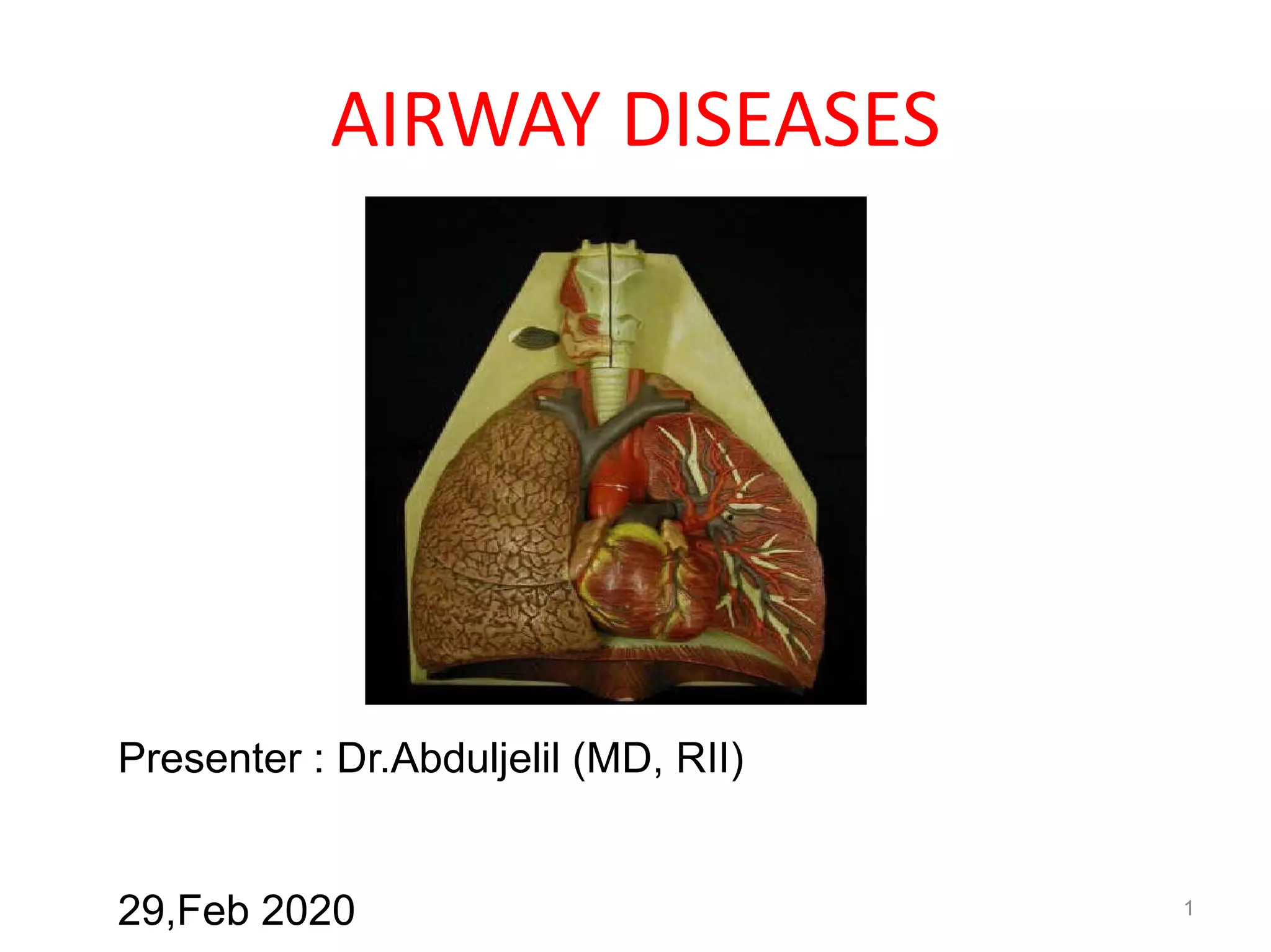
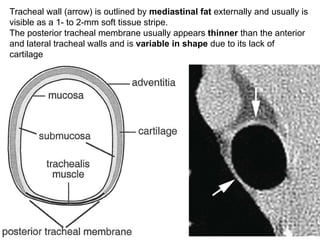
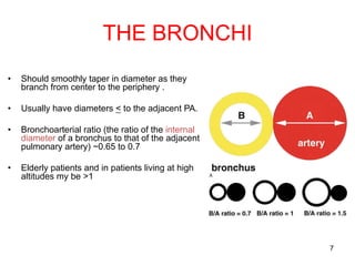
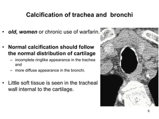
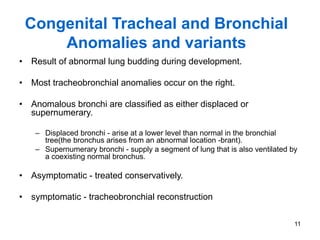
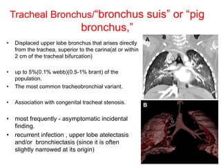
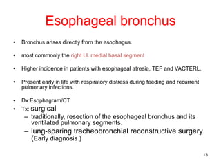
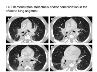
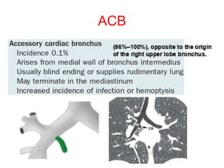
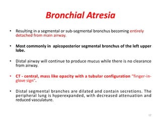
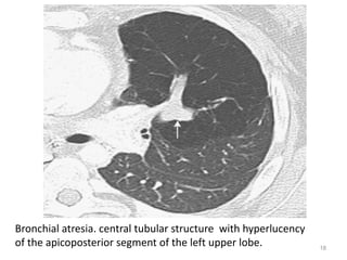
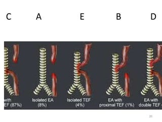
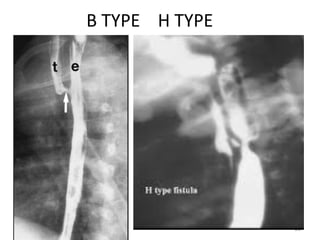
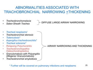
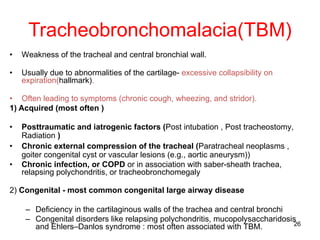
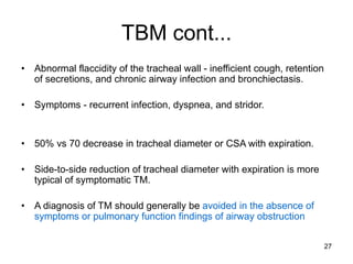
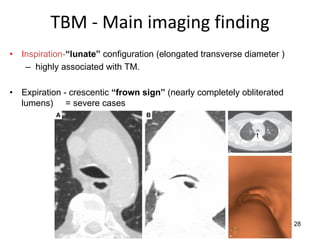
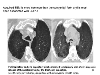
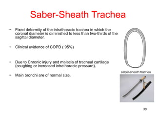
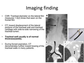
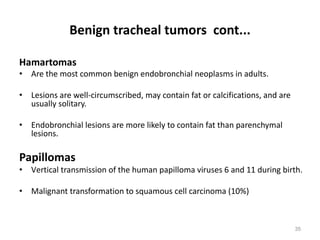
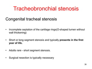
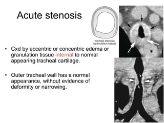
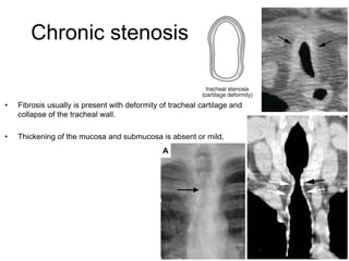
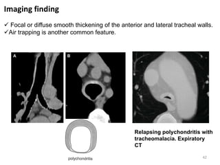
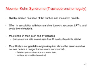
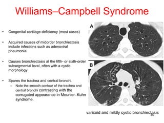
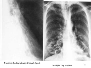
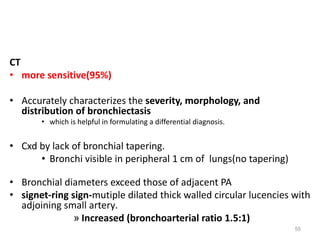
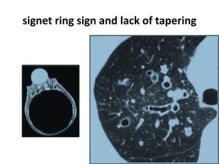
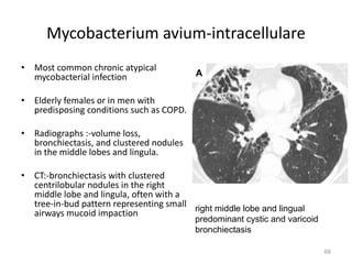
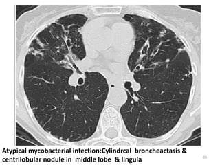
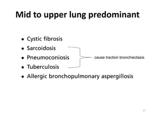
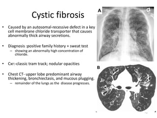
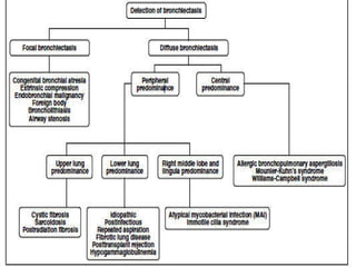
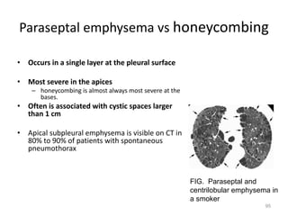
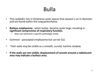
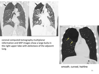
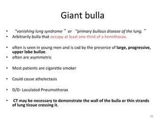
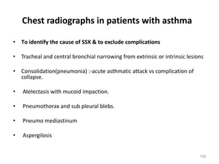
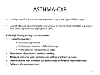
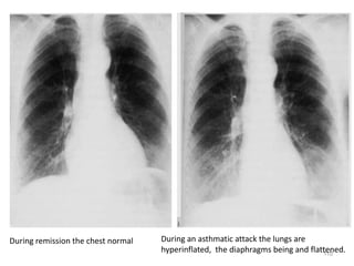
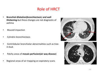
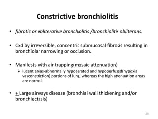
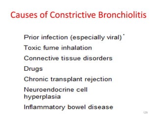
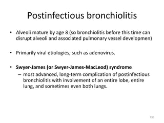
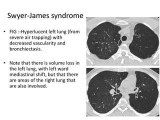
1
AIRWAY DISEASES
Presenter : Dr.Abduljelil (MD, RII)
29,Feb 2020
2
OUTLINE
• INTRODUCTION
• CONGENITAL TRACHEAL AND BRONCHIAL ANOMALIES AND VARIANTS
• ABNORMALITIES ASSOCIATED WITH TRACHOBRONCHIAL NARROWING
+THICKENING
• ABNORMALITIES ASSOCIATED WITH INCREASED TRACHOBRONCHIAL
DIAMETER
• BRONCHIECTASIS
• EMPHYSEMA
• CHRONIC BRONCHITIS
• BRONCHIAL ASTHMA
• BRONCHIOLITIS
3
INTRODUCTION
• Typical morphologies, locations, and distributions airways disease is
central to an accurate imaging differential diagnosis.
• Volumetric, spiral acquisition with thin collimation (3 mm) and thin
slice reconstructions (1-2 mm) in the axial plane are preferred
– detection and evaluation of airway thickening, nodularity, masses, or other
abnormalities.
• Expiratory series obtained during maximum forced expiration
– to maximize sensitivity for air trapping, and
– to allow for assessment of expiratory airway collapse : eg TBM
• Bronchoscopic endolumenal views( “virtual bronchoscopy” )
– have high sensitivity similar to bronchoscopy for detection of lesions of the
large airways such as tracheal stenosis
4
The trachea
• Inferior aspect of the cricoid cartilage (C6) ->
carina (T5).
• 10 to 12 cm in length.
• Extrathoracic(2 to 4 cm) and intrathoracic(6 to 9
cm) portions at the level it passes posterior to
the manubrium
• Anterior 2/3- incomplete ring and post- non straited
muscle and fibrous tissue
5
Tracheal wall (arrow) is outlined by mediastinal fat externally and usually is
visible as a 1- to 2-mm soft tissue stripe.
The posterior tracheal membrane usually appears thinner than the anterior
and lateral tracheal walls and is variable in shape due to its lack of
cartilage
6
Tracheal diameter
• Inspiration - circular or ovoid shape
with smooth walls measuring 1 to 3
mm
• Forced expiration - cross-sectional
area diminish by more than 50% in
normal subjects.
• Similar in size from the thoracic inlet to
the carina.
• Coronal diameter:
– Men 13 to 25 mm
– Women 10 to 21 mm.
• Sagittal diameter:
– Men 13 to 27 mm
– Women 10 to 23 mm
• Tracheal index : coronal /sagittal
diameter .
Ø Normal value is ~ 1(0.6-1.0)
7
THE BRONCHI
• Should smoothly taper in diameter as they
branch from center to the periphery .
• Usually have diameters < to the adjacent PA.
• Bronchoarterial ratio (the ratio of the internal
diameter of a bronchus to that of the adjacent
pulmonary artery) ~0.65 to 0.7
• Elderly patients and in patients living at high
altitudes my be >1
8
• Expiratory CXR the trachea often buckles.
• Always buckles in the direction opposite to
the location of the aortic arch.
• Sharp angles of the (buckled trachea) vs
more rounded displacement (masses).
• preserved tracheal diameter is also a clue to
normal buckling.
Pediatric airways
9
Calcification of trachea and bronchi
• old, women or chronic use of warfarin.
• Normal calcification should follow
the normal distribution of cartilage
– incomplete ringlike appearance in the trachea
and
– more diffuse appearance in the bronchi.
• Little soft tissue is seen in the tracheal
wall internal to the cartilage.
10
Functions of large airways
• More than simple tubes
• Cilia within the pseudostratified columnar
epithelium help
• clear inhaled toxins, organisms, and debris.
• Secretory glands ( submucosal layer) - secretions
that assist clearance and serve a protective role.
11
Congenital Tracheal and Bronchial
Anomalies and variants
• Result of abnormal lung budding during development.
• Most tracheobronchial anomalies occur on the right.
• Anomalous bronchi are classified as either displaced or
supernumerary.
– Displaced bronchi - arise at a lower level than normal in the bronchial
tree(the bronchus arises from an abnormal location -brant).
– Supernumerary bronchi - supply a segment of lung that is also ventilated by
a coexisting normal bronchus.
• Asymptomatic - treated conservatively.
• symptomatic - tracheobronchial reconstruction
12
Tracheal Bronchus/“bronchus suis” or “pig
bronchus,”
• Displaced upper lobe bronchus that arises directly
from the trachea, superior to the carina(at or within
2 cm of the tracheal bifurcation)
• up to 5%(0.1% webb)(0.5-1% brant) of the
population.
• The most common tracheobronchial variant.
• Association with congenital tracheal stenosis.
• most frequently - asymptomatic incidental
finding.
• recurrent infection , upper lobe atelectasis
and/or bronchiectasis (since it is often
slightly narrowed at its origin)
13
Esophageal bronchus
• Bronchus arises directly from the esophagus.
• most commonly the right LL medial basal segment
• Higher incidence in patients with esophageal atresia, TEF and VACTERL.
• Present early in life with respiratory distress during feeding and recurrent
pulmonary infections.
• Dx:Esophagram/CT
• Tx: surgical
– traditionally, resection of the esophageal bronchus and its
ventilated pulmonary segments.
– lung-sparing tracheobronchial reconstructive surgery
(Early diagnosis )
14
• CT demonstrates atelectasis and/or consolidation in the
affected lung segment
15
ACB
16
Bridging bronchus (BB)
• Very rare anomaly
• Displaced BI arising from the medial aspect of the
left mainstem bronchus and crossing the midline to
ventilate portions of the right lung.
• Two types.
• BB vs tracheal bronchus
– carina -T4-T5 level and midline
– pseudocarina of BB is typically located at ~T5-T7 level and
left of midline
17
Bronchial Atresia
• Resulting in a segmental or sub-segmental bronchus becoming entirely
detached from main airway.
• Most commonly in apicoposterior segmental bronchus of the left upper
lobe.
• Distal airway will continue to produce mucus while there is no clearance
from airway.
• CT - central, mass like opacity with a tubular configuration “finger-in-
glove sign".
• Distal segmental branches are dilated and contain secretions. The
peripheral lung is hyperexpanded, with decreased attenuation and
reduced vasculature.
18
Bronchial atresia. central tubular structure with hyperlucency
of the apicoposterior segment of the left upper lobe.
19
Tracheo-esophageal fistula
• Absence in continuity of the esophagus due to inappropriate
division of primitive foregut.
• The recurrence risk among parents of one affected child is
0.5% to 2%, increasing to 20% when more than one child is
affected.
• Most common Congenital malformation.
• Often seen with other congenital anomalies, of which the
VACTERL is most commonly known
• Antenatal Diagnosis:-Current ultrasound technology does not
allow for a defnite diagnosis of EA/TEF. prenatal diagnosis of
EA/TEF relies, in principle, on two nonspecifc signs:
polyhydramnios and an absent or small stomach bubble.
20
C A E B D
21
Classification (Gross's Anatomical
Classification)
• Type A: isolated Esophageal atresia(8%).
• Type B: proximal fistula with distal atresia.
• Type C: proximal atresia with distal
tracheoesophageal fistula (most common
type) (85%).
• Type D: Esophageal atresia with proximal and
distal fistula.
• Type E: isolated fistula without atresia(4%)
22
The absence of air in the stomach points to EA
without distal fstula
23
B TYPE H TYPE
24
Esophageal gap length**
• Usually not known preoperatively.
• Absence of air in the stomach has been linked with a long gap
– but has also been described in association with a distal fstula occluded with mucus.
• measured radiologically, either with metal bougies or with a contrast agent
at the time of the gastrostomy.
• less than two vertebrae - primary anastomosis
• Two to six vertebrae - delayed primary anastomosis
• More than six vertebrae - esophageal replacement.
• In EA/TEF, a longer gap between the two esophageal ends should be
expected when the distal fistula arises from the carina.
** source : Ashcraft’s Pediatric Surgery Copyright © 2010
25
ABNORMALITIES ASSOCIATED WITH
TRACHOBRONCHIAL NARROWING +THICKENING
• Tracheobronchomalacia
• Saber-Sheath Trachea
• Tracheal neoplasms*
• Tracheobronchial stenosis
• Tuberculosis*
• Aspergillus tracheobronchitis*
• Tracheal scleroma*
• Relapsing Polychondritis
• Tracheobronchopathia
Osteochondroplastica
• Granulomatosis with Polyangiitis
(Wegener Granulomatosis)
• Tracheobronchial amyloidosis
DIFFUSE LARGE AIRWAY NARROWING
AIRWAY NARROWING AND THICKENING
* Further will be covered on pulmonary infections and neoplasms
26
Tracheobronchomalacia(TBM)
• Weakness of the tracheal and central bronchial wall.
• Usually due to abnormalities of the cartilage- excessive collapsibility on
expiration(hallmark).
• Often leading to symptoms (chronic cough, wheezing, and stridor).
1) Acquired (most often )
• Posttraumatic and iatrogenic factors (Post intubation , Post tracheostomy,
Radiation )
• Chronic external compression of the tracheal (Paratracheal neoplasms ,
goiter congenital cyst or vascular lesions (e.g., aortic aneurysm))
• Chronic infection, or COPD or in association with saber-sheath trachea,
relapsing polychondritis, or tracheobronchomegaly
2) Congenital - most common congenital large airway disease
– Deficiency in the cartilaginous walls of the trachea and central bronchi
– Congenital disorders like relapsing polychondritis, mucopolysaccharidosis
and Ehlers–Danlos syndrome : most often associated with TBM.
27
TBM cont...
• Abnormal flaccidity of the tracheal wall - inefficient cough, retention
of secretions, and chronic airway infection and bronchiectasis.
• Symptoms - recurrent infection, dyspnea, and stridor.
• 50% vs 70 decrease in tracheal diameter or CSA with expiration.
• Side-to-side reduction of tracheal diameter with expiration is more
typical of symptomatic TM.
• A diagnosis of TM should generally be avoided in the absence of
symptoms or pulmonary function findings of airway obstruction
28
TBM - Main imaging finding
• Inspiration-“lunate” configuration (elongated transverse diameter )
– highly associated with TM.
• Expiration - crescentic “frown sign” (nearly completely obliterated
lumens) = severe cases
29
Acquired TBM is more common than the congenital form and is most
often associated with COPD
End inspiratory and end expiratory axial computed tomography scan shows excessive
collapse of the posterior wall of the trachea in expiration.
Note the extensive changes consistent with emphysema in both lungs.
30
Saber-Sheath Trachea
• Fixed deformity of the intrathoracic trachea in which the
coronal diameter is diminished to less than two-thirds of the
sagittal diameter.
• Clinical evidence of COPD ( 95%)
• Due to Chronic injury and malacia of tracheal cartilage
(coughing or increased intrathoracic pressure).
• Main bronchi are of normal size.
31
• CXR: Tracheal diameter on the lateral film
measures 1.5(2) times that seen on the
frontal film
• CT: Inward displacement of the lateral
portions of the tracheal wall and tracheal
cartilage with side-to-side narrowing of the
tracheal lumen.
• Tracheal wall usually is of normal
thickness(webb).
• During forced expiration, CT
demonstrates further inward bowing of the
tracheal walls in many patients.
Imaging finding
32
Tracheal neoplasms
• Uncommon(1% - 2% of all respiratory tract tumors), with 90% being malignant.
• Majority of airways neoplasms are seen adults
• Arise from tracheal epithelium or mucous glands (90%)
• Mesenchymal elements of the tracheal wall (10%).
• Two major types of tracheal carcinomas
– Squamous cell (55%)
– Adenocystic (18%)..
• Malignant neoplasms :
• CT as an eccentric irregular soft tissue mass within lumen, most typically arising from posterior
and lateral wall
• Masses >2 cm in diameter are likely to be malignant, while those <2 cm are more likely benign.
Calcification is uncommon.
NB: Most tumors involving the central airways are caused by direct invasion by an adjacent tumor
(thyroid, lung, or esophagus).
33
A: Lateral chest radiograph in a 68- year-old man : mass in the midtrachea (black arrows).
B: CT scan demonstrates an enhancing mass (arrow) in the posterior trachea with narrowing
of the tracheal lumen
Bronchoscopic biopsy showed squamous cell carcinoma
ØMiddle-aged male smokers
ØAssociated with laryngeal, bronchogenic, or esophageal malignancies ( 25%)
34
• Benign neoplasms of the airway are more common in children
(70%– 80%) and uncommon in adults (10%).
 Squamous papilloma (young children),
 Pleomorphic adenoma
 Mesenchymal hamartomathos of cartilaginous origin.
 Well circumscribed/smoothly marginated/< 2 cm in diameter.
Benign tracheal tumors
35
Benign tracheal tumors cont...
Hamartomas
• Are the most common benign endobronchial neoplasms in adults.
• Lesions are well-circumscribed, may contain fat or calcifications, and are
usually solitary.
• Endobronchial lesions are more likely to contain fat than parenchymal
lesions.
Papillomas
• Vertical transmission of the human papilloma viruses 6 and 11 during birth.
• Malignant transformation to squamous cell carcinoma (10%)
36
Tracheobronchial stenosis
Congenital tracheal stenosis
• Incomplete septation of the cartilage rings(O-shaped lumen without
wall thickening)
• Short or long segment stenosis and typically presents in the first
year of life.
• Adults rare - short segment stenosis.
• Surgical resection is typically necessary
37
Postintubation or tracheostomy stenosis
• Extrathoracic trachea most often is involved.
• Intrathoracic - endotracheal intubation
• Postintubation stenosis is rare with the low-pressure, high-volume
endotracheal tube cuffs in current use.
• Most frequent reason for tracheal surgery.
• Stenosis—typically 1.5 to 2.5 cm in length—commonly occurs at the:-
– level of the tube balloon , 1 to 1.5 cm proximal to the tube tip; or
– tracheostomy stoma.
38
Acute stenosis
• Cxd by eccentric or concentric edema or
granulation tissue internal to normal
appearing tracheal cartilage.
• Outer tracheal wall has a normal
appearance, without evidence of
deformity or narrowing.
39
Chronic stenosis
• Fibrosis usually is present with deformity of tracheal cartilage and
collapse of the tracheal wall.
• Thickening of the mucosa and submucosa is absent or mild,
40
Infectious cause of tracheobronchial
narrowing + thickening
• Tuberculosis
– The most common infectious cause of airway stenosis.
– Granulomatous involvement / extrinsic compression LAP.
– Endoluminal tuberculoses are usually associated with
cavitary TB.
– left mainstem bronchus is most frequently.
– TB stricture vs carcinoma :long segment circumferential
involvement and the absence of an intraluminal mass.
• Aspergillus tracheobronchitis
– ~5% of immunosuppressed patients with invasive
aspergillosis
– Chest radiography and CT are often normal but focal
• Tracheal scleroma
– Granulomatous infection caused by Klebsiella
rhinoscleromatis, which is localized in the upper
airways(typically the nasal and sinus cavities).
– Laryngeal and trachea involvement (30%)
41
Relapsing Polychondritis
• Autoimmune disorder that affects the cartilage of the peripheral joints, nose, ear,
larynx, tracheobronchial tree and large elastic arteries.
• Airway involvement (50% )
• The diagnosis is made by noting recurrent inflammation at two or more cartilaginous
sites, most commonly
– pinnae of the ear (producing cauliflower ears) and
– bridge of the nose (saddle nose deformity)
• more symptomatic and have poorer prognosis owing to associated tracheomalacia
• Treatment often requires the use of tracheal and bronchial stents to maintain airway
patency
42
Relapsing polychondritis with
tracheomalacia. Expiratory
CT
Imaging finding
ü Focal or diffuse smooth thickening of the anterior and lateral tracheal walls.
üAir trapping is another common feature.
43
Granulomatosis with Polyangiitis
(Wegener Granulomatosis)
• systemic necrotizing granulomatous vasculitis that involves the upper airways
(sinusitis, nasal mucosal ulcers, bone deformities and subglottic stenosis), lung,
and kidneys.
• Thoracic involvement most commonly presents as nodules or masses in the
lung
– but can affect the trachea (14%) and the main bronchi ( 22%)
• Focal disease of subglottic trachea is most common affectied
• Nodular or smooth circumferential wall thickening
– relapsing polychondritis- spare the posterior membrane.
• Malacia is not typically seen
• Diagnosis confirmed by increased circulating ANCA (+ in 90% )
44
Chest radiograph shows bilateral lung nodules (arrows). A tracheostomy is in place.
CT shows tracheal narrowing associated with concentric thickening of the tracheal wall
45
ABNORMALITIES ASSOCIATED WITH
INCREASED TRACHOBRONCHIAL DIAMETER
• Tracheal Diverticulum
• Mounier-Kuhn Syndrome (Tracheobronchomegaly)
• Williams–Campbell Syndrome
46
Tracheal Diverticulum
• 1% of population.
• Congenital:Contains layers of tracheal wall.
• Acquired: only respiratory epitilium(no cartilage/muscle).
– Associated with COPD.
• It usually is asymptomatic and is detected incidentally.
• Almost always occurs near the thoracic inlet(4-5cm
below vocal cord), along the posterolateral right
trachea, between the cartilaginous and muscular
portions of the tracheal wall
• It can appear as an isolated paratracheal air cyst or as
an air-filled structure communicating with the tracheal
lumen
47
Mounier-Kuhn Syndrome (Tracheobronchomegaly)
• Cxd by marked dilatation of the trachea and mainstem bronchi.
• Often in association with tracheal diverticulosis, recurrent LRTIs, and
cystic bronchiectasis.
• Most often in men in 3rd and 4th decades
– (can present in a wide range of ages, from 18 months of age to the elderly).
• Most likely is congenital in origin(Acquired should be entertained as
causes before a congenital source is considered).
• Deficiency of smooth muscle and elastic fibers.
• cartilage abnormality - is acquired
48
Diagnosis: Mounier-kuhn disease
• Diameter of trachea >3cm and bronchi>2.5cm-on xray,
in coronal diameter(brant)
• On CT, a tracheal diameter > 3 cm (measured 2 cm
above the aortic arch) and diameters of 2.4 and 2.3
cm for the right and left main bronchi.
• Bronchiectasis (cystic and/or varicoid ) of segmental
and proximal subsegmental bronchi commonly is
present.
– Tends to involve the central bronchi; peripheral airways
appear normal.
• An irregular “corrugated” appearance of the
trachea is noted, in keeping with deficiency of elastic
and muscular fibers.
minIP
49
Note the presence of characteristic diverticula along the central airways (arrows)
and concomitant varicose bronchiectasis within the right upper lobe (arrowheads)
50
Williams–Campbell Syndrome
• Congenital cartilage deficiency (most cases)
• Acquired causes of midorder bronchiectasis
include infections such as adenoviral
pneumonia.
• Causes bronchiectasis at the fifth- or sixth-order
subsegmental level, often with a cystic
morphology
• Spares the trachea and central bronchi.
– Note the smooth contour of the trachea and
central bronchi contrasting with the
corrugated appearance in Mounier–Kuhn
syndrome.
varicoid and mildly cystic bronchiectasis
51
BRONCHIECTASIS
• Irreversible localized or diffuse permanent dilatation of cartilage
containing airways or bronchi
Causes :
1) Local
• Infection (Chronic)– bacteria, mycobacteria, fungus, virus
• Acquired bronchial obstruction (neoplasm, foreign body, bronchholith)
• Extrinsic bronchial obstruction (lymph node enlargement, neoplasm)
52
2) Diffuse
•Inherited molecular and cellular defects -(cystic fibrosis, a1 antitrypsin deficiency)
•Inherited bronchial structural deficiencies-(bronchial atresia,congenital
tracheobronchomegaly, Williams-campell syndrome)
•Asthma
•Kartagener syndrome-Primary ciliary dyskinesia
• triad of sinusitis, situs inversus, and bronchiectasis
•Pulmonary fibrosis -results in traction bronchiectasis
•Immunodeficincy
•Chronic aspiration*
53
Detection Bronchiectasis
Chest radiographs: non specific
• lack sensitivity for detecting mild or even moderate disease.
• Scaring ,volume loss and loss of sharp definition of broncho vascular
marking
moderate to severe bronchiectasis
• Tram tracks
– parallel branching lines representing dilated bronchi radiating from the hila.
• Ring opacities
– Bronchial wall thickening (best seen endon)
• Increase in bronchovascular markings.
• If cystic: multiple peripheral thin walled cyst with/wo air fluid
level
54
• X ray photo
Multiple ring shadow
Tramline shadow visuble through heart
55
CT
• more sensitive(95%)
• Accurately characterizes the severity, morphology, and
distribution of bronchiectasis
• which is helpful in formulating a differential diagnosis.
• Cxd by lack of bronchial tapering.
• Bronchi visible in peripheral 1 cm of lungs(no tapering)
• Bronchial diameters exceed those of adjacent PA
• signet-ring sign-mutiple dilated thick walled circular lucencies with
adjoining small artery.
» Increased (bronchoarterial ratio 1.5:1)
56
signet ring sign and lack of tapering
57
58
TYPES -BASED ON MORPHOLOGY
• Although at times a dominant morphology can suggest a particular cause, 2 or more
morphologies are frequently seen in the same patient
59
Cylindrical (smooth and tubular)
• Uniform mild dilatation with loss
of normal tapering of bronchi.
• Bronchus identification with 1cm
of costal pleura.
• The most common form.
• Usually present to some degree in
most cases of bronchiectasis.
60
Varicose (irregular or undulating)
• cystic bronchial dilatation
interrupted by focal areas of
narrowing.
• suggests the presence of external
bronchial traction.
– common in diseases such as sarcoidosis,
pneumoconioses, and UIP or fibrotic NSIP patterns
of pulmonary fibrosis.
Cxd by a beaded appearance, mimicking
a “string of pearls”
61
Cystic (saccular)
• Cxd by thin-walled cystic spaces that connect with proximal airways, with or
without fluid levels
• Often present in cystic fibrosis.
• but is neither present in all cases nor specific to this disease.
62
Distribution of Bronchiectasis
• Craniocaudal and anteroposterior
– often the most helpful clues to an etiology.
• Central or peripheral predominance.
• Symmetric or asymmetric distribution.
63
Lower lung predominant
64
1) Aspiration and prior infection
• Chronic aspiration bedridden and neurologically
impaired,chronic GERD.
• Damage to the airways.
• acidic gastric contents, and a foreign body reaction
• Relatively symmetric, basilar distribution.
• but some cases can be asymmetric, presumably owing to
favoring of one decubitus position over another.
65
2) Pulmonary fibrosis
• Often causes traction bronchiectasis that is lower lobe predominant.
• UIP/IPF ,rheumatoid lung disease - moderate amount of varicoid
lower lobe traction bronchiectasis.
• Fibrotic forms of NSIP in CTD such as scleroderma - much more
striking lower lobe central traction bronchiectasis accompanied by
adjacent bronchocentric GGO.
66
3) Alpha-1 antitrypsin deficiency
• Often causes a panlobular pattern of lower lobe emphysema.
• However, clinically significant bronchiectasis is a finding in(27%)
– can appear in the lower lobes before emphysema is noticeable.
67
Anterior, often middle lobe and lingula
predominance
• Chronic mycobacterium avium-intracellulare
infection,
• Primary ciliary dyskinesia
• Acute respiratory distress syndrome fibrosis
68
Mycobacterium avium-intracellulare
• Most common chronic atypical
mycobacterial infection
• Elderly females or in men with
predisposing conditions such as COPD.
• Radiographs :-volume loss,
bronchiectasis, and clustered nodules
in the middle lobes and lingula.
• CT:-bronchiectasis with clustered
centrilobular nodules in the right
middle lobe and lingula, often with a
tree-in-bud pattern representing small
airways mucoid impaction right middle lobe and lingual
predominant cystic and varicoid
bronchiectasis
69
Atypical mycobacterial infection:Cylindrcal broncheactasis &
centrilobular nodule in middle lobe & lingula
70
Acute respiratory distress syndrome fibrosis
• Gravitationally dependent consolidation in ARDS-relative
protection of the posterior lungs from ventilatory barotrauma
• Unlike chronic Mycobacterium avium intracellulare infection,
tree-in-bud nodules and mucoid impaction are usually not
prominent features.
71
Mid to upper lung predominant
cause traction bronchiectasis
72
Cystic fibrosis
• Caused by an autosomal-recessive defect in a key
cell membrane chloride transporter that causes
abnormally thick airway secretions.
• Diagnosis :positive family history + sweat test
– showing an abnormally high concentration of
chloride.
• Cxr:-classic tram track; nodular opacities
• Chest CT- upper lobe predominant airway
thickening, bronchiectasis, and mucous plugging.
– remainder of the lungs as the disease progresses.
73
Sarcoidosis
• Bilateral, symmetric, upper lobe varicoid bronchiectasis and bronchial
retraction
• Perilymphatic pattern of bronchiectasis.
• In the fibrotic stages of sarcoidosis and silicosis, perilymphatic nodules can
coalesce into large central upper lobe masses, termed “progressive massive
fibrosis.”
minIP
74
Tuberculosis
• Traction bronchiectasis is frequently
more asymmetric
• unlike sarcoidosis or pneumoconioses.
• postprimary latent/inactive
tuberculosis infections(71% )
• Active infection(56%)
• tree-in-bud nodularity, consolidation,
or cavities more often found in active
tuberculosis infection.
active tuberculosis - upper lobe
cystic bronchiectasis and bronchial
wall thickening with upper lobe
volume loss and cavitation.
Numerous tree-in-bud nodules
represent endobronchial spread of
infection
75
Allergic bronchopulmonary aspergillosis
• Chronic airways disease caused by immune
reaction to aspergillus.
• Cxr : show branchial tubular opacities
representing mucoid impaction of bronchi,
the “finger-in-glove sign.”
• upper lobe predominance, central
bronchiectasis, bronchial wall thickening, and
at times pronounced mucoid impaction.
• high attenuation of mucous within bronchi is
seen ~ 30%
central mid to upper lung branching
opacities representing bronchiectasis
and mucoid impaction, called the
“finger-in glove” sign.
76
ABPA -CT
B,central bronchiectasis in the upper lobes with mucoid impaction.
C, high attenuation mucoid impaction within the bronchi owing to the iron and
manganese content of mucous plugs in this condition.
77
Aspeilosis. impacted bronchi in the left upper lobe producing a
“gloved finger” appearance.
78
Asymmetric or focal distribution
Swyer–James syndrome
• Asymmetric bronchiolitis obliterans primarily affecting 1 lung.
• Usually due to a severe pneumonia in early life.
• Lung with primary involvement is usually smaller(volume loss) and more lucent than the
contralateral lung.
• Fig: small volume left lung with cylindrical bronchiectasis and bronchial wall thickening; the right
lung is grossly normal in appearance.
79
80
EMPHYSEMA
v *“A condition of the lung characterized by permanent, abnormal
enlargement of airspaces distal to the terminal bronchiole,
accompanied by the destruction of their walls,” but “without obvious
fibrosis.”
Types of emphysema - based on the anatomic distribution of the areas of
lung destruction
1. Centrilobular
2. Panlobular
3. Paraseptal
4. Paracicatricial
• Bullous emphysema is a nonspecific term used to describe
emphysema with a predominant bullous component
* American Thoracic Society
81
• imbalance in the dynamic relationship between elastolytic and antielastolytic factors in the lung.
• respiratory bronchioles and the adjacent alveolar spaces, which are located in the central portion of
the SPL, are progressively enlarged and destroyed.
• Lung tissue in the periphery of the lobule is spared initially but may become involved in the later
stages of the disease
• Airflow obstruction in patients with emphysema is due to airway collapse on expiration.
Risk factors
• smoking: by far the most common
• Alpha 1 anti trypsin deficency
• intravenous injection of methylphenidate
PATHOGENESIS
.
82
Main radiological sign - CXR
1) increased lung volume or overinflation and
2) lung destruction (bullae or reduced vascularity)(40%sen).
• Although the likelihood of a positive diagnosis depends on the severity of
disease
• moderate to severe emphysema can be diagnosed radiographically, whereas
mild emphysema is difficult to detect
80 %sen
83
• Hyperinflation of lung –increased lucency, flat
diapragm.
• Retrosterrnal air space depth.
• Heart appears long and narrow,wide rib and
sternal bowing.
• `Barrel chest' : increase AP diameter
Main radiological sign :
84
vascular changes
• paucity of blood vessels, often
distorted
– Pulmonary artery hypertension
• pruning of peripheral vessels.
• increased caliber of central arteries.
– cor pulmonale :cardiomegaly and
right ventricular enlargement
85
HRCT Findings
• Highly accurate in diagnosing emphysema, and findings correlate closely
with pathology.
• Cxd by the presence of areas of abnormally low attenuation
• Normal lung a enua on usually varies from −770 to −875 HU
• Emphysema is considered to be present if attenuation measures less
than −950 HU.
• Focal areas of emphysema can be easily distinguished from lung cysts or
honeycombing; with the exception of paraseptal emphysema or bullae,
focal areas of emphysema lack distinct walls.
86
Centriobular Emphysema
• centriacinar or proximal acinar
emphysema
• Destruction & dilatation of respiratory
bronchioles(central portion of secondary lobules).
– Emphysematous spaces lie near the center of SPL
and the lung tissue distal to the emphysematous
spaces is often normal . alveolar ducts, sacs and
alveoli are spared until a late stage.
• Upper lung zones
• It is usually found in smokers, frequently
in association with chronic bronchitis.
87
• The presence of bullae on chest radiographs is the
only specific sign of lung destruction caused by
emphysema and usually means that paraseptal or
severe CLE is present
• Severe centrilobular emphysema.
• Chest radiograph shows lucency in the upper lobes,
with vessels being invisible. This appearance is
diagnostic of severe emphysema or bullae. Vessels
appear displaced inferiorly (arrows)
• CLE usually shows an upper lobe predominance of
increased lucency and decreased vascularity
CLE- CXR
88
CLE-HRCT
• Mild to moderate degree - multiple small, round areas of
abnormally low attenuation.
• With more severe CLE- confluent.
– centrilobular distribution of abnormalities is no longer
recognizable on HRCT (or on pathology)
– mimic panlobular emphysema
• distinction between these is of little clinical significance
vNo walls are visible. Some areas of emphysema are
seen to surround small centrilobular arteries (arrows)
89
CLE: large irregular areas of lucency, without any definable walls, with a paucity of pulmonary
vessels
90
Criteria for reporting the extent of CLE - recent
Fleischner Society statement
1) Trace CLE, minimal centrilobular lucencies( < 0.5% of a lung zone).
2) Mild CLE, scattered centrilobular lucencies ( 0.5% to 5% ).
3) Moderate CLE, many centrilobular lucencies( > 5% of any lung zone).
4) Confluent CLE, coalescent centrilobular or lobular lucencies, with lucencies spanning
several secondary lobules.
5) Advanced destructive emphysema (ADE), with panlobular lucencies and
hyperexpansion of pulmonary lobules.
91
Panlobular Emphysema
• panacinar or diffuse emphysema
• Affects entire SPL producing diffuse destruction and enlarged airspaces
throughout the lung.
• Lower lobe predominance in most cases, presumably due to the greater blood
flow in this region especially in chronic bronchitis.
• classically associated with alfa -1-antitrypsin deficiency.
92
• lucency and decreased vascularity usually
appear to involve the lung uniformly or have
a basal predominance
FIG:
• lung volumes are increased.
• Lungs appear more lucent at the lung bases,
and vessel size is greatest in the upper lobes.
• Diaphragmatic slips are visible
PLE-CXR
93
PLE-HRCT
• Almost always appears generalized or most
severe in the lower lobes.
• difficult to distinguish from diffuse small airway
obstruction and air trapping resulting from bronchiolitis
obliterans.
• Focal lucencies, which are more typical of
CLE or paraseptal emphysema, and bullae
are relatively uncommon.
• but may be seen in less abnormal lung regions
• About 40% of patients with alpha-1-antitrypsin
deficiency show bronchiectasis or bronchial
wall thickening
minIP MIP
94
Paraseptal Emphysema
• distal acinar
• Involves the distal portion of the lobule(predominantly involves
the alveolar ducts and sacs)
• Adjacent to the visceral pleura and interlobular septa, within
otherwise normal lung
• isolated phenomenon in young adults, often associated with
spontaneous pneumothorax-rupture of a subpleural bulla, or
• older patients with CLE.
• classified as mild or substantial, depending on the presence of
predominant small (≤1 cm) or large (>1 cm) subpleural lucencies
• When these paraseptal cysts exceed 1 cm in size, with an
exceedingly thin wall - bullae.
95
• Occurs in a single layer at the pleural surface
• Most severe in the apices
– honeycombing is almost always most severe at the
bases.
• Often is associated with cystic spaces larger
than 1 cm
• Apical subpleural emphysema is visible on CT in
80% to 90% of patients with spontaneous
pneumothorax
Paraseptal emphysema vs honeycombing
FIG. Paraseptal and
centrilobular emphysema in
a smoker
96
Bulla
• Thin-walled(1 mm in thickness) cystic spaces that exceed 1 cm in diameter
and are found within the lung parenchyma.
• Bullous emphysema :-when bullae become quite large, resulting in
significant compromise of respiratory function.
– does not represent a specific pathologic entity
• Common - paraseptal emphysema but can be CLE.
• Their walls may be visible as a smooth, curved, hairline shadow.
• If the walls are not visible, displacement of vessels around a radiolucent
area may indicate a buIlous area.
97
• Typically, bullae increase progressively in size over time.
• Rarely, may spontaneously decrease in size or disappear.
– usually as a result of secondary infection or obstruction of the
proximal airway.
• Spontaneous pneumothorax is common
Bulla cont..
fig:‘Both upper zones are occupied by large bullae which are
compressing upper lobes.
No evidence of generalised emphysema or air trapping.
The level and shape of the diaphragm are normal
98
coronal computed tomography multiplanar
reformation and MIP images show a large bulla in
the right upper lobe with atelectasis of the adjacent
lung.
smooth, curved, hairline
99
• “vanishing lung syndrome” or “primary bullous disease of the lung.”
• Arbitrarily bulla that occupy at least one third of a hemithorax.
• often is seen in young men and is cxd by the presence of large, progressive,
upper lobe bullae.
• often are asymmetric
• Most patients are cigarette smoker
• Could cause athelectasis
• D/D- Loculated Pneumothorax
• CT may be necessary to demonstrate the wall of the bulla or thin strands
of lung tissue crossing it.
Giant bulla
100
Atelectatic pseudomass. Computed tomography demonstrates a right paraspinal mass
(arrow) with central air bronchograms. A very large bulla almost completely fills the right
hemithorax. The mass represented collapsed normal lung that re-expanded following
resection of the bulla.
101
•Normal right lung is compressed and atelectatic (arrow).
•The contour of this air collection indicates that it is a bulla instead of a
pneumothorax; a pneumothorax would be concentric rather than rounded.
102
Paracicatrieial Emphysema
• irregular or scar emphysema
• Distension and destruction of terminal air spaces
adjacent to fibrotic lesions.
• Causes
– Tuberculosis (MC), silicosis (pneumoconioses associated
with progressive massive fibrosis).
• Often associated traction bronchiectasis and
honeycomb lung.
103
(PCE) from progressive massive fibrosis caused by silicosis: conglomerate masses
with surrounding low attenuation (arrows) indicating PCE
104
COPD ??
• Defined on the basis of pulmonary function test findings of airway obstruction.
• COPD is a functional diagnosis, whereas chest radiography depicts anatomy only.
• Usually associated with cigarette smoking
• Forced expiratory volume in 1 second (FEV1) to forced vital capacity (FVC) of less than 0.7.
• Nonspecific entity - emphysema, chronic bronchitis, and reversible or (partially) irreversible small
airway obstruction.
• Although some increase in lung markings may be seen on chest radiographs in patients with COPD,
these may be due to mild lung fibrosis, bronchial wall thickening or impaction, or small airway disease:-
“dirty chest,” because of its nonspecific appearance.
• Using CT, morphologic abnormalities, such as emphysema, bronchial wall thickening, increased lung
volume, and expiratory air trapping, may be assessed and quantitated.
• The differing appearances of CT abnormalities- COPD phenotypes.
(a) Emphysema-predominant COPD,
(b) Airway-predominant COPD, and
(c) mixed.
105
CHRONIC BRINCHITIS
• Clinical than radiologic
• Defined as the excess production and expectoration of sputum that occurs on most days for at least 3
consecutive months in at least 2 consecutive years.
• cigarette smoking is responsible for 85% to 90%.
• 50%- normal chest radiographs.
• Mostly lower lobe bronchi with thickened wall from mucous gland hyperplasia.
• complications :
Ø Pulmonary emphysema
Ø superimposed infection or possibly bronchiectasis.
Ø cor pulmonale.
l “dirty chest,” in which the peripheral lung markings are accentuated.
l may represent thickened airway walls,
l smoking related small airways disease (i.e., respiratory bronchiolitis), or
l prominent PAs from PAH complicating associated centrilobular emphysema
106
a) Hyperinflation with increased parenchymal markings
b)Chronic bronchitis small poorly define opacities are present throughout both
lungs, producing the 'dirty chest bronchial thickening ( arrows ), centrilobular
tree-in-bud opacities ( arrowheads ) and mosaic attenuation, the latter likely due
to airways disease
107
ASTHMA
• Clinical syndrome result from hyper-reactivity of larger airways to a
variety of stimuli, causing narrowing of the bronchi, wheezing and often
dyspnoea
• Bronchial smooth muscle contraction, bronchial wall inflammation, and
excessive mucus production.
• Narrowing of the airways is usually reversible.
– but in some patients with chronic asthma there may be an element of irreversible
airflow obstruction
• Extrinsic or atopic asthma is usually associated with a history of allergy
and raised plasma IgE.
• Intrinsic or non-atopic asthma may be precipitated by a variety of factors
such as exercise, emotion and infection.
108
• To identify the cause of SSX & to exclude complications
• Tracheal and central bronchial narrowing from extrinsic or intrinsic lesions
• Consolidation(pneumonia) :-acute asthmatic attack vs complication of
collapse.
• Atelectasis with mucoid impaction.
• Pneumothorax and sub pleural blebs.
• Pneumo mediastinum
• Aspergilosis
Chest radiographs in patients with asthma
109
• Usually normal but in more severe patients may show hyperinflated lungs.
• Lung shadowing usually indicates pneumonia or eosinophilic infiltrates in patients
with bronchopulmonary aspergillosis (BPA).
Radiologic finding during attack may sows:
• Hyperinflation signs:
• increase lung volume
• Flattening or inversion of the diaphragm
• Prominance of retrosternal air space
• Attenuation of peripheral vascular marking.
• Thickend bronchial wall- peribronchial cuffing and tram tracking.
• Prominant hila with transiant pul. HTN caused by hypoxic vasoconstriction.
• Evidence of a pneumothorax.
ASTHMA-CXR
110
During remission the chest normal During an asthmatic attack the lungs are
hyperinflated, the diaphragms being and flattened.
111
Role of HRCT
• Bronchial dilatation(bronchiectasis) and wall
thickening-but these changes are not diagnostic of
asthma
• Mucoid impaction
• Cylindric bronchiectasis
• Centrilobular bronchiolar abnormalities such as tree-
in-bud.
• Patchy areas of mosaic perfusion(air way disease)
• Regional areas of air-trapping on expiratory scans
112
COPD vs Asthma
• In COPD symptoms show less variability, never completely
remit, and show much less (or no) reversibility to
bronchodilators.
• ~ 15% of COPD patients have features of asthma
– increased sputum eosinophils and a response to OCS
– these patients probably have both diseases concomitantly.
113
BRONCHIOLITIS
• An spectrum of inflammatory and fibrosing disorders that predominantly
affect the small airways <2mm(terminal and respiratory bronchioles)
• Current pathologic classification includes three main categories :
1. Cellular bronchiolitis.
2. Bronchiolitis obliterans with intraluminal polyps.
3. Constrictive (obliterative) bronchiolitis.
114
BRONCHIOLITIS-HRCT
• Used in conjunction with clinical data and pathologic findings to solidify a diagnosis
of small airways disease
• Often appears similar, despite etiology, so clinical history is crucial!!
• Normal bronchioles are below the size threshold for HRCT.
• Cellular bronchiolitis (inflammatory process)-centrilobular nodules (direct finding
of the disease)
– May vary in size and attenuation.
• Constrictive bronchiolitis (fibrotic process )-mosaic attenuation from air trapping
(indirect finding of airway obstruction)
• Mimics of small airways disease
– Centrilobular nodules -arteriolar disease or aerogenous spread of adenocarcinoma.
– Mosaic attenuation -secondary to small vessels disease or ground-glass opacity.
115
centrilobular nodules should spare the pleural and fissural surfaces(note
the 1- to 2-mm gap between the nodules and these surfaces), thus
distinguishing them from perilymphatic nodules or random .
miliary TBsarcoidosistuberculosis
116
Systematic approach -bronchiolitis
bronchiolitis obliterans with organizing pneumonia
117
Cellular bronchiolitis
• Inflammatory or proliferative bronchiolitis.
• Result from inflammation in the peribronchiolar alveoli.
• At HRCT, the primary manifestation is centrilobular nodules.
v may be 1- to 2-mm nodules, or
v coalesce into nodular foci of consolidation or ground-glass opacities that occupy nearly the entire SPL
Ø Tree-in-bud : most frequently encountered in infectious bronchiolitis or
aspiration.
Ø ill-defined ground-glass centrilobular nodules are frequently seen in either
v hypersensitivity pneumonitis or
v respiratory bronchiolitis (RB)
nespecially when they are bilateral and symmetric.
118
Axial CT image demonstrates diffuse centrilobular nodular and
branching opacities (arrows) with tree-in-bud configuration.
119
Causes of Cellular Bronchiolitis
120
1)Tree-in-bud pattern: pus in air ways
• Multiple, branching, soft tissue attenuation
opacities originating from a single stalk.
• Sub type of centrilobular nodule
• Bronchiolar impaction.
• Endobroncial spread.
• Almost always infection or aspiration
• Sputum Dx
121
Tree-in-bud con....
Infectious(almost always)
• Bacterial, mycobacterial, viral, fungal
• Infectious varient(cystic fibrosis, panbronchiolithis immune
deficency…)
Non infectious
• Aspiration
• Mucous plug in astma
• Folicular bronchiolitis(rare)
• Lymphatic process(rare)
122
Aspiration Bronchiolitis
• Elderly or bedridden with neurologic
conditions or dementia predisposing to
oropharyngeal dysphagia.
• Head and neck cancer
• Tree-in-bud opacities + bronchocentric
consolidation.
• Often dependent areas.
123
Infectious bronchiolitis
1) Acute infectious bronchiolitis
• most often -infants and children with viral (parainfluenza, RSV,
adenovirus) or mycoplasma.
• in adults, patients are often immunocompromised;
viral ,bacterial and fungal infections, such as airway invasive
aspergillosis .
2) Chronic infective bronchiolitis
• commonly often results Mycobacterium tuberculosis and
nontuberculous mycobacteria.
124
Infectious bronchiolitis con....
• Often patchy and asymmetric tree-in-bud opacities and centrilobular nodules
• TB : tree-in-bud and cavitation upper lobes or superior segments of lower lobes
• Nontuberculous mycobacterial(NTM) infection:middle lobe and lingua with tree-in-
bud, bronchiectasis, and volume loss
TB
125
2) Ground glass and diffuse nodules (hx very
important)
• Hypersensitivity pneumonitis:birds, farmer
• Respiratory bronchiolithis :smoker
• Folicular broncolithiasis: ct disease
• Viral infection
• Vascular- edema and hemorrhage
126
Hypersensitivity pneumonitis
• Usually nonsmoker unlike RB.
• History of exposure.
• Pathologically- inflammation and
granulomatous infiltration of the
interstitium and bronchioles.
• Diffuse or lower lobe-predominant,
symmetric, poorly defined centrilobular
nodules + larger ground-glass opacities
•
• +Air trapping - helpful feature that can
distinguish HP from other entities with
diffuse ground-glass centrilobular nodules.
v component of constrictive bronchiolitis
diffuse centrilobular nodules
and some areas of more ill-
defined ground-glass opacity
127
Respiratory Bronchiolitis
• Most patients are asymptomatic with
incidental imaging and histologic findings.
• Bilateral, diffuse, upper-lobe predominant
ground-glass nodules
• + Other smoking related diseases
(emphysema, DIP, LCH)
• Pathology:brown-pigmented macrophages
filling respiratory bronchiolar lumens and
alveoli.
Diffuse centrilobular GG nodules
128
Constrictive bronchiolitis
• fibrotic or obliterative bronchiolitis /bronchiolitis obliterans.
• Cxd by irreversible, concentric submucosal fibrosis resulting in
bronchiolar narrowing or occlusion.
• Manifests with air trapping(mosaic attenuation)
Ø lucent areas-abnormally hypoaerated and hypoperfused(hypoxia
vasconstriction) portions of lung, whereas the high attenuation areas
are normal.
• + Large airways disease (bronchial wall thickening and/or
bronchiectasis)
129
Causes of Constrictive Bronchiolitis
*
130
Postinfectious bronchiolitis
• Alveoli mature by age 8 (so bronchiolitis before this time can
disrupt alveoli and associated pulmonary vessel developmen)
• Primarily viral etiologies, such as adenovirus.
• Swyer-James (or Swyer-James-MacLeod) syndrome
– most advanced, long-term complication of postinfectious
bronchiolitis with involvement of an entire lobe, entire
lung, and sometimes even both lungs.
131
Swyer-James syndrome
• FIG :-Hyperlucent left lung (from
severe air trapping) with
decreased vascularity and
bronchiectasis.
• Note that there is volume loss in
the left lung, with left ward
mediastinal shift, but that there
are areas of the right lung that
are also involved.
132
Role of expiratory imaging (AIR TRAPPING
MIMICS)
• Mosaic attenuation does (Heterogeneous areas of differing lung
attenuation)
• Air trapping
– Air trapping is accentuated with expiratory imaging, lung attenuation becomes
more uniform on expiratory imaging in small vessels disease or GGO
• Vessel disease (eg, chronic PTE,PAH)
– peripheral pulmonary vasculature is attenuated in areas of decreased attenuation.
• GGO (eg, Pneumocystis pneumonia)
– size and number of the segmental and subsegmental pulmonary arteries is
uniform throughout.
133
134
no air trapping; the entire right lung becomes more uniform with increased attenuation
INS EXP
Chronic PTE
135
Flowchart for diagnosis of mosaic attenuation
136
References
• Fundamentals of diagnostic radiology 5thed(2019)
• Thoracic imaging, pulmonary and cardiovascular
radiography 3rd ed(2017)
• Imaging of small airway disease ,Radiol Clin N Am (2016)
• Imaging of large air way disease ,Radiol Clin N Am (2016)
• Imaging of diseases of the chest 5th ed.(2010)
• Diagnostic radiology Grainger & Allison 5th Ed.
• Imaging of small air way disease Radiographics may
1996.
137
THANK YOU

Airways disease abduljelil final

  • 1.
    1 AIRWAY DISEASES Presenter :Dr.Abduljelil (MD, RII) 29,Feb 2020
  • 2.
    2 OUTLINE • INTRODUCTION • CONGENITALTRACHEAL AND BRONCHIAL ANOMALIES AND VARIANTS • ABNORMALITIES ASSOCIATED WITH TRACHOBRONCHIAL NARROWING +THICKENING • ABNORMALITIES ASSOCIATED WITH INCREASED TRACHOBRONCHIAL DIAMETER • BRONCHIECTASIS • EMPHYSEMA • CHRONIC BRONCHITIS • BRONCHIAL ASTHMA • BRONCHIOLITIS
  • 3.
    3 INTRODUCTION • Typical morphologies,locations, and distributions airways disease is central to an accurate imaging differential diagnosis. • Volumetric, spiral acquisition with thin collimation (3 mm) and thin slice reconstructions (1-2 mm) in the axial plane are preferred – detection and evaluation of airway thickening, nodularity, masses, or other abnormalities. • Expiratory series obtained during maximum forced expiration – to maximize sensitivity for air trapping, and – to allow for assessment of expiratory airway collapse : eg TBM • Bronchoscopic endolumenal views( “virtual bronchoscopy” ) – have high sensitivity similar to bronchoscopy for detection of lesions of the large airways such as tracheal stenosis
  • 4.
    4 The trachea • Inferioraspect of the cricoid cartilage (C6) -> carina (T5). • 10 to 12 cm in length. • Extrathoracic(2 to 4 cm) and intrathoracic(6 to 9 cm) portions at the level it passes posterior to the manubrium • Anterior 2/3- incomplete ring and post- non straited muscle and fibrous tissue
  • 5.
    5 Tracheal wall (arrow)is outlined by mediastinal fat externally and usually is visible as a 1- to 2-mm soft tissue stripe. The posterior tracheal membrane usually appears thinner than the anterior and lateral tracheal walls and is variable in shape due to its lack of cartilage
  • 6.
    6 Tracheal diameter • Inspiration- circular or ovoid shape with smooth walls measuring 1 to 3 mm • Forced expiration - cross-sectional area diminish by more than 50% in normal subjects. • Similar in size from the thoracic inlet to the carina. • Coronal diameter: – Men 13 to 25 mm – Women 10 to 21 mm. • Sagittal diameter: – Men 13 to 27 mm – Women 10 to 23 mm • Tracheal index : coronal /sagittal diameter . Ø Normal value is ~ 1(0.6-1.0)
  • 7.
    7 THE BRONCHI • Shouldsmoothly taper in diameter as they branch from center to the periphery . • Usually have diameters < to the adjacent PA. • Bronchoarterial ratio (the ratio of the internal diameter of a bronchus to that of the adjacent pulmonary artery) ~0.65 to 0.7 • Elderly patients and in patients living at high altitudes my be >1
  • 8.
    8 • Expiratory CXRthe trachea often buckles. • Always buckles in the direction opposite to the location of the aortic arch. • Sharp angles of the (buckled trachea) vs more rounded displacement (masses). • preserved tracheal diameter is also a clue to normal buckling. Pediatric airways
  • 9.
    9 Calcification of tracheaand bronchi • old, women or chronic use of warfarin. • Normal calcification should follow the normal distribution of cartilage – incomplete ringlike appearance in the trachea and – more diffuse appearance in the bronchi. • Little soft tissue is seen in the tracheal wall internal to the cartilage.
  • 10.
    10 Functions of largeairways • More than simple tubes • Cilia within the pseudostratified columnar epithelium help • clear inhaled toxins, organisms, and debris. • Secretory glands ( submucosal layer) - secretions that assist clearance and serve a protective role.
  • 11.
    11 Congenital Tracheal andBronchial Anomalies and variants • Result of abnormal lung budding during development. • Most tracheobronchial anomalies occur on the right. • Anomalous bronchi are classified as either displaced or supernumerary. – Displaced bronchi - arise at a lower level than normal in the bronchial tree(the bronchus arises from an abnormal location -brant). – Supernumerary bronchi - supply a segment of lung that is also ventilated by a coexisting normal bronchus. • Asymptomatic - treated conservatively. • symptomatic - tracheobronchial reconstruction
  • 12.
    12 Tracheal Bronchus/“bronchus suis”or “pig bronchus,” • Displaced upper lobe bronchus that arises directly from the trachea, superior to the carina(at or within 2 cm of the tracheal bifurcation) • up to 5%(0.1% webb)(0.5-1% brant) of the population. • The most common tracheobronchial variant. • Association with congenital tracheal stenosis. • most frequently - asymptomatic incidental finding. • recurrent infection , upper lobe atelectasis and/or bronchiectasis (since it is often slightly narrowed at its origin)
  • 13.
    13 Esophageal bronchus • Bronchusarises directly from the esophagus. • most commonly the right LL medial basal segment • Higher incidence in patients with esophageal atresia, TEF and VACTERL. • Present early in life with respiratory distress during feeding and recurrent pulmonary infections. • Dx:Esophagram/CT • Tx: surgical – traditionally, resection of the esophageal bronchus and its ventilated pulmonary segments. – lung-sparing tracheobronchial reconstructive surgery (Early diagnosis )
  • 14.
    14 • CT demonstratesatelectasis and/or consolidation in the affected lung segment
  • 15.
  • 16.
    16 Bridging bronchus (BB) •Very rare anomaly • Displaced BI arising from the medial aspect of the left mainstem bronchus and crossing the midline to ventilate portions of the right lung. • Two types. • BB vs tracheal bronchus – carina -T4-T5 level and midline – pseudocarina of BB is typically located at ~T5-T7 level and left of midline
  • 17.
    17 Bronchial Atresia • Resultingin a segmental or sub-segmental bronchus becoming entirely detached from main airway. • Most commonly in apicoposterior segmental bronchus of the left upper lobe. • Distal airway will continue to produce mucus while there is no clearance from airway. • CT - central, mass like opacity with a tubular configuration “finger-in- glove sign". • Distal segmental branches are dilated and contain secretions. The peripheral lung is hyperexpanded, with decreased attenuation and reduced vasculature.
  • 18.
    18 Bronchial atresia. centraltubular structure with hyperlucency of the apicoposterior segment of the left upper lobe.
  • 19.
    19 Tracheo-esophageal fistula • Absencein continuity of the esophagus due to inappropriate division of primitive foregut. • The recurrence risk among parents of one affected child is 0.5% to 2%, increasing to 20% when more than one child is affected. • Most common Congenital malformation. • Often seen with other congenital anomalies, of which the VACTERL is most commonly known • Antenatal Diagnosis:-Current ultrasound technology does not allow for a defnite diagnosis of EA/TEF. prenatal diagnosis of EA/TEF relies, in principle, on two nonspecifc signs: polyhydramnios and an absent or small stomach bubble.
  • 20.
  • 21.
    21 Classification (Gross's Anatomical Classification) •Type A: isolated Esophageal atresia(8%). • Type B: proximal fistula with distal atresia. • Type C: proximal atresia with distal tracheoesophageal fistula (most common type) (85%). • Type D: Esophageal atresia with proximal and distal fistula. • Type E: isolated fistula without atresia(4%)
  • 22.
    22 The absence ofair in the stomach points to EA without distal fstula
  • 23.
  • 24.
    24 Esophageal gap length** •Usually not known preoperatively. • Absence of air in the stomach has been linked with a long gap – but has also been described in association with a distal fstula occluded with mucus. • measured radiologically, either with metal bougies or with a contrast agent at the time of the gastrostomy. • less than two vertebrae - primary anastomosis • Two to six vertebrae - delayed primary anastomosis • More than six vertebrae - esophageal replacement. • In EA/TEF, a longer gap between the two esophageal ends should be expected when the distal fistula arises from the carina. ** source : Ashcraft’s Pediatric Surgery Copyright © 2010
  • 25.
    25 ABNORMALITIES ASSOCIATED WITH TRACHOBRONCHIALNARROWING +THICKENING • Tracheobronchomalacia • Saber-Sheath Trachea • Tracheal neoplasms* • Tracheobronchial stenosis • Tuberculosis* • Aspergillus tracheobronchitis* • Tracheal scleroma* • Relapsing Polychondritis • Tracheobronchopathia Osteochondroplastica • Granulomatosis with Polyangiitis (Wegener Granulomatosis) • Tracheobronchial amyloidosis DIFFUSE LARGE AIRWAY NARROWING AIRWAY NARROWING AND THICKENING * Further will be covered on pulmonary infections and neoplasms
  • 26.
    26 Tracheobronchomalacia(TBM) • Weakness ofthe tracheal and central bronchial wall. • Usually due to abnormalities of the cartilage- excessive collapsibility on expiration(hallmark). • Often leading to symptoms (chronic cough, wheezing, and stridor). 1) Acquired (most often ) • Posttraumatic and iatrogenic factors (Post intubation , Post tracheostomy, Radiation ) • Chronic external compression of the tracheal (Paratracheal neoplasms , goiter congenital cyst or vascular lesions (e.g., aortic aneurysm)) • Chronic infection, or COPD or in association with saber-sheath trachea, relapsing polychondritis, or tracheobronchomegaly 2) Congenital - most common congenital large airway disease – Deficiency in the cartilaginous walls of the trachea and central bronchi – Congenital disorders like relapsing polychondritis, mucopolysaccharidosis and Ehlers–Danlos syndrome : most often associated with TBM.
  • 27.
    27 TBM cont... • Abnormalflaccidity of the tracheal wall - inefficient cough, retention of secretions, and chronic airway infection and bronchiectasis. • Symptoms - recurrent infection, dyspnea, and stridor. • 50% vs 70 decrease in tracheal diameter or CSA with expiration. • Side-to-side reduction of tracheal diameter with expiration is more typical of symptomatic TM. • A diagnosis of TM should generally be avoided in the absence of symptoms or pulmonary function findings of airway obstruction
  • 28.
    28 TBM - Mainimaging finding • Inspiration-“lunate” configuration (elongated transverse diameter ) – highly associated with TM. • Expiration - crescentic “frown sign” (nearly completely obliterated lumens) = severe cases
  • 29.
    29 Acquired TBM ismore common than the congenital form and is most often associated with COPD End inspiratory and end expiratory axial computed tomography scan shows excessive collapse of the posterior wall of the trachea in expiration. Note the extensive changes consistent with emphysema in both lungs.
  • 30.
    30 Saber-Sheath Trachea • Fixeddeformity of the intrathoracic trachea in which the coronal diameter is diminished to less than two-thirds of the sagittal diameter. • Clinical evidence of COPD ( 95%) • Due to Chronic injury and malacia of tracheal cartilage (coughing or increased intrathoracic pressure). • Main bronchi are of normal size.
  • 31.
    31 • CXR: Trachealdiameter on the lateral film measures 1.5(2) times that seen on the frontal film • CT: Inward displacement of the lateral portions of the tracheal wall and tracheal cartilage with side-to-side narrowing of the tracheal lumen. • Tracheal wall usually is of normal thickness(webb). • During forced expiration, CT demonstrates further inward bowing of the tracheal walls in many patients. Imaging finding
  • 32.
    32 Tracheal neoplasms • Uncommon(1%- 2% of all respiratory tract tumors), with 90% being malignant. • Majority of airways neoplasms are seen adults • Arise from tracheal epithelium or mucous glands (90%) • Mesenchymal elements of the tracheal wall (10%). • Two major types of tracheal carcinomas – Squamous cell (55%) – Adenocystic (18%).. • Malignant neoplasms : • CT as an eccentric irregular soft tissue mass within lumen, most typically arising from posterior and lateral wall • Masses >2 cm in diameter are likely to be malignant, while those <2 cm are more likely benign. Calcification is uncommon. NB: Most tumors involving the central airways are caused by direct invasion by an adjacent tumor (thyroid, lung, or esophagus).
  • 33.
    33 A: Lateral chestradiograph in a 68- year-old man : mass in the midtrachea (black arrows). B: CT scan demonstrates an enhancing mass (arrow) in the posterior trachea with narrowing of the tracheal lumen Bronchoscopic biopsy showed squamous cell carcinoma ØMiddle-aged male smokers ØAssociated with laryngeal, bronchogenic, or esophageal malignancies ( 25%)
  • 34.
    34 • Benign neoplasmsof the airway are more common in children (70%– 80%) and uncommon in adults (10%).  Squamous papilloma (young children),  Pleomorphic adenoma  Mesenchymal hamartomathos of cartilaginous origin.  Well circumscribed/smoothly marginated/< 2 cm in diameter. Benign tracheal tumors
  • 35.
    35 Benign tracheal tumorscont... Hamartomas • Are the most common benign endobronchial neoplasms in adults. • Lesions are well-circumscribed, may contain fat or calcifications, and are usually solitary. • Endobronchial lesions are more likely to contain fat than parenchymal lesions. Papillomas • Vertical transmission of the human papilloma viruses 6 and 11 during birth. • Malignant transformation to squamous cell carcinoma (10%)
  • 36.
    36 Tracheobronchial stenosis Congenital trachealstenosis • Incomplete septation of the cartilage rings(O-shaped lumen without wall thickening) • Short or long segment stenosis and typically presents in the first year of life. • Adults rare - short segment stenosis. • Surgical resection is typically necessary
  • 37.
    37 Postintubation or tracheostomystenosis • Extrathoracic trachea most often is involved. • Intrathoracic - endotracheal intubation • Postintubation stenosis is rare with the low-pressure, high-volume endotracheal tube cuffs in current use. • Most frequent reason for tracheal surgery. • Stenosis—typically 1.5 to 2.5 cm in length—commonly occurs at the:- – level of the tube balloon , 1 to 1.5 cm proximal to the tube tip; or – tracheostomy stoma.
  • 38.
    38 Acute stenosis • Cxdby eccentric or concentric edema or granulation tissue internal to normal appearing tracheal cartilage. • Outer tracheal wall has a normal appearance, without evidence of deformity or narrowing.
  • 39.
    39 Chronic stenosis • Fibrosisusually is present with deformity of tracheal cartilage and collapse of the tracheal wall. • Thickening of the mucosa and submucosa is absent or mild,
  • 40.
    40 Infectious cause oftracheobronchial narrowing + thickening • Tuberculosis – The most common infectious cause of airway stenosis. – Granulomatous involvement / extrinsic compression LAP. – Endoluminal tuberculoses are usually associated with cavitary TB. – left mainstem bronchus is most frequently. – TB stricture vs carcinoma :long segment circumferential involvement and the absence of an intraluminal mass. • Aspergillus tracheobronchitis – ~5% of immunosuppressed patients with invasive aspergillosis – Chest radiography and CT are often normal but focal • Tracheal scleroma – Granulomatous infection caused by Klebsiella rhinoscleromatis, which is localized in the upper airways(typically the nasal and sinus cavities). – Laryngeal and trachea involvement (30%)
  • 41.
    41 Relapsing Polychondritis • Autoimmunedisorder that affects the cartilage of the peripheral joints, nose, ear, larynx, tracheobronchial tree and large elastic arteries. • Airway involvement (50% ) • The diagnosis is made by noting recurrent inflammation at two or more cartilaginous sites, most commonly – pinnae of the ear (producing cauliflower ears) and – bridge of the nose (saddle nose deformity) • more symptomatic and have poorer prognosis owing to associated tracheomalacia • Treatment often requires the use of tracheal and bronchial stents to maintain airway patency
  • 42.
    42 Relapsing polychondritis with tracheomalacia.Expiratory CT Imaging finding ü Focal or diffuse smooth thickening of the anterior and lateral tracheal walls. üAir trapping is another common feature.
  • 43.
    43 Granulomatosis with Polyangiitis (WegenerGranulomatosis) • systemic necrotizing granulomatous vasculitis that involves the upper airways (sinusitis, nasal mucosal ulcers, bone deformities and subglottic stenosis), lung, and kidneys. • Thoracic involvement most commonly presents as nodules or masses in the lung – but can affect the trachea (14%) and the main bronchi ( 22%) • Focal disease of subglottic trachea is most common affectied • Nodular or smooth circumferential wall thickening – relapsing polychondritis- spare the posterior membrane. • Malacia is not typically seen • Diagnosis confirmed by increased circulating ANCA (+ in 90% )
  • 44.
    44 Chest radiograph showsbilateral lung nodules (arrows). A tracheostomy is in place. CT shows tracheal narrowing associated with concentric thickening of the tracheal wall
  • 45.
    45 ABNORMALITIES ASSOCIATED WITH INCREASEDTRACHOBRONCHIAL DIAMETER • Tracheal Diverticulum • Mounier-Kuhn Syndrome (Tracheobronchomegaly) • Williams–Campbell Syndrome
  • 46.
    46 Tracheal Diverticulum • 1%of population. • Congenital:Contains layers of tracheal wall. • Acquired: only respiratory epitilium(no cartilage/muscle). – Associated with COPD. • It usually is asymptomatic and is detected incidentally. • Almost always occurs near the thoracic inlet(4-5cm below vocal cord), along the posterolateral right trachea, between the cartilaginous and muscular portions of the tracheal wall • It can appear as an isolated paratracheal air cyst or as an air-filled structure communicating with the tracheal lumen
  • 47.
    47 Mounier-Kuhn Syndrome (Tracheobronchomegaly) •Cxd by marked dilatation of the trachea and mainstem bronchi. • Often in association with tracheal diverticulosis, recurrent LRTIs, and cystic bronchiectasis. • Most often in men in 3rd and 4th decades – (can present in a wide range of ages, from 18 months of age to the elderly). • Most likely is congenital in origin(Acquired should be entertained as causes before a congenital source is considered). • Deficiency of smooth muscle and elastic fibers. • cartilage abnormality - is acquired
  • 48.
    48 Diagnosis: Mounier-kuhn disease •Diameter of trachea >3cm and bronchi>2.5cm-on xray, in coronal diameter(brant) • On CT, a tracheal diameter > 3 cm (measured 2 cm above the aortic arch) and diameters of 2.4 and 2.3 cm for the right and left main bronchi. • Bronchiectasis (cystic and/or varicoid ) of segmental and proximal subsegmental bronchi commonly is present. – Tends to involve the central bronchi; peripheral airways appear normal. • An irregular “corrugated” appearance of the trachea is noted, in keeping with deficiency of elastic and muscular fibers. minIP
  • 49.
    49 Note the presenceof characteristic diverticula along the central airways (arrows) and concomitant varicose bronchiectasis within the right upper lobe (arrowheads)
  • 50.
    50 Williams–Campbell Syndrome • Congenitalcartilage deficiency (most cases) • Acquired causes of midorder bronchiectasis include infections such as adenoviral pneumonia. • Causes bronchiectasis at the fifth- or sixth-order subsegmental level, often with a cystic morphology • Spares the trachea and central bronchi. – Note the smooth contour of the trachea and central bronchi contrasting with the corrugated appearance in Mounier–Kuhn syndrome. varicoid and mildly cystic bronchiectasis
  • 51.
    51 BRONCHIECTASIS • Irreversible localizedor diffuse permanent dilatation of cartilage containing airways or bronchi Causes : 1) Local • Infection (Chronic)– bacteria, mycobacteria, fungus, virus • Acquired bronchial obstruction (neoplasm, foreign body, bronchholith) • Extrinsic bronchial obstruction (lymph node enlargement, neoplasm)
  • 52.
    52 2) Diffuse •Inherited molecularand cellular defects -(cystic fibrosis, a1 antitrypsin deficiency) •Inherited bronchial structural deficiencies-(bronchial atresia,congenital tracheobronchomegaly, Williams-campell syndrome) •Asthma •Kartagener syndrome-Primary ciliary dyskinesia • triad of sinusitis, situs inversus, and bronchiectasis •Pulmonary fibrosis -results in traction bronchiectasis •Immunodeficincy •Chronic aspiration*
  • 53.
    53 Detection Bronchiectasis Chest radiographs:non specific • lack sensitivity for detecting mild or even moderate disease. • Scaring ,volume loss and loss of sharp definition of broncho vascular marking moderate to severe bronchiectasis • Tram tracks – parallel branching lines representing dilated bronchi radiating from the hila. • Ring opacities – Bronchial wall thickening (best seen endon) • Increase in bronchovascular markings. • If cystic: multiple peripheral thin walled cyst with/wo air fluid level
  • 54.
    54 • X rayphoto Multiple ring shadow Tramline shadow visuble through heart
  • 55.
    55 CT • more sensitive(95%) •Accurately characterizes the severity, morphology, and distribution of bronchiectasis • which is helpful in formulating a differential diagnosis. • Cxd by lack of bronchial tapering. • Bronchi visible in peripheral 1 cm of lungs(no tapering) • Bronchial diameters exceed those of adjacent PA • signet-ring sign-mutiple dilated thick walled circular lucencies with adjoining small artery. » Increased (bronchoarterial ratio 1.5:1)
  • 56.
    56 signet ring signand lack of tapering
  • 57.
  • 58.
    58 TYPES -BASED ONMORPHOLOGY • Although at times a dominant morphology can suggest a particular cause, 2 or more morphologies are frequently seen in the same patient
  • 59.
    59 Cylindrical (smooth andtubular) • Uniform mild dilatation with loss of normal tapering of bronchi. • Bronchus identification with 1cm of costal pleura. • The most common form. • Usually present to some degree in most cases of bronchiectasis.
  • 60.
    60 Varicose (irregular orundulating) • cystic bronchial dilatation interrupted by focal areas of narrowing. • suggests the presence of external bronchial traction. – common in diseases such as sarcoidosis, pneumoconioses, and UIP or fibrotic NSIP patterns of pulmonary fibrosis. Cxd by a beaded appearance, mimicking a “string of pearls”
  • 61.
    61 Cystic (saccular) • Cxdby thin-walled cystic spaces that connect with proximal airways, with or without fluid levels • Often present in cystic fibrosis. • but is neither present in all cases nor specific to this disease.
  • 62.
    62 Distribution of Bronchiectasis •Craniocaudal and anteroposterior – often the most helpful clues to an etiology. • Central or peripheral predominance. • Symmetric or asymmetric distribution.
  • 63.
  • 64.
    64 1) Aspiration andprior infection • Chronic aspiration bedridden and neurologically impaired,chronic GERD. • Damage to the airways. • acidic gastric contents, and a foreign body reaction • Relatively symmetric, basilar distribution. • but some cases can be asymmetric, presumably owing to favoring of one decubitus position over another.
  • 65.
    65 2) Pulmonary fibrosis •Often causes traction bronchiectasis that is lower lobe predominant. • UIP/IPF ,rheumatoid lung disease - moderate amount of varicoid lower lobe traction bronchiectasis. • Fibrotic forms of NSIP in CTD such as scleroderma - much more striking lower lobe central traction bronchiectasis accompanied by adjacent bronchocentric GGO.
  • 66.
    66 3) Alpha-1 antitrypsindeficiency • Often causes a panlobular pattern of lower lobe emphysema. • However, clinically significant bronchiectasis is a finding in(27%) – can appear in the lower lobes before emphysema is noticeable.
  • 67.
    67 Anterior, often middlelobe and lingula predominance • Chronic mycobacterium avium-intracellulare infection, • Primary ciliary dyskinesia • Acute respiratory distress syndrome fibrosis
  • 68.
    68 Mycobacterium avium-intracellulare • Mostcommon chronic atypical mycobacterial infection • Elderly females or in men with predisposing conditions such as COPD. • Radiographs :-volume loss, bronchiectasis, and clustered nodules in the middle lobes and lingula. • CT:-bronchiectasis with clustered centrilobular nodules in the right middle lobe and lingula, often with a tree-in-bud pattern representing small airways mucoid impaction right middle lobe and lingual predominant cystic and varicoid bronchiectasis
  • 69.
    69 Atypical mycobacterial infection:Cylindrcalbroncheactasis & centrilobular nodule in middle lobe & lingula
  • 70.
    70 Acute respiratory distresssyndrome fibrosis • Gravitationally dependent consolidation in ARDS-relative protection of the posterior lungs from ventilatory barotrauma • Unlike chronic Mycobacterium avium intracellulare infection, tree-in-bud nodules and mucoid impaction are usually not prominent features.
  • 71.
    71 Mid to upperlung predominant cause traction bronchiectasis
  • 72.
    72 Cystic fibrosis • Causedby an autosomal-recessive defect in a key cell membrane chloride transporter that causes abnormally thick airway secretions. • Diagnosis :positive family history + sweat test – showing an abnormally high concentration of chloride. • Cxr:-classic tram track; nodular opacities • Chest CT- upper lobe predominant airway thickening, bronchiectasis, and mucous plugging. – remainder of the lungs as the disease progresses.
  • 73.
    73 Sarcoidosis • Bilateral, symmetric,upper lobe varicoid bronchiectasis and bronchial retraction • Perilymphatic pattern of bronchiectasis. • In the fibrotic stages of sarcoidosis and silicosis, perilymphatic nodules can coalesce into large central upper lobe masses, termed “progressive massive fibrosis.” minIP
  • 74.
    74 Tuberculosis • Traction bronchiectasisis frequently more asymmetric • unlike sarcoidosis or pneumoconioses. • postprimary latent/inactive tuberculosis infections(71% ) • Active infection(56%) • tree-in-bud nodularity, consolidation, or cavities more often found in active tuberculosis infection. active tuberculosis - upper lobe cystic bronchiectasis and bronchial wall thickening with upper lobe volume loss and cavitation. Numerous tree-in-bud nodules represent endobronchial spread of infection
  • 75.
    75 Allergic bronchopulmonary aspergillosis •Chronic airways disease caused by immune reaction to aspergillus. • Cxr : show branchial tubular opacities representing mucoid impaction of bronchi, the “finger-in-glove sign.” • upper lobe predominance, central bronchiectasis, bronchial wall thickening, and at times pronounced mucoid impaction. • high attenuation of mucous within bronchi is seen ~ 30% central mid to upper lung branching opacities representing bronchiectasis and mucoid impaction, called the “finger-in glove” sign.
  • 76.
    76 ABPA -CT B,central bronchiectasisin the upper lobes with mucoid impaction. C, high attenuation mucoid impaction within the bronchi owing to the iron and manganese content of mucous plugs in this condition.
  • 77.
    77 Aspeilosis. impacted bronchiin the left upper lobe producing a “gloved finger” appearance.
  • 78.
    78 Asymmetric or focaldistribution Swyer–James syndrome • Asymmetric bronchiolitis obliterans primarily affecting 1 lung. • Usually due to a severe pneumonia in early life. • Lung with primary involvement is usually smaller(volume loss) and more lucent than the contralateral lung. • Fig: small volume left lung with cylindrical bronchiectasis and bronchial wall thickening; the right lung is grossly normal in appearance.
  • 79.
  • 80.
    80 EMPHYSEMA v *“A conditionof the lung characterized by permanent, abnormal enlargement of airspaces distal to the terminal bronchiole, accompanied by the destruction of their walls,” but “without obvious fibrosis.” Types of emphysema - based on the anatomic distribution of the areas of lung destruction 1. Centrilobular 2. Panlobular 3. Paraseptal 4. Paracicatricial • Bullous emphysema is a nonspecific term used to describe emphysema with a predominant bullous component * American Thoracic Society
  • 81.
    81 • imbalance inthe dynamic relationship between elastolytic and antielastolytic factors in the lung. • respiratory bronchioles and the adjacent alveolar spaces, which are located in the central portion of the SPL, are progressively enlarged and destroyed. • Lung tissue in the periphery of the lobule is spared initially but may become involved in the later stages of the disease • Airflow obstruction in patients with emphysema is due to airway collapse on expiration. Risk factors • smoking: by far the most common • Alpha 1 anti trypsin deficency • intravenous injection of methylphenidate PATHOGENESIS .
  • 82.
    82 Main radiological sign- CXR 1) increased lung volume or overinflation and 2) lung destruction (bullae or reduced vascularity)(40%sen). • Although the likelihood of a positive diagnosis depends on the severity of disease • moderate to severe emphysema can be diagnosed radiographically, whereas mild emphysema is difficult to detect 80 %sen
  • 83.
    83 • Hyperinflation oflung –increased lucency, flat diapragm. • Retrosterrnal air space depth. • Heart appears long and narrow,wide rib and sternal bowing. • `Barrel chest' : increase AP diameter Main radiological sign :
  • 84.
    84 vascular changes • paucityof blood vessels, often distorted – Pulmonary artery hypertension • pruning of peripheral vessels. • increased caliber of central arteries. – cor pulmonale :cardiomegaly and right ventricular enlargement
  • 85.
    85 HRCT Findings • Highlyaccurate in diagnosing emphysema, and findings correlate closely with pathology. • Cxd by the presence of areas of abnormally low attenuation • Normal lung a enua on usually varies from −770 to −875 HU • Emphysema is considered to be present if attenuation measures less than −950 HU. • Focal areas of emphysema can be easily distinguished from lung cysts or honeycombing; with the exception of paraseptal emphysema or bullae, focal areas of emphysema lack distinct walls.
  • 86.
    86 Centriobular Emphysema • centriacinaror proximal acinar emphysema • Destruction & dilatation of respiratory bronchioles(central portion of secondary lobules). – Emphysematous spaces lie near the center of SPL and the lung tissue distal to the emphysematous spaces is often normal . alveolar ducts, sacs and alveoli are spared until a late stage. • Upper lung zones • It is usually found in smokers, frequently in association with chronic bronchitis.
  • 87.
    87 • The presenceof bullae on chest radiographs is the only specific sign of lung destruction caused by emphysema and usually means that paraseptal or severe CLE is present • Severe centrilobular emphysema. • Chest radiograph shows lucency in the upper lobes, with vessels being invisible. This appearance is diagnostic of severe emphysema or bullae. Vessels appear displaced inferiorly (arrows) • CLE usually shows an upper lobe predominance of increased lucency and decreased vascularity CLE- CXR
  • 88.
    88 CLE-HRCT • Mild tomoderate degree - multiple small, round areas of abnormally low attenuation. • With more severe CLE- confluent. – centrilobular distribution of abnormalities is no longer recognizable on HRCT (or on pathology) – mimic panlobular emphysema • distinction between these is of little clinical significance vNo walls are visible. Some areas of emphysema are seen to surround small centrilobular arteries (arrows)
  • 89.
    89 CLE: large irregularareas of lucency, without any definable walls, with a paucity of pulmonary vessels
  • 90.
    90 Criteria for reportingthe extent of CLE - recent Fleischner Society statement 1) Trace CLE, minimal centrilobular lucencies( < 0.5% of a lung zone). 2) Mild CLE, scattered centrilobular lucencies ( 0.5% to 5% ). 3) Moderate CLE, many centrilobular lucencies( > 5% of any lung zone). 4) Confluent CLE, coalescent centrilobular or lobular lucencies, with lucencies spanning several secondary lobules. 5) Advanced destructive emphysema (ADE), with panlobular lucencies and hyperexpansion of pulmonary lobules.
  • 91.
    91 Panlobular Emphysema • panacinaror diffuse emphysema • Affects entire SPL producing diffuse destruction and enlarged airspaces throughout the lung. • Lower lobe predominance in most cases, presumably due to the greater blood flow in this region especially in chronic bronchitis. • classically associated with alfa -1-antitrypsin deficiency.
  • 92.
    92 • lucency anddecreased vascularity usually appear to involve the lung uniformly or have a basal predominance FIG: • lung volumes are increased. • Lungs appear more lucent at the lung bases, and vessel size is greatest in the upper lobes. • Diaphragmatic slips are visible PLE-CXR
  • 93.
    93 PLE-HRCT • Almost alwaysappears generalized or most severe in the lower lobes. • difficult to distinguish from diffuse small airway obstruction and air trapping resulting from bronchiolitis obliterans. • Focal lucencies, which are more typical of CLE or paraseptal emphysema, and bullae are relatively uncommon. • but may be seen in less abnormal lung regions • About 40% of patients with alpha-1-antitrypsin deficiency show bronchiectasis or bronchial wall thickening minIP MIP
  • 94.
    94 Paraseptal Emphysema • distalacinar • Involves the distal portion of the lobule(predominantly involves the alveolar ducts and sacs) • Adjacent to the visceral pleura and interlobular septa, within otherwise normal lung • isolated phenomenon in young adults, often associated with spontaneous pneumothorax-rupture of a subpleural bulla, or • older patients with CLE. • classified as mild or substantial, depending on the presence of predominant small (≤1 cm) or large (>1 cm) subpleural lucencies • When these paraseptal cysts exceed 1 cm in size, with an exceedingly thin wall - bullae.
  • 95.
    95 • Occurs ina single layer at the pleural surface • Most severe in the apices – honeycombing is almost always most severe at the bases. • Often is associated with cystic spaces larger than 1 cm • Apical subpleural emphysema is visible on CT in 80% to 90% of patients with spontaneous pneumothorax Paraseptal emphysema vs honeycombing FIG. Paraseptal and centrilobular emphysema in a smoker
  • 96.
    96 Bulla • Thin-walled(1 mmin thickness) cystic spaces that exceed 1 cm in diameter and are found within the lung parenchyma. • Bullous emphysema :-when bullae become quite large, resulting in significant compromise of respiratory function. – does not represent a specific pathologic entity • Common - paraseptal emphysema but can be CLE. • Their walls may be visible as a smooth, curved, hairline shadow. • If the walls are not visible, displacement of vessels around a radiolucent area may indicate a buIlous area.
  • 97.
    97 • Typically, bullaeincrease progressively in size over time. • Rarely, may spontaneously decrease in size or disappear. – usually as a result of secondary infection or obstruction of the proximal airway. • Spontaneous pneumothorax is common Bulla cont.. fig:‘Both upper zones are occupied by large bullae which are compressing upper lobes. No evidence of generalised emphysema or air trapping. The level and shape of the diaphragm are normal
  • 98.
    98 coronal computed tomographymultiplanar reformation and MIP images show a large bulla in the right upper lobe with atelectasis of the adjacent lung. smooth, curved, hairline
  • 99.
    99 • “vanishing lungsyndrome” or “primary bullous disease of the lung.” • Arbitrarily bulla that occupy at least one third of a hemithorax. • often is seen in young men and is cxd by the presence of large, progressive, upper lobe bullae. • often are asymmetric • Most patients are cigarette smoker • Could cause athelectasis • D/D- Loculated Pneumothorax • CT may be necessary to demonstrate the wall of the bulla or thin strands of lung tissue crossing it. Giant bulla
  • 100.
    100 Atelectatic pseudomass. Computedtomography demonstrates a right paraspinal mass (arrow) with central air bronchograms. A very large bulla almost completely fills the right hemithorax. The mass represented collapsed normal lung that re-expanded following resection of the bulla.
  • 101.
    101 •Normal right lungis compressed and atelectatic (arrow). •The contour of this air collection indicates that it is a bulla instead of a pneumothorax; a pneumothorax would be concentric rather than rounded.
  • 102.
    102 Paracicatrieial Emphysema • irregularor scar emphysema • Distension and destruction of terminal air spaces adjacent to fibrotic lesions. • Causes – Tuberculosis (MC), silicosis (pneumoconioses associated with progressive massive fibrosis). • Often associated traction bronchiectasis and honeycomb lung.
  • 103.
    103 (PCE) from progressivemassive fibrosis caused by silicosis: conglomerate masses with surrounding low attenuation (arrows) indicating PCE
  • 104.
    104 COPD ?? • Definedon the basis of pulmonary function test findings of airway obstruction. • COPD is a functional diagnosis, whereas chest radiography depicts anatomy only. • Usually associated with cigarette smoking • Forced expiratory volume in 1 second (FEV1) to forced vital capacity (FVC) of less than 0.7. • Nonspecific entity - emphysema, chronic bronchitis, and reversible or (partially) irreversible small airway obstruction. • Although some increase in lung markings may be seen on chest radiographs in patients with COPD, these may be due to mild lung fibrosis, bronchial wall thickening or impaction, or small airway disease:- “dirty chest,” because of its nonspecific appearance. • Using CT, morphologic abnormalities, such as emphysema, bronchial wall thickening, increased lung volume, and expiratory air trapping, may be assessed and quantitated. • The differing appearances of CT abnormalities- COPD phenotypes. (a) Emphysema-predominant COPD, (b) Airway-predominant COPD, and (c) mixed.
  • 105.
    105 CHRONIC BRINCHITIS • Clinicalthan radiologic • Defined as the excess production and expectoration of sputum that occurs on most days for at least 3 consecutive months in at least 2 consecutive years. • cigarette smoking is responsible for 85% to 90%. • 50%- normal chest radiographs. • Mostly lower lobe bronchi with thickened wall from mucous gland hyperplasia. • complications : Ø Pulmonary emphysema Ø superimposed infection or possibly bronchiectasis. Ø cor pulmonale. l “dirty chest,” in which the peripheral lung markings are accentuated. l may represent thickened airway walls, l smoking related small airways disease (i.e., respiratory bronchiolitis), or l prominent PAs from PAH complicating associated centrilobular emphysema
  • 106.
    106 a) Hyperinflation withincreased parenchymal markings b)Chronic bronchitis small poorly define opacities are present throughout both lungs, producing the 'dirty chest bronchial thickening ( arrows ), centrilobular tree-in-bud opacities ( arrowheads ) and mosaic attenuation, the latter likely due to airways disease
  • 107.
    107 ASTHMA • Clinical syndromeresult from hyper-reactivity of larger airways to a variety of stimuli, causing narrowing of the bronchi, wheezing and often dyspnoea • Bronchial smooth muscle contraction, bronchial wall inflammation, and excessive mucus production. • Narrowing of the airways is usually reversible. – but in some patients with chronic asthma there may be an element of irreversible airflow obstruction • Extrinsic or atopic asthma is usually associated with a history of allergy and raised plasma IgE. • Intrinsic or non-atopic asthma may be precipitated by a variety of factors such as exercise, emotion and infection.
  • 108.
    108 • To identifythe cause of SSX & to exclude complications • Tracheal and central bronchial narrowing from extrinsic or intrinsic lesions • Consolidation(pneumonia) :-acute asthmatic attack vs complication of collapse. • Atelectasis with mucoid impaction. • Pneumothorax and sub pleural blebs. • Pneumo mediastinum • Aspergilosis Chest radiographs in patients with asthma
  • 109.
    109 • Usually normalbut in more severe patients may show hyperinflated lungs. • Lung shadowing usually indicates pneumonia or eosinophilic infiltrates in patients with bronchopulmonary aspergillosis (BPA). Radiologic finding during attack may sows: • Hyperinflation signs: • increase lung volume • Flattening or inversion of the diaphragm • Prominance of retrosternal air space • Attenuation of peripheral vascular marking. • Thickend bronchial wall- peribronchial cuffing and tram tracking. • Prominant hila with transiant pul. HTN caused by hypoxic vasoconstriction. • Evidence of a pneumothorax. ASTHMA-CXR
  • 110.
    110 During remission thechest normal During an asthmatic attack the lungs are hyperinflated, the diaphragms being and flattened.
  • 111.
    111 Role of HRCT •Bronchial dilatation(bronchiectasis) and wall thickening-but these changes are not diagnostic of asthma • Mucoid impaction • Cylindric bronchiectasis • Centrilobular bronchiolar abnormalities such as tree- in-bud. • Patchy areas of mosaic perfusion(air way disease) • Regional areas of air-trapping on expiratory scans
  • 112.
    112 COPD vs Asthma •In COPD symptoms show less variability, never completely remit, and show much less (or no) reversibility to bronchodilators. • ~ 15% of COPD patients have features of asthma – increased sputum eosinophils and a response to OCS – these patients probably have both diseases concomitantly.
  • 113.
    113 BRONCHIOLITIS • An spectrumof inflammatory and fibrosing disorders that predominantly affect the small airways <2mm(terminal and respiratory bronchioles) • Current pathologic classification includes three main categories : 1. Cellular bronchiolitis. 2. Bronchiolitis obliterans with intraluminal polyps. 3. Constrictive (obliterative) bronchiolitis.
  • 114.
    114 BRONCHIOLITIS-HRCT • Used inconjunction with clinical data and pathologic findings to solidify a diagnosis of small airways disease • Often appears similar, despite etiology, so clinical history is crucial!! • Normal bronchioles are below the size threshold for HRCT. • Cellular bronchiolitis (inflammatory process)-centrilobular nodules (direct finding of the disease) – May vary in size and attenuation. • Constrictive bronchiolitis (fibrotic process )-mosaic attenuation from air trapping (indirect finding of airway obstruction) • Mimics of small airways disease – Centrilobular nodules -arteriolar disease or aerogenous spread of adenocarcinoma. – Mosaic attenuation -secondary to small vessels disease or ground-glass opacity.
  • 115.
    115 centrilobular nodules shouldspare the pleural and fissural surfaces(note the 1- to 2-mm gap between the nodules and these surfaces), thus distinguishing them from perilymphatic nodules or random . miliary TBsarcoidosistuberculosis
  • 116.
    116 Systematic approach -bronchiolitis bronchiolitisobliterans with organizing pneumonia
  • 117.
    117 Cellular bronchiolitis • Inflammatoryor proliferative bronchiolitis. • Result from inflammation in the peribronchiolar alveoli. • At HRCT, the primary manifestation is centrilobular nodules. v may be 1- to 2-mm nodules, or v coalesce into nodular foci of consolidation or ground-glass opacities that occupy nearly the entire SPL Ø Tree-in-bud : most frequently encountered in infectious bronchiolitis or aspiration. Ø ill-defined ground-glass centrilobular nodules are frequently seen in either v hypersensitivity pneumonitis or v respiratory bronchiolitis (RB) nespecially when they are bilateral and symmetric.
  • 118.
    118 Axial CT imagedemonstrates diffuse centrilobular nodular and branching opacities (arrows) with tree-in-bud configuration.
  • 119.
  • 120.
    120 1)Tree-in-bud pattern: pusin air ways • Multiple, branching, soft tissue attenuation opacities originating from a single stalk. • Sub type of centrilobular nodule • Bronchiolar impaction. • Endobroncial spread. • Almost always infection or aspiration • Sputum Dx
  • 121.
    121 Tree-in-bud con.... Infectious(almost always) •Bacterial, mycobacterial, viral, fungal • Infectious varient(cystic fibrosis, panbronchiolithis immune deficency…) Non infectious • Aspiration • Mucous plug in astma • Folicular bronchiolitis(rare) • Lymphatic process(rare)
  • 122.
    122 Aspiration Bronchiolitis • Elderlyor bedridden with neurologic conditions or dementia predisposing to oropharyngeal dysphagia. • Head and neck cancer • Tree-in-bud opacities + bronchocentric consolidation. • Often dependent areas.
  • 123.
    123 Infectious bronchiolitis 1) Acuteinfectious bronchiolitis • most often -infants and children with viral (parainfluenza, RSV, adenovirus) or mycoplasma. • in adults, patients are often immunocompromised; viral ,bacterial and fungal infections, such as airway invasive aspergillosis . 2) Chronic infective bronchiolitis • commonly often results Mycobacterium tuberculosis and nontuberculous mycobacteria.
  • 124.
    124 Infectious bronchiolitis con.... •Often patchy and asymmetric tree-in-bud opacities and centrilobular nodules • TB : tree-in-bud and cavitation upper lobes or superior segments of lower lobes • Nontuberculous mycobacterial(NTM) infection:middle lobe and lingua with tree-in- bud, bronchiectasis, and volume loss TB
  • 125.
    125 2) Ground glassand diffuse nodules (hx very important) • Hypersensitivity pneumonitis:birds, farmer • Respiratory bronchiolithis :smoker • Folicular broncolithiasis: ct disease • Viral infection • Vascular- edema and hemorrhage
  • 126.
    126 Hypersensitivity pneumonitis • Usuallynonsmoker unlike RB. • History of exposure. • Pathologically- inflammation and granulomatous infiltration of the interstitium and bronchioles. • Diffuse or lower lobe-predominant, symmetric, poorly defined centrilobular nodules + larger ground-glass opacities • • +Air trapping - helpful feature that can distinguish HP from other entities with diffuse ground-glass centrilobular nodules. v component of constrictive bronchiolitis diffuse centrilobular nodules and some areas of more ill- defined ground-glass opacity
  • 127.
    127 Respiratory Bronchiolitis • Mostpatients are asymptomatic with incidental imaging and histologic findings. • Bilateral, diffuse, upper-lobe predominant ground-glass nodules • + Other smoking related diseases (emphysema, DIP, LCH) • Pathology:brown-pigmented macrophages filling respiratory bronchiolar lumens and alveoli. Diffuse centrilobular GG nodules
  • 128.
    128 Constrictive bronchiolitis • fibroticor obliterative bronchiolitis /bronchiolitis obliterans. • Cxd by irreversible, concentric submucosal fibrosis resulting in bronchiolar narrowing or occlusion. • Manifests with air trapping(mosaic attenuation) Ø lucent areas-abnormally hypoaerated and hypoperfused(hypoxia vasconstriction) portions of lung, whereas the high attenuation areas are normal. • + Large airways disease (bronchial wall thickening and/or bronchiectasis)
  • 129.
  • 130.
    130 Postinfectious bronchiolitis • Alveolimature by age 8 (so bronchiolitis before this time can disrupt alveoli and associated pulmonary vessel developmen) • Primarily viral etiologies, such as adenovirus. • Swyer-James (or Swyer-James-MacLeod) syndrome – most advanced, long-term complication of postinfectious bronchiolitis with involvement of an entire lobe, entire lung, and sometimes even both lungs.
  • 131.
    131 Swyer-James syndrome • FIG:-Hyperlucent left lung (from severe air trapping) with decreased vascularity and bronchiectasis. • Note that there is volume loss in the left lung, with left ward mediastinal shift, but that there are areas of the right lung that are also involved.
  • 132.
    132 Role of expiratoryimaging (AIR TRAPPING MIMICS) • Mosaic attenuation does (Heterogeneous areas of differing lung attenuation) • Air trapping – Air trapping is accentuated with expiratory imaging, lung attenuation becomes more uniform on expiratory imaging in small vessels disease or GGO • Vessel disease (eg, chronic PTE,PAH) – peripheral pulmonary vasculature is attenuated in areas of decreased attenuation. • GGO (eg, Pneumocystis pneumonia) – size and number of the segmental and subsegmental pulmonary arteries is uniform throughout.
  • 133.
  • 134.
    134 no air trapping;the entire right lung becomes more uniform with increased attenuation INS EXP Chronic PTE
  • 135.
    135 Flowchart for diagnosisof mosaic attenuation
  • 136.
    136 References • Fundamentals ofdiagnostic radiology 5thed(2019) • Thoracic imaging, pulmonary and cardiovascular radiography 3rd ed(2017) • Imaging of small airway disease ,Radiol Clin N Am (2016) • Imaging of large air way disease ,Radiol Clin N Am (2016) • Imaging of diseases of the chest 5th ed.(2010) • Diagnostic radiology Grainger & Allison 5th Ed. • Imaging of small air way disease Radiographics may 1996.
  • 137.