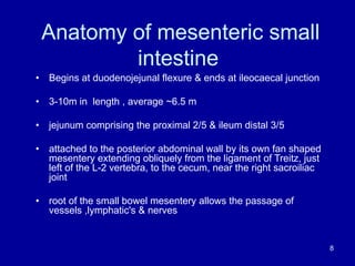
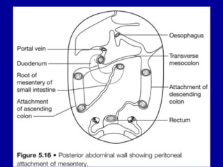
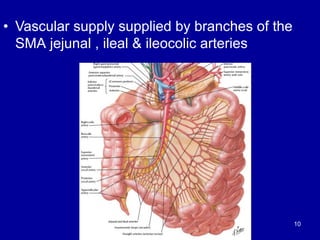
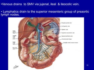
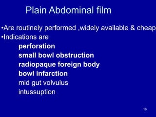
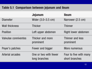
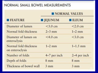
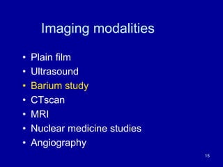
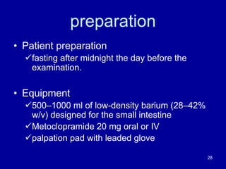
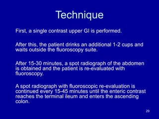
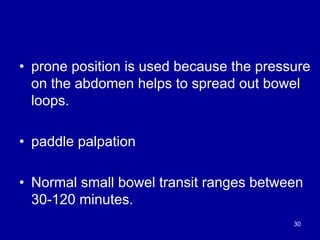
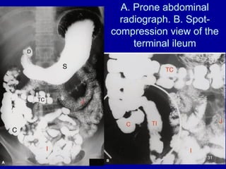
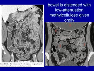
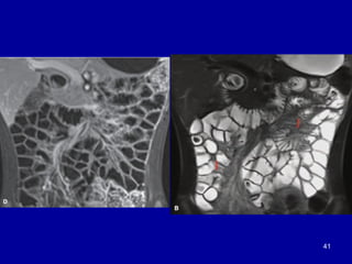
Radiologic anatomy of
Mesenteric small bowel
Presenter: Dr. Abduljelil N (R1)
. 1
OUTLINE
•Embryology of small bowel
•Anatomy of the small bowel
•Imaging modalities of small bowel
•References
2
Development of the small bowel
• Development of the midgut is
characterized by rapid elongation of the
gut and its mesentery, resulting in
formation of the primary intestinal loop
• physiological umbilical herniation
starting at sixth week
3
4
• Coincident with growth in length,the primary intestinal loop
rotates around an axis formed by the SMA
• Rotation occurs during herniation (about 90◦) as well as during
return of the intestinal loops into the abdominal cavity
(remaining 180◦)
• During the 10th week, herniated intestinal loops begin to return
to the abdominal cavity
5
6
7
Anatomy of mesenteric small
intestine
• Begins at duodenojejunal flexure & ends at ileocaecal junction
• 3-10m in length , average ~6.5 m
• jejunum comprising the proximal 2/5 & ileum distal 3/5
• attached to the posterior abdominal wall by its own fan shaped
mesentery extending obliquely from the ligament of Treitz, just
left of the L-2 vertebra, to the cecum, near the right sacroiliac
joint
• root of the small bowel mesentery allows the passage of
vessels ,lymphatic's & nerves
8
9
• Vascular supply supplied by branches of the
SMA jejunal , ileal & ileocolic arteries
10
•Venous drains to SMV via jujenal, ileal & ileocolic vein.
• Lymphatics drain to the superior mesenteric group of preaortic
lymph nodes.
11
12
13
14
Imaging modalities
• Plain film
• Ultrasound
• Barium study
• CTscan
• MRI
• Nuclear medicine studies
• Angiography
15
Plain Abdominal film
•Are routinely performed ,widely available & cheap
•Indications are
perforation
small bowl obstruction
radiopaque foreign body
bowl infarction
mid gut volvulus
intussuption
16
Gas pattern
17
Normal fluid levels
18
Small bowel
Central
Plicae circularis
Pliability(“bent finger”
appearance)
19
Large bowel
haustra
peripherally
stippled appearance of fecal
matter
20
Ultrasound
•Gas content within the gut lumen can make visibility difficult
• allows assessement of content , diameter & motility of GIT
•Ultrasound is superior to both CT & MRI for resolution of the gut wall
layers
•Normal gut is compressible and gas pockets displaced away
from the region of interest
• thickened abnormal bowel loops are non compressible &
remain unchanged
•The normal gut wall is uniform with an average thickness of
3mm if distended & 5mm if not distended
21
22
23
24
Barium study
• Barium studies remain the cornerstone of small bowel
imaging
• And still provide the best radiological assessment
when subtle alterations of mucosal morphology are
being sought
• Provide information about small bowel caliber , its
disposition , the wall thickness , and distribution of the
valvulae conniventes. 25
SMALL BOWEL FOLLOW-THROUGH
single contrast examination of the esophagus,
stomach, and small intestine
Indications
ü strictures
ü Partial obstruction
ü diverticula
ü masses
ü extraluminal tethering
ü Malabsorption 26
Contraindications
1.Complete or high-grade obstruction.
üThis is usually better evaluated by CT
examination (without oral contrast) using the
intraluminal fluid caused by the obstruction as
a natural contrast agent.
2.Suspected perforation (unless a water-
soluble contrast medium is used).
27
preparation
• Patient preparation
üfasting after midnight the day before the
examination.
• Equipment
ü500–1000 ml of low-density barium (28–42%
w/v) designed for the small intestine
üMetoclopramide 20 mg oral or IV
üpalpation pad with leaded glove
28
Technique
First, a single contrast upper GI is performed.
After this, the patient drinks an additional 1-2 cups and
waits outside the fluoroscopy suite.
After 15-30 minutes, a spot radiograph of the abdomen
is obtained and the patient is re-evaluated with
fluoroscopy.
A spot radiograph with fluoroscopic re-evaluation is
continued every 15-45 minutes until the enteric contrast
reaches the terminal ileum and enters the ascending
colon.
29
• prone position is used because the pressure
on the abdomen helps to spread out bowel
loops.
• paddle palpation
• Normal small bowel transit ranges between
30-120 minutes.
30
A. Prone abdominal
radiograph. B. Spot-
compression view of the
terminal ileum
31
Per-oral pneumocolon
• technique that can be used during SBFT
to better visualize the ascending colon and
terminal ileum
• gas (ideally CO2) is insufflated into the
colon through the rectum in the majority of
patients(85-90%), the gas refluxes through
the ileocecal valve into the terminal ileum
• to create a double contrast
32
Limitations of the SBFT
• overlap of bowel loops,
• poor distension,
• flocculation of barium,
• intermittent barium filling, and
• unpredictable transit time
– little control over the degree of small bowel
filling and distention
– less severe strictures and small masses may
be difficult to see 33
Small bowel enema (enteroclysis )
• The study is performed by passing a specially
designed 12 to 14 French enteroclysis catheter
through the mouth or nose and into the distal
duodenum or proximal jejunum
• performed in one of three main ways:
1.single-contrast enteroclysis: low density barium (20-
40% w/v),volume of 600-1200 mL at an initial rate of
75 mL/min
2.air-contrast enteroclysis: medium density barium
(40-80% w/v),volume of 300-600 mL
ü Room air or CO2 is introduced via a pump when the barium
column reaches the distal small bowel
34
3. methylcellulose enteroclysis: high density
barium (80% w/v),
ü volume of 220-300 mL of barium infused at 60-80
mL through a syringe until half of the expected
intestinal loops are visualized.
ü 1000 mL of Methylcellulose is instilled through an
electric pump
ü Enteric contrast coats the bowel wall and
methylcellulose distends the small bowel
35
enteroclysis cont..
Advantage
– Is more sensitive study for detail small bowel examination
– provides more uniform distension of the bowel, even
distribution of barium, superior anatomic detail, and shorter
overall examination time
Disadvantage
– substantial discomfort associated with tube placement
– Patients preparation is extensive, requires jejunal
intubation, laxative,???sedation
– Need skilled radiologist for intubation 36
37
CT and MRI enterography
• Improves up on barium study by demonstrating extra luminal
compartment, mesentery, solid organs, peritoneum &
retroperitonium
• MRE is used mostly in the initial diagnosis and follow-up of
inflammatory bowel disease; CTE is used for small bowel
lesions
• Contrast administration can be performed perorally (i.e., CT /
MR enterography) or by infusion via nasoenteric intubation
(i.e., CT/MR enteroclysis)
• Neutral or positive oral contrast can be used
38
CT enterography cont..
• Patients drink approximately 1.5–2 L of oral contrast over
45–60 min
• 150 ml of Iohexol (Omnipaque 300) administered intravenously
• Neutral or low-density oral contrast media are a prerequisite
for good-quality CT enterography
ü Water–methylcellulose solution,
ü lactulose solution,
ü polyethylene glycol,
ü low-density barium, 0.1% w/v Volumen and
ü milk
39
bowel is distended with
low-attenuation
methylcellulose given
orally
40
41
Nuclear medicine studies
• are useful alternative and adjunctive methods in
the investigation of small bowel pathology
üinflammatory bowel disease
ülocalisation of intestinal bleeding
üMeckel’s diverticulum
üCarcinoid tumours of the small bowel 42
Radionuclide scintigraphy, using
99mTc-pertechnetate
43
Angiography
• to detect the site of bleeding
• vascular malformation
• mesentric ischeamia
44
References
• Langman’s Medical Embryology 8 th ed
• Applied Radiologic anatomy 1st ed, published 1999
• Applied Radiologic anatomy 2nd ed, published 2012
• Fundamentals of diagnostic radiology 4th ed ,published 2012
• Practical fluoroscopy of GI & GU Tracts First ,published 2012
• Anatomy for Diagnositc Imaging 3rd edition ,published © 2011
• Grainger and Allison`s Diagnostic Radiology 6th ed,published
© 2015
• CAROL DIAGNOSTIC ULTRASOUND 5th ed,published © 2018
45
Thank you
46

radiological anatomy of Small intestine abdul final

  • 1.
    Radiologic anatomy of Mesentericsmall bowel Presenter: Dr. Abduljelil N (R1) . 1
  • 2.
    OUTLINE •Embryology of smallbowel •Anatomy of the small bowel •Imaging modalities of small bowel •References 2
  • 3.
    Development of thesmall bowel • Development of the midgut is characterized by rapid elongation of the gut and its mesentery, resulting in formation of the primary intestinal loop • physiological umbilical herniation starting at sixth week 3
  • 4.
  • 5.
    • Coincident withgrowth in length,the primary intestinal loop rotates around an axis formed by the SMA • Rotation occurs during herniation (about 90◦) as well as during return of the intestinal loops into the abdominal cavity (remaining 180◦) • During the 10th week, herniated intestinal loops begin to return to the abdominal cavity 5
  • 6.
  • 7.
  • 8.
    Anatomy of mesentericsmall intestine • Begins at duodenojejunal flexure & ends at ileocaecal junction • 3-10m in length , average ~6.5 m • jejunum comprising the proximal 2/5 & ileum distal 3/5 • attached to the posterior abdominal wall by its own fan shaped mesentery extending obliquely from the ligament of Treitz, just left of the L-2 vertebra, to the cecum, near the right sacroiliac joint • root of the small bowel mesentery allows the passage of vessels ,lymphatic's & nerves 8
  • 9.
  • 10.
    • Vascular supplysupplied by branches of the SMA jejunal , ileal & ileocolic arteries 10
  • 11.
    •Venous drains toSMV via jujenal, ileal & ileocolic vein. • Lymphatics drain to the superior mesenteric group of preaortic lymph nodes. 11
  • 12.
  • 13.
  • 14.
  • 15.
    Imaging modalities • Plainfilm • Ultrasound • Barium study • CTscan • MRI • Nuclear medicine studies • Angiography 15
  • 16.
    Plain Abdominal film •Areroutinely performed ,widely available & cheap •Indications are perforation small bowl obstruction radiopaque foreign body bowl infarction mid gut volvulus intussuption 16
  • 17.
  • 18.
  • 19.
  • 20.
  • 21.
    Ultrasound •Gas content withinthe gut lumen can make visibility difficult • allows assessement of content , diameter & motility of GIT •Ultrasound is superior to both CT & MRI for resolution of the gut wall layers •Normal gut is compressible and gas pockets displaced away from the region of interest • thickened abnormal bowel loops are non compressible & remain unchanged •The normal gut wall is uniform with an average thickness of 3mm if distended & 5mm if not distended 21
  • 22.
  • 23.
  • 24.
  • 25.
    Barium study • Bariumstudies remain the cornerstone of small bowel imaging • And still provide the best radiological assessment when subtle alterations of mucosal morphology are being sought • Provide information about small bowel caliber , its disposition , the wall thickness , and distribution of the valvulae conniventes. 25
  • 26.
    SMALL BOWEL FOLLOW-THROUGH singlecontrast examination of the esophagus, stomach, and small intestine Indications ü strictures ü Partial obstruction ü diverticula ü masses ü extraluminal tethering ü Malabsorption 26
  • 27.
    Contraindications 1.Complete or high-gradeobstruction. üThis is usually better evaluated by CT examination (without oral contrast) using the intraluminal fluid caused by the obstruction as a natural contrast agent. 2.Suspected perforation (unless a water- soluble contrast medium is used). 27
  • 28.
    preparation • Patient preparation üfastingafter midnight the day before the examination. • Equipment ü500–1000 ml of low-density barium (28–42% w/v) designed for the small intestine üMetoclopramide 20 mg oral or IV üpalpation pad with leaded glove 28
  • 29.
    Technique First, a singlecontrast upper GI is performed. After this, the patient drinks an additional 1-2 cups and waits outside the fluoroscopy suite. After 15-30 minutes, a spot radiograph of the abdomen is obtained and the patient is re-evaluated with fluoroscopy. A spot radiograph with fluoroscopic re-evaluation is continued every 15-45 minutes until the enteric contrast reaches the terminal ileum and enters the ascending colon. 29
  • 30.
    • prone positionis used because the pressure on the abdomen helps to spread out bowel loops. • paddle palpation • Normal small bowel transit ranges between 30-120 minutes. 30
  • 31.
    A. Prone abdominal radiograph.B. Spot- compression view of the terminal ileum 31
  • 32.
    Per-oral pneumocolon • techniquethat can be used during SBFT to better visualize the ascending colon and terminal ileum • gas (ideally CO2) is insufflated into the colon through the rectum in the majority of patients(85-90%), the gas refluxes through the ileocecal valve into the terminal ileum • to create a double contrast 32
  • 33.
    Limitations of theSBFT • overlap of bowel loops, • poor distension, • flocculation of barium, • intermittent barium filling, and • unpredictable transit time – little control over the degree of small bowel filling and distention – less severe strictures and small masses may be difficult to see 33
  • 34.
    Small bowel enema(enteroclysis ) • The study is performed by passing a specially designed 12 to 14 French enteroclysis catheter through the mouth or nose and into the distal duodenum or proximal jejunum • performed in one of three main ways: 1.single-contrast enteroclysis: low density barium (20- 40% w/v),volume of 600-1200 mL at an initial rate of 75 mL/min 2.air-contrast enteroclysis: medium density barium (40-80% w/v),volume of 300-600 mL ü Room air or CO2 is introduced via a pump when the barium column reaches the distal small bowel 34
  • 35.
    3. methylcellulose enteroclysis:high density barium (80% w/v), ü volume of 220-300 mL of barium infused at 60-80 mL through a syringe until half of the expected intestinal loops are visualized. ü 1000 mL of Methylcellulose is instilled through an electric pump ü Enteric contrast coats the bowel wall and methylcellulose distends the small bowel 35
  • 36.
    enteroclysis cont.. Advantage – Ismore sensitive study for detail small bowel examination – provides more uniform distension of the bowel, even distribution of barium, superior anatomic detail, and shorter overall examination time Disadvantage – substantial discomfort associated with tube placement – Patients preparation is extensive, requires jejunal intubation, laxative,???sedation – Need skilled radiologist for intubation 36
  • 37.
  • 38.
    CT and MRIenterography • Improves up on barium study by demonstrating extra luminal compartment, mesentery, solid organs, peritoneum & retroperitonium • MRE is used mostly in the initial diagnosis and follow-up of inflammatory bowel disease; CTE is used for small bowel lesions • Contrast administration can be performed perorally (i.e., CT / MR enterography) or by infusion via nasoenteric intubation (i.e., CT/MR enteroclysis) • Neutral or positive oral contrast can be used 38
  • 39.
    CT enterography cont.. •Patients drink approximately 1.5–2 L of oral contrast over 45–60 min • 150 ml of Iohexol (Omnipaque 300) administered intravenously • Neutral or low-density oral contrast media are a prerequisite for good-quality CT enterography ü Water–methylcellulose solution, ü lactulose solution, ü polyethylene glycol, ü low-density barium, 0.1% w/v Volumen and ü milk 39
  • 40.
    bowel is distendedwith low-attenuation methylcellulose given orally 40
  • 41.
  • 42.
    Nuclear medicine studies •are useful alternative and adjunctive methods in the investigation of small bowel pathology üinflammatory bowel disease ülocalisation of intestinal bleeding üMeckel’s diverticulum üCarcinoid tumours of the small bowel 42
  • 43.
  • 44.
    Angiography • to detectthe site of bleeding • vascular malformation • mesentric ischeamia 44
  • 45.
    References • Langman’s MedicalEmbryology 8 th ed • Applied Radiologic anatomy 1st ed, published 1999 • Applied Radiologic anatomy 2nd ed, published 2012 • Fundamentals of diagnostic radiology 4th ed ,published 2012 • Practical fluoroscopy of GI & GU Tracts First ,published 2012 • Anatomy for Diagnositc Imaging 3rd edition ,published © 2011 • Grainger and Allison`s Diagnostic Radiology 6th ed,published © 2015 • CAROL DIAGNOSTIC ULTRASOUND 5th ed,published © 2018 45
  • 46.